Acridine Orange Staining Kit - BioReagent, Biological Stain, for microscopy, high purity

Cat. No.: A1456513
AVAILABLE TO ORDER
GRADE & PURITY BioReagent ? BioReagent grade — tested suitable for life-science and molecular-biology use. Use for cell culture, assays, and biochemical work needing biological compatibility. Biological Stain ? Biological stain grade — dyes characterized for staining cells and tissues. Use in histology and microscopy where staining consistency matters. for Microscopy ? Microscopy grade — reagents/stains suited to sample prep and imaging. Use in microscopy where clarity and low background are needed.
★
Size
Germany (EU)
USA*
Price
Qty
100T
A1456513-100T
Made to order · 8–12 wks
€17.27
500T
A1456513-500T
Made to order · 8–12 wks
€60.65
Enter a quantity for the sizes you want to add.
🧪
Why this grade

BioReagent, Biological Stain, for microscopy Biological Stain,BioReagent,for Microscopy for sensitive chromatographic and analytical workflows requiring minimal baseline interference.

🌡

Storage & shipping

Store at 2-8°C,Protected from light,Do not freeze Ships Wet ice,Do not freeze Check lot-specific COA for exact specifications.

📋

Quality documents

SDS, COA, datasheet, and spec sheet available for download. Lot-specific COA accessible via lot number lookup.

📚

Literature proof

Cited in 0 peer-reviewed publications across chromatography, organic synthesis, and cross-coupling reactions.

Overview

Acridine Orange (AO) is a metachromatic fluorescent dye whose emission color varies depending on the target it binds to:

l   When binding to double-stranded DNA: It intercalates between base pairs and emits green fluorescence upon excitation (Ex 488 nm, Em 530 nm).

l   When binding to single-stranded RNA or lysosomes: It attaches via electrostatic interactions and emits orange-red fluorescence (Em>640 nm).

Under a fluorescence microscope, Acridine Orange permeates the membranes of normal cells, staining the nucleus with uniform green or yellow-green fluorescence. In apoptotic cells, due to chromatin condensation and fragmentation into apoptotic bodies, AO stains them with intense, condensed yellow-green fluorescence or fragmented yellow-green particles. In necrotic cells, the yellow-green fluorescence is reduced or absent.

Acridine Orange is often used in combination with Propidium Iodide (PI) for dual staining. Since PI stains only dead cells, producing orange-red fluorescence, this method allows differentiation among normal, apoptotic, and necrotic cells.

Components

A1456513

Component

100 Test

500 Test

Storage Condition

Quantity Per Test

A1456513A

AO Dilution Buffer

10 mL

50 mL

2-8℃

0.1 mL per  0.5-1.0 × 10⁶  cells

A1456513B

AO Staining Solution

100 μL

500 μL

2-8℃, Protect from light. Do not freeze

1 μL per  0.5-1.0 × 10⁶  cells

Note: The recommended number of cells to stain per test is  0.5-1.0 × 10⁶ cells.

Procedure

1. Preparation of Acridine Orange Staining Solution

   Mix the AO Staining Solution with the Dilution Buffer at a ratio of 1:1000 to prepare the working solution. For example, add 10 μL of AO Staining Solution to 10 mL of Dilution Buffer to obtain 10 mL of Acridine Orange staining solution. 

2. Staining with Acridine Orange

a. For adherent cells: 

(a) Gently aspirate the culture medium from the plate. Rinse with PBS for about 10 seconds, then remove PBS. 

(b) Add Acridine Orange staining solution and incubate at room temperature for 5 minutes. Remove the staining solution and rinse with PBS for about 10 seconds. Repeat the rinse once. 

Note: For adherent cells cultured in a 6-well plate with a confluence exceeding 80%, it is recommended to add the staining working solution at a volume of 1 mL per well. This volume can be optimized based on the specific experimental system.

(c) Add an appropriate amount of cell culture medium, staining buffer, or other suitable solution to cover the well bottom. Observe under a microscope. Depending on the detection requirements, green fluorescence can be observed at Ex/Em = 488/530 nm, and red fluorescence can be observed at Ex/Em = 540/640 nm. Alternatively, measure fluorescence intensity using a fluorescence microplate reader with bottom-reading capability.

b. For suspension cells: 

(a) Take 1 mL of cell suspension. Centrifuge at 500×g for 5 minutes at room temperature. Gently aspirate the medium, resuspend in PBS, and centrifuge again at  500×g for 5 minutes. Remove PBS. 

(b) Add an appropriate amount of Acridine Orange staining solution to achieve a cell density of approximately 10⁶ cells/mL .

(c) Incubate at room temperature for 5 minutes. 

(d) A drop of the sample was directly applied onto a glass slide, covered with a coverslip, and examined under a microscope. Depending on the detection requirements, green fluorescence can be observed at Ex/Em = 488/530 nm, and red fluorescence can be observed at Ex/Em = 540/640 nm. Alternatively, after staining, analyze directly by flow cytometry or measure fluorescence with a microplate reader.

Note: Centrifugation to remove staining solution can reduce background fluorescence. For suspension cells or adherent cells in suspension, consider reducing the AO staining solution concentration by 2–5 times and shortening the staining time to 2 minutes.

Precautions

1. AO Staining Solution is toxic. Handle with care. 

2. For your safety and health, wear a lab coat and disposable gloves. 

3. Fluorescent dyes are susceptible to quenching. It is recommended to complete detection on the same day after staining.

Specifications

Synonyms
AO Stain kit | Cell Apoptosis Acridine Orange Detection Kit
Specifications & Purity
BioReagent, Biological Stain, for microscopy
Grade
Biological Stain, BioReagent, for Microscopy
Note
This product can replace A774791.
Application
Flow Cytometry, for microscopy
Sensitivity
Light-sensitive
Storage and Shipping
Storage
Store at 2-8°C,Protected from light,Do not freeze
Shipped In
Wet ice,Do not freeze
Stability And Storage
store at 2-8°C long term (12 months). Store in the dark. Do not freeze.
Images

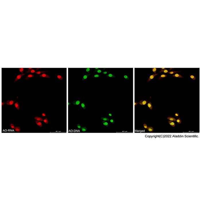
Acridine Orange Staining Kit (A1456513)
HeLa cells were stained using the Acridine Orange Staining Kit (A1456513), with the working solution diluted at a ratio of 1/1000 prior to application. Following 10 minutes of staining, the cells were observed under laser scanning confocal microscopy.
Fluorescence micrograph of acridine orange (AO)-stained HeLa cells captured under laser scanning confocal microscopy. The distinct metachromatic emission of AO is observed: nuclei exhibit green fluorescence (Ex=488 nm, Em=500-550 nm) due to AO intercalation into double-stranded DNA, while nucleoli and cytoplasm exhibit red fluorescence (Em=640-700 nm) due to AO stacking on single-stranded RNA. The yellow-orange regions in the merged image result from the spatial overlap of the green fluorescence from the nucleoplasm and the intense red fluorescence from the RNA-rich nucleoli.

Documentation

📋 Safety Data Sheet (SDS)

Comprehensive hazard, handling, storage, and regulatory compliance document.

Download SDS →

✅ Certificate of Analysis (COA)

Lot-specific quality data. Enter your lot number to retrieve the exact COA.

Look up COA →

📊 Datasheet

Quick-reference summary of product specifications and applications.

View datasheet →

🔬 Specification Sheet

Full quality attributes and acceptance criteria for this grade.

View spec sheet →

Advanced Data

Certificates(CoA,COO,BSE/TSE and Analysis Chart)
C of A & Other Certificates(BSE/TSE, COO):
Analytical Chart:

Find and download the COA for your product by matching the lot number on the packaging.

2 results found

Lot NumberCertificate TypeDateItem
ZJ26F0838347Certificate of AnalysisAug 21, 2026 A1456513
ZJ26F0838346Certificate of AnalysisAug 21, 2026 A1456513
FAQs and Articles
Solution Calculators
Reviews

Customer Reviews

Application Protocols

The kit’s exact components and working concentrations are Not specified for this item; refer to CoA/Spec Sheet and the kit insert for authoritative instructions. Below are general, literature-based protocols to aid planning. Validate and adapt to the provided kit.

A. Live-cell nucleic acid staining (general)

  • Prepare working solution in serum-free PBS or HBSS (protect from light).
  • Incubate cells 5–20 min at RT or 37°C; avoid CO2-dependent pH drift.
  • Wash 2–3× with dye-free buffer.
  • Image promptly. Typical filter sets: excitation 470–500 nm; collect green (520–540 nm) and orange/red (600–650 nm).

B. Fixed-cell/tissue sections (general)

  • Fix specimen (e.g., paraformaldehyde or alcohol; fixation method affects staining pattern). Rinse thoroughly.
  • Incubate in working dye solution for 5–15 min at RT (protected from light).
  • Wash 2–3×; mount in aqueous medium; image as above.

C. Bacterial total counts (general)

  • Deposit sample on filter or slide; air dry.
  • Stain briefly (1–5 min), rinse, and image under epifluorescence.

Controls and optimization

  • Include unstained and single-channel controls.
  • Titrate dye concentration and time to balance signal/background and minimize phototoxicity.

Note: Dispose of dye solutions as hazardous waste. Always follow the kit insert where it differs from these general guidelines.

Biological Roles

Acridine orange itself does not play a physiological role; it is an exogenous small-molecule fluorophore used to probe cellular components. The following describes its interaction biology (literature/general):

  • Nucleic acid binding:
    • Intercalates between base pairs in double-stranded DNA, enhancing green fluorescence. Associates electrostatically with single-stranded nucleic acids, often producing orange-red emission due to aggregation/metachromasia.
  • pH-dependent behavior:
    • As a cationic weak base, it accumulates in acidic organelles (lysosomes, late endosomes). Protonation and high local concentration promote red-shifted emission.
  • Chromatin and RNA contrast:
    • Differential emission enables qualitative assessment of chromatin condensation (green) versus RNA-rich cytoplasm (orange-red) in microscopy.
  • Photophysics in cells:
    • The dye’s quantum yield and emission color are sensitive to microenvironment (viscosity, pH, binding mode), which can be exploited in ratiometric imaging but requires careful calibration.

Considerations for biological experiments:

  • Cytotoxicity/phototoxicity risk increases with dose and illumination; minimize concentration and exposure time.
  • Efflux transporters can reduce intracellular accumulation in some cell types; incubation conditions may require optimization.
  • Fixation/permeabilization steps can alter distribution and metachromatic response; validate for fixed vs live protocols separately.

Note: All uses are for research only; no diagnostic or therapeutic claims are made.

Buffer Applications

Buffer choice strongly influences acridine orange staining performance due to pH- and ionic-strength–dependent binding and emission.

General guidance (literature/practice):

  • Neutral physiological buffers (e.g., PBS, HBSS):
    • Typical for live-cell DNA/RNA contrast; maintain near-neutral pH (7.2–7.4) and isotonicity. Avoid serum during staining to reduce background.
  • Slightly acidic buffers (pH 4–5):
    • Can emphasize red emission in acidic vesicles. Use only if your experimental goal is to highlight lysosomes; prolonged exposure at low pH can stress cells.
  • Tris vs phosphate:
    • Tris can weakly interact with cationic dyes; PBS often yields more consistent metachromatic behavior.
  • Ionic strength:
    • Excess salt can screen electrostatic interactions, decreasing cytoplasmic/RNA signal. Maintain standard physiological ionic strength unless protocol indicates otherwise.
  • Detergents and alcohols:
    • Even low levels of nonionic detergents or alcohols can alter permeability and distribution. Reserve for controlled permeabilization/fixation steps, followed by thorough buffer washes.

Practical tips:

  • Filter buffers (0.22 µm) to reduce particulates and background.
  • Warm buffers to ambient/37°C (as appropriate) to maintain cell physiology in live-cell workflows.
  • Always match the pH/ionic strength of wash buffers to staining buffer to minimize dye redistribution during rinsing.

Item-specific buffer recipes and working concentrations: Not specified for this item; refer to CoA/Spec Sheet.

Green Alternatives

Acridine orange is a DNA/RNA-interactive dye and should be handled as a potential mutagen. Depending on your application, safer or greener alternatives may be considered.

Potential alternatives (literature/general):

  • Gel and nucleic acid stains (in vitro):
    • SYBR Safe, GelGreen/GelRed (lower mutagenicity profiles than ethidium bromide; tailored for gels, some used in cells with caution).
  • Live-cell DNA dyes:
    • Hoechst 33342 or DAPI (cell-permeant/impermeant blue DNA dyes; not metachromatic but widely used; note UV excitation and potential cytotoxicity with prolonged exposure).
  • Lysosomal probes:
    • LysoTracker series (acidic compartment labeling with defined spectra; designed for live-cell imaging).

Comparison highlights (general):

| Criterion | Acridine Orange | Hoechst/DAPI | SYBR Safe/GelGreen | |---|---|---|---| | Emission versatility | Green (DNA) + orange/red (RNA/acidic) | Blue only | Green (primarily gels) | | Excitation | Blue-green (488–500 nm) | UV/violet (350–405 nm) | Blue (470–500 nm) | | Mutagenicity concern | Intercalator; handle as potential mutagen | DNA-binding; caution advised | Engineered for reduced hazard vs EtBr | | Live-cell compatibility | Yes, with optimization | Often yes (Hoechst 33342) | Primarily gels; limited live-cell use |

Trade-offs:

  • AO provides unique metachromatic contrast not replicated by many “safer” dyes. Alternatives may offer improved safety profiles but reduced functional readouts. Select based on spectral needs, live-cell compatibility, and institutional safety policies.
Pharmaceutical Uses

This product is a research-use-only microscopy staining kit and is not intended for pharmaceutical formulation or clinical use.

Context (general information):

  • Acridine orange and related dyes are laboratory reagents used to visualize nucleic acids and acidic organelles in research settings.
  • They are not typical excipients in drug products and are not included in pharmacopeial monographs for formulation use.

Implications for users in regulated environments:

  • For any use in GMP-adjacent analytics (e.g., cell bank characterization), ensure appropriate qualification and method validation with this specific lot.
  • Documentation such as CoA and SDS should be retained for audit trails.

Item-specific regulatory status, pharmacopeial references, and acceptance criteria: Not specified for this item; refer to CoA/Spec Sheet.

Physical Properties

Item-specific physicochemical specifications for this kit are not provided. Consult the CoA/Spec Sheet for exact values relevant to the supplied reagents.

  • Appearance: Not specified for this item; refer to CoA/Spec Sheet.
  • Concentration(s) and composition of solutions: Not specified for this item; refer to CoA/Spec Sheet.
  • pH of working solutions/buffers: Not specified for this item; refer to CoA/Spec Sheet.

Literature/general properties of acridine orange dyes (for method planning only; not product specifications):

  • Solubility: Readily soluble in water and aqueous buffers when provided as a protonated salt; soluble in polar organics (e.g., methanol, ethanol).
  • Acid–base: Cationic dye; fluorescence and binding behavior are pH-dependent due to protonation and aggregation.
  • Photophysics (typical, bound-state; literature):
    • Excitation maxima: ~460–502 nm (blue-green excitation)
    • Emission: DNA-bound green emission ~520–535 nm; RNA/single-stranded/acidic compartment aggregated state orange-red ~620–650 nm (metachromasia)
  • Light sensitivity: Photo-bleaches under strong illumination; protect from light during handling.

Note: Do not treat the above as kit specifications. Actual solvent, pH, concentration, and stabilizers (if any) for the supplied reagents are Not specified for this item; refer to CoA/Spec Sheet.

Quality and Grades
  • Grade/Purity: BioReagent, Biological Stain, for Microscopy

What this implies for this item:

  • BioReagent: Produced with attention to impurities that could interfere with biological assays; suitable for research workflows but not intended for clinical/diagnostic use.
  • Biological Stain, for Microscopy: The reagents are intended for fluorescent staining of biological specimens (cells, tissues, microorganisms) and to provide consistent optical performance under microscopy.

Interpretation and best practices:

  • Low background: Biological-stain grade typically emphasizes minimized fluorescent impurities to reduce background.
  • Batch-to-batch consistency: Critical for qualitative/quantitative imaging; always verify with a control sample when opening a new lot.
  • Documentation: Certificates of Analysis (CoA) usually report appearance and performance checks (e.g., staining quality on reference samples). Item-specific acceptance criteria are Not specified for this item; refer to CoA/Spec Sheet.
  • Stabilization: If stabilizers, preservatives, or surfactants are present in the kit, their identity/levels are Not specified for this item; refer to CoA/Spec Sheet. Their presence can influence compatibility with live vs fixed cells.

Recommendation:

  • For quantitative imaging or flow cytometry, validate instrument settings and spectral overlap using the same lot. For chromatographic or spectroscopy QC, verify absorbance/emission profiles against your method’s reference standards.
Reaction and Applications

As a microscopy staining kit, this product is not used as a reagent in synthetic organic reactions. Its primary applications are analytical/biological staining. Below are common research uses of acridine orange (literature/practice; not item-specific):

  • Nucleic acid staining in cells, tissues, and microorganisms:
    • Intercalates into double-stranded DNA (green emission) and associates with single-stranded DNA/RNA or aggregates in acidic organelles (orange-red emission), enabling ratiometric contrast.
  • Assessment of cell status:
    • Qualitative visualization of chromatin condensation, RNA content, or acidic vesicular organelles in cell biology experiments.
  • Microscopy modalities:
    • Widefield epifluorescence (FITC/GFP-like excitation for green channel; TRITC/Cy3-like for red). Confocal and flow cytometry are also commonly used with AO (spectral settings must be validated).
  • Microbiology:
    • Rapid staining of bacteria and protozoa for total cell counts under fluorescence microscopy.

Practical tips (general):

  • Protect from light throughout preparation and staining; minimize exposure to intense excitation to reduce photobleaching.
  • Control samples: Include unstained and single-color controls to set detector gains and compensate spectral overlap.
  • Rinses: Thorough washing reduces unbound dye and background; use dye-free buffer of the same ionic strength.
  • Concentration/time: Optimize empirically for specimen type; over-staining increases background and can obscure metachromatic contrast.
Reaction Conditions

Not applicable to chemical synthesis. However, for experimental planning, the following operational conditions relate to staining performance (literature/practice; not item-specific specifications):

  • Temperature: Room temperature to 37°C commonly used for live cells; room temperature for fixed specimens. Elevated temperatures can alter uptake and photophysics.
  • Incubation time: Typically minutes to tens of minutes; optimize to balance signal intensity and background.
  • Illumination: Excite with blue-green light (e.g., 488–500 nm). Minimize exposure duration and intensity to limit photobleaching/phototoxicity.
  • pH: Neutral buffers for general DNA/RNA contrast; mildly acidic conditions accentuate lysosomal red emission.
  • Washes: Multiple brief rinses in dye-free buffer reduce non-specific background.

Expected outcomes (qualitative):

  • Green nuclear emission associated with dsDNA; orange-red cytoplasmic/lysosomal signal reflecting RNA/acidic compartments. Relative intensities vary with cell type, fixation, and buffer.

Item-specific incubation times, concentrations, and buffer formulations: Not specified for this item; refer to CoA/Spec Sheet.

Safety and Handling

Follow standard practices for handling fluorescent nucleic-acid dyes and microscopy stains. Acridine dyes can intercalate with DNA/RNA and should be handled as potential mutagens.

Item-specific hazard data:

  • Signal word: Not specified for this item; refer to SDS.
  • GHS classification/pictograms: Not specified for this item; refer to SDS.
  • H-statements: Not specified for this item; refer to SDS.

General safety guidance (literature/practice; defer to SDS for authority):

  • PPE: Lab coat, nitrile gloves, safety glasses; consider double-gloving and splash protection when preparing stock solutions.
  • Engineering controls: Use a chemical fume hood for weighing/dilution to minimize aerosol exposure; avoid UV/blue light exposure to reduce photodegradation.
  • Avoid: Skin/eye contact; inhalation/ingestion; contamination of benches/instruments (dyes stain surfaces persistently).
  • Incompatibilities: Strong oxidizers may degrade the dye; avoid high-pH hypochlorite in presence of organics. Store away from light and reducing/oxidizing agents.
  • First aid (overview):
    • Skin: Immediately wash with soap and water; remove contaminated clothing.
    • Eyes: Rinse cautiously with water for several minutes; seek medical attention.
    • Inhalation: Move to fresh air; seek medical attention if symptoms occur.
    • Ingestion: Rinse mouth; do not induce vomiting; seek medical attention.
  • Waste: Collect dye-containing liquids and disposables as hazardous chemical waste; do not discharge to drain.

Always consult the product-specific SDS for authoritative safety and regulatory information.

Solvent Selection

This kit is designed for aqueous staining workflows.

  • Primary medium: Aqueous buffers (e.g., PBS, HBSS) are typically used for dilution, incubation, and washes. Item-specific buffer composition is Not specified for this item; refer to CoA/Spec Sheet.
  • Polarity/miscibility (general): Acridine orange salts are highly water-soluble; solutions are also miscible with lower alcohols (methanol/ethanol). High organic content can alter cell permeability and staining patterns.

Practical selection guidance (general):

  • Live-cell staining: Use physiological, serum-free buffered saline at neutral pH to minimize background and maintain viability. Avoid organic cosolvents and detergents unless validated.
  • Fixed-cell/tissue staining: Alcohols (e.g., 70% ethanol) may be used in fixation steps; subsequent staining is usually in aqueous buffer to preserve metachromatic emission behavior.
  • Acidic compartment labeling (lysosomes): Slightly acidic buffers (pH ~4.0–5.0) can accentuate red-shifted emission due to protonation/aggregation; adjust only if your protocol specifies.
  • Avoid: Strongly basic buffers (>pH 9) that can reduce cationic binding; oxidizing rinse solutions that can quench fluorescence.

Comparison (general):

  • PBS vs Tris: Tris may weakly interact with some dyes and can alter apparent pKa; PBS is often preferred for reproducibility.
  • Serum/proteins: Binding to proteins can increase background; consider BSA-free buffers during staining and introduce serum only after final washes.
Storage and Reconstitution

Item-specific storage and shipping conditions:

  • Storage conditions: Store at 2–8°C, protected from light, do not freeze.
  • Shipped in: Wet ice, do not freeze.

Reconstitution and preparation:

  • Reconstitution protocol (if applicable): Not specified for this item; refer to CoA/Spec Sheet.
  • If supplied as a concentrate or solid, prepare working solutions using high-purity water or appropriate buffer; filter if necessary (0.22 µm). Since the exact form is not specified, defer to the kit insert.

Handling and stability (general guidance):

  • Allow components to equilibrate to room temperature before opening to minimize condensation; return promptly to 2–8°C after use.
  • Protect all components and working solutions from light (use amber vials/foil wrap).
  • Avoid repeated freeze–thaw; aliquot into light-protected microtubes if frequent use is expected.
  • Label aliquots with opening date and lot number; monitor for precipitation, color change, or loss of fluorescence as signs of degradation.

Shelf life:

  • Item-specific expiry and retest periods are Not specified for this item; refer to CoA/Spec Sheet and the product label.
Structure and Identity

This product is a ready-to-use staining kit built around the fluorescent nucleic-acid dye acridine orange (AO) for microscopy applications.

  • SKU: A1456513
  • Product type: Acridine Orange Staining Kit (research-use only)
  • Kit composition: Not specified for this item; refer to CoA/Spec Sheet.
  • Molecular formula (of dye component): Not specified for this item; refer to CoA/Spec Sheet.
    • Literature note: Acridine orange dyes are typically based on a 3,6-bis(dimethylamino)acridine scaffold; commercial forms are often protonated salts.
  • Molecular weight: Not specified for this item; refer to CoA/Spec Sheet.
  • SMILES / InChIKey: Not specified for this item; refer to CoA/Spec Sheet.

Structural features (general, literature):

  • Core chromophore: planar tricyclic acridine ring system enabling intercalation into nucleic acids.
  • Substituents: dialkylamino groups (commonly at positions 3 and 6) that modulate pKa and fluorescence response to environment/pH.
  • Ionic state: commonly used as a protonated salt (e.g., hydrochloride) for water solubility; exists as a cation at neutral/acidic pH.

2D structure description (general):

  • Three fused benzene/azine rings (acridine) with an endocyclic tertiary nitrogen; para-orientated dimethylamino groups on the outer rings. The cationic, planar polycyclic system promotes DNA intercalation; protonation state and aggregation influence emission color (metachromasia).
Synthetic Utility

Not applicable. This item is a microscopy staining kit and is not intended as a building block or reagent for chemical synthesis.

General note (for completeness):

  • Acridine orange is a functional dye rather than a synthetic intermediate. While acridine scaffolds are of interest in dye chemistry, this kit is optimized for staining performance, not for use in chemical transformations.
Target Specificity

This product is not an antibody or affinity reagent. No antigen/epitope/clone/isotype information applies.

Functional binding (general, literature):

  • Acridine orange exhibits non-covalent interactions with nucleic acids (intercalation with dsDNA; electrostatic and stacking interactions with ssDNA/RNA) and accumulates in acidic organelles due to proton trapping. These are physicochemical interactions rather than specific biochemical recognition.

Item-specific target validation data (e.g., tested cell lines, control slides): Not specified for this item; refer to CoA/Spec Sheet.

Need help choosing the grade?

Our grade selection guide covers purity, stabilizer status, and application suitability for all variants in our catalog.

View BioReagent grade guide → View Biological Stain grade guide → View for Microscopy grade guide →

Shall we send you a message when we have discounts available?

Remind me later

Thank you! Please check your email inbox to confirm.

Oops! Notifications are disabled.