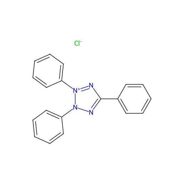
2,3,5-Triphenyltetrazolium chloride (TTC) has a molecular weight of 334.80, a molecular formula of C₁₉H₁₅ClN₄, and a CAS number of 298-96-4. TTC is a lipid-soluble, light-sensitive compound initially used to assess seed viability and later employed to stain and detect ischemic infarcts in mammalian tissues. TTC acts as a proton acceptor for the pyridine nucleotide-linked enzyme system in the respiratory chain. It reacts with respiratory enzymes in normal tissue, producing a red color. In ischemic tissue, where respiratory enzyme activity is reduced, the reaction does not occur, leaving the tissue unstained (pale). This staining is a gross staining method used to evaluate dehydrogenase activity within tissues.
TTC Staining Solution (1%) can detect myocardial infarction and cerebral necrosis 3–6 hours earlier than electron microscopy and 24 hours earlier than light microscopy. It is commonly used for staining fresh heart and brain tissue from autopsies, as well as early infarcted tissue in experimental animal models. It can also be used to assess seed and pollen viability. Features of this product include: ① TTC staining can be performed directly on fresh tissue without fixation, embedding, or other processing steps; ② TTC staining is rapid, typically taking 20–30 minutes; ③ TTC staining solution can be reused; 10 mL is sufficient for staining approximately 20 tissue sections.
Instructions for Use
1. Staining of Brain Tissue
1.1 Collect the fresh brain tissue sample (typically from anesthetized animals, either directly or after perfusion with physiological saline). After removal, quickly freeze at -20°C for 20–30 minutes to facilitate sectioning.
1.2 Slice the brain tissue. For animal samples, the recommended thickness is 2–3 mm; for human samples, 3–5 mm. Typically, 4–5 consecutive slices can be obtained. The first cut is made at the midpoint between the frontal pole and the optic chiasm; the second at the level of the optic chiasm; the third at the infundibular stalk; and the fourth between the infundibular stalk and the caudal pole of the posterior lobe (Reference: Zhang Juntian, Ed., Modern Pharmacological Experimental Methods).
1.3 Immerse the tissue slices in TTC Staining Solution (1%) and stain in the dark for 30–35 minutes.
1.4 Fix the stained slices in 4% paraformaldehyde or 10% neutral formalin for 4–24 hours.
1.5 Blot excess liquid from the tissue surface. Use an image analysis system (e.g., IPP) to measure the infarct area and calculate the cerebral infarct volume.
2. Staining of Seeds
2.1 Take seeds with varying viability. Cut them transversely or make a precise longitudinal cut through the center of the embryo. Use one half of each seed for staining.
2.2 Place the seed halves in TTC Staining Solution (1%) and incubate at 37°C in the dark for 20 minutes.
2.3 Pour off the staining solution, rinse the seeds 2–3 times with tap water, and immediately observe the coloration of the embryo.
Precautions
TTC Staining Solution (1%) is slightly irritating to humans. Please handle with care and take appropriate protective measures.
When removing the brain, take care to preserve its structural integrity.
If staining results are suboptimal, appropriately extend the staining time.
The fresher the sample, the better. To prevent the weakening or loss of enzyme activity in normal myocardial or brain tissue, perform staining as soon as possible.
For your safety and health, please wear a lab coat and disposable gloves during operation.
Staining Results
Tissue Type
| Color
|
Normal myocardium or brain tissue
| Red
|
Myocardial or cerebral infarct necrotic area
| Pale (unstained)
|
Ischemic brain tissue
| Intermediate between red and pale |

 ZJ26F0939580