
Lifecycle Functions, Immunological Characteristics, and Diagnostic and Therapeutic Research Applications of Key Monkeypox Virus Antigens
Lifecycle Functions, Immunological Characteristics, and Diagnostic and Therapeutic Research Applications of Key Monkeypox Virus Antigens
Monkeypox virus can form mature virions and enveloped virions. Different surface antigens participate in viral attachment, membrane fusion, and cell-to-cell spread and are also important targets for nucleic acid detection, immunodiagnosis, vaccine design, and antiviral drug research.
Keywords: monkeypox virus; Orthopoxvirus; mature virion; enveloped virion; A29L; M1R; A35R; H3L; immune evasion; nucleic acid detection; neutralizing antibody
1 Genome Structure and Virological Characteristics of Monkeypox Virus
1.1 Classification and Basic Structure of Monkeypox Virus
Monkeypox virus (MPXV) belongs to the genus Orthopoxvirus in the family Poxviridae and is an enveloped double-stranded DNA virus. Viral particles are brick-shaped or ovoid and consist internally of a nucleoprotein-like core, lateral bodies, and lipoprotein membranes. Monkeypox virus shares substantial genetic and antigenic homology with vaccinia virus, variola virus, and other orthopoxviruses, allowing a degree of cross-immunity among different orthopoxviruses.
1.2 Double-Stranded DNA Genome Structure
The monkeypox virus genome is linear double-stranded DNA with covalently closed hairpin structures and inverted terminal repeats at both ends. The central genomic region mainly encodes conserved functional proteins involved in DNA replication, transcription, virion assembly, and membrane fusion, whereas the terminal regions are enriched in genes associated with host range, virulence, and immune regulation. Deletions, duplications, or variations in terminal genes can affect tissue adaptation, immune evasion, and transmission characteristics.
1.3 Cytoplasmic Replication
Unlike most DNA viruses, monkeypox virus transcription and genome replication occur primarily in the host-cell cytoplasm. Viral particles carry their own DNA-dependent RNA polymerase, transcription factors, and mRNA-processing enzymes, allowing rapid initiation of early gene expression after cell entry without relying on the host nuclear transcription system.
1.4 Early, Intermediate, and Late Gene Expression
Monkeypox virus gene expression follows a defined temporal sequence. Early genes mainly encode DNA replication enzymes, immune-evasion proteins, and host-regulatory factors. After DNA replication begins, intermediate genes are expressed and promote late transcription. Late genes mainly encode core proteins, membrane proteins, assembly factors, and virion structural components. Continuous regulation among these stages ensures that viral replication and morphogenesis occur in the correct sequence.
1.5 Viral Clades and Antigen Conservation
Different monkeypox virus clades show certain differences in gene sequence and virulence, but most core replication proteins, membrane-fusion proteins, and major structural antigens are relatively conserved. Antigens such as A29L, M1R, A35R, and H3L generally retain stable structures and functions and are important candidate targets for vaccines, antibodies, and immunological detection.
Table 1 Major Functional Proteins of Monkeypox Virus and Their Research Positioning
Functional Stage | Representative Proteins or Orthologs | Major Functions | Major Research Applications |
Viral attachment | A29L, H3L, E8L | Bind cell-surface glycosaminoglycans or extracellular matrix molecules | Attachment inhibition, neutralizing antibody, and vaccine research |
Membrane fusion and entry | M1R and members of the entry-fusion complex | Mediate fusion between viral and host membranes | Entry-mechanism and neutralizing-antibody evaluation |
DNA replication | E9L, D5R, A20R, D4R | DNA polymerization, helicase activity, and replication elongation | Replication-mechanism and inhibitor research |
Virion assembly | D13L, A14L, A17L, A10L | Membrane formation, scaffold assembly, and core maturation | Viral morphogenesis research |
Envelopment and release | F13L/VP37, A35R | Envelopment, transport, and extracellular spread | Antiviral-drug and transmission-blocking research |
Immune evasion | Interferon-, complement-, and cytokine-regulatory proteins | Suppress innate immunity and inflammatory responses | Virulence, host-response, and drug-target research |
2 Formation and Spread of Mature and Enveloped Virions
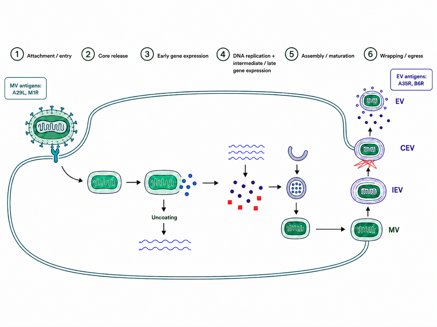
Figure 1. Cytoplasmic replication of monkeypox virus and formation of mature and enveloped virions
2.1 Two Infectious Virion Forms
Monkeypox virus mainly forms two infectious forms: mature virions (MVs) and enveloped virions (EVs). MVs have relatively strong environmental stability and are suited for transmission between hosts. EVs possess an additional membrane outside the MV and are more effective in cell-to-cell and long-distance spread within tissues.
2.2 Formation of Mature Virions
After viral replication, crescent-shaped membrane structures first form within cytoplasmic viral factories and gradually enclose viral core components to produce immature virions. Removal of scaffold proteins, proteolytic processing of core proteins, and condensation of the nucleoprotein-like structure convert immature particles into infectious MVs. MVs are an important form released through cell lysis and involved in environmental transmission.
2.3 Formation of Wrapped Virions
Some MVs acquire additional membranes derived from the Golgi apparatus or endosomal system to form intracellular wrapped virions. This process depends on F13L/VP37 and other membrane-transport proteins. Wrapped particles are subsequently transported along microtubules toward the cell periphery and fuse with the plasma membrane, exposing the outer viral particle at the cell surface.
2.4 Cell-Associated and Extracellular Enveloped Virions
Enveloped virions reaching the cell surface may remain attached to the plasma membrane as cell-associated enveloped virions or may be released extracellularly as extracellular enveloped virions. Cell-associated virions can induce actin-tail formation and propel viral spread toward neighboring cells, whereas extracellular enveloped virions participate in long-distance dissemination within tissues.
2.5 Functional Differences Between MV and EV Spread
The MV membrane is relatively stable and supports host-to-host transmission and prolonged environmental persistence. The EV outer membrane is more fragile but helps the virus evade some neutralizing antibodies and improves dissemination in vivo. Antibodies directed against only one virion form may not completely block infection, so vaccine and antibody combinations generally need to cover both MV and EV antigens.
Table 2 Structural and Functional Differences Between the Two Infectious Forms of Monkeypox Virus
Comparison Item | Mature Virion (MV) | Enveloped Virion (EV) |
Membrane structure | Contains the mature viral membrane | Contains an additional envelope outside the MV |
Major site of formation | Cytoplasmic viral factories | Golgi- or endosome-associated membrane systems |
Major mode of spread | Host-to-host transmission and release by cell lysis | Cell-to-cell and long-distance spread within tissues |
Environmental stability | Relatively high | Outer membrane is relatively fragile |
Representative antigens | A29L, M1R, H3L, E8L | Enveloped-virion antigens such as A35R |
Main focus of antibody research | Blocking attachment and membrane fusion | Blocking cell-to-cell spread and in vivo dissemination |
3 Proteins Associated With Monkeypox Virus Attachment, Entry, and Membrane Fusion
3.1 Multiprotein Cooperation in Viral Attachment
Monkeypox virus does not depend on a single receptor for attachment. Instead, multiple surface proteins interact with host-cell glycosaminoglycans, extracellular matrix molecules, and membrane proteins. This functional redundancy broadens viral cell tropism and reduces the impact of mutation in any single attachment protein on infectivity.
3.2 A29L Attachment Protein
A29L is an important attachment antigen on the MV surface and is homologous to vaccinia virus A27. It participates in binding between viral particles and cell-surface molecules. A29L has favorable immunogenicity, and anti-A29L antibodies can be used to study virion recognition, attachment inhibition, and antibody-mediated immune responses.
3.3 H3L and Glycosaminoglycan Binding
H3L is located on the MV surface and can bind glycosaminoglycans such as heparan sulfate on host cells, promoting viral enrichment at the plasma membrane. H3L can serve as an antibody target and can also be used to analyze attachment efficiency and infection susceptibility in different cell types.
3.4 E8L and Extracellular Matrix Recognition
Monkeypox virus E8L is homologous to vaccinia virus D8 and can recognize chondroitin sulfate and other cell-surface or extracellular matrix components. Together with other attachment proteins, it stabilizes virus-cell contact. Because attachment proteins have partially overlapping functions, blocking one protein alone generally produces only a partial reduction in viral attachment.
3.5 M1R and Viral Membrane Fusion
M1R is a highly conserved antigen on the MV membrane and is homologous to vaccinia virus L1. M1R participates in processes associated with viral entry and membrane fusion, and its conformation depends on correct disulfide-bond formation and membrane localization. Antibodies targeting M1R can be used to investigate entry blockade and represent an important component of multivalent vaccines and antibody combinations.
3.6 Entry-Fusion Complex
Monkeypox virus membrane fusion is jointly mediated by an entry-fusion complex composed of multiple highly conserved transmembrane proteins. This complex includes orthopoxvirus homologs such as A16, A21, A28, G4, G9, H2, J5, L1, L5, O3, and F9. Loss of any essential component may prevent membrane fusion or core release after viral attachment.
3.7 Different Entry Pathways
Depending on the cell type and virion form, monkeypox virus can enter cells through direct fusion with the plasma membrane or through endocytic pathways. After endocytosis, the virus is affected by acidification, membrane rearrangement, and host factors, after which the entry-fusion complex mediates release of the viral core into the cytoplasm.
4 Proteins Associated With Monkeypox Virus Replication, Assembly, and Release
4.1 Early Transcription Complex
After the viral core enters the cytoplasm, prepackaged RNA polymerase and early transcription factors immediately initiate early gene expression. Early proteins promote core uncoating, suppress host immunity, and establish an environment suitable for DNA replication, providing the basis for subsequent genome amplification and structural-protein expression.
4.2 E9L DNA Polymerase
E9L encodes the viral DNA polymerase and is a core enzyme for monkeypox virus genome replication. E9L forms a replication complex with the processivity factors A20R and D4R, increasing the speed and continuity of DNA synthesis. Nucleotide analogs can inhibit replication by interfering with viral DNA synthesis, making E9L and its associated complex important targets for antiviral-drug research.
4.3 D5R Helicase-Primase
D5R has helicase- and nucleoside triphosphatase-related functions and participates in replication initiation and replication-fork progression. Impairment of D5R blocks viral DNA amplification and consequently suppresses intermediate and late gene expression, making it a suitable target for studies of replication mechanisms and novel inhibitors.
4.4 Viral Membrane Formation and Immature Virion Assembly
Membrane proteins such as A14L and A17L participate in formation and stabilization of crescent membranes, whereas D13L forms a scaffold on the outside of the membrane and controls the geometry of immature virions. The viral genome, core proteins, and enzyme complexes are subsequently incorporated into the membrane structure to complete immature virion assembly.
4.5 Core Maturation and Proteolysis
After immature virions form, viral proteases process multiple core precursor proteins, allowing the core structure to condense and acquire infectivity. Correct cleavage of core proteins such as A10L and A3L is essential for mature virion formation. Disruption of protein processing can generate morphologically recognizable particles that lack full infectivity.
4.6 F13L and Viral Envelopment
The VP37 protein encoded by F13L is located on membranes associated with wrapped virions and participates in acquisition of additional membranes by MVs and formation of wrapped particles. Inhibition of F13L/VP37 does not completely prevent intracellular MV formation but reduces production and release of enveloped virions, thereby limiting viral dissemination within the host.
4.7 A35R-Mediated Enveloped-Virion Spread
A35R is a key antigen on the EV surface and is homologous to vaccinia virus A33. It participates in envelope stability, cell-surface spread, and immune recognition. Antibodies targeting A35R can be used to study inhibition of EV dissemination. In combination with MV-targeted antibodies, they can cover different forms of viral spread throughout the lifecycle.
5 Mechanisms of Monkeypox Virus Immune Evasion and Host-Response Regulation
5.1 Suppression of Interferon Pathways
Monkeypox virus encodes multiple intracellular antagonists and secreted binding proteins that can suppress type I interferon production, block interferon-receptor signaling, or neutralize extracellular interferons. Some viral proteins also inhibit PKR, the OAS-RNase L pathway, and interferon-stimulated gene responses, allowing infected cells to continue viral protein synthesis.
5.2 Evasion of cGAS-STING Signaling
After monkeypox virus DNA enters the cytoplasm, it can be recognized by cGAS, promoting formation of the second messenger cGAMP and activation of STING, TBK1, and IRF3 signaling, which induces type I interferon production. The virus can reduce innate immune responses by interfering with DNA sensing, STING-complex formation, or IRF3 activation, thereby extending the replication window.
5.3 TBK1 and IRF3 Activation
After STING activation, TBK1 is recruited and phosphorylated. Activated TBK1 subsequently phosphorylates IRF3. IRF3 dimerizes and enters the nucleus, where it initiates expression of interferons and related antiviral genes. Detection of total TBK1 and phosphorylation at Ser172 helps distinguish changes in pathway-protein abundance from actual pathway activation.
5.4 JAK-STAT Antiviral Signaling
Binding of type I interferons to their receptors activates JAK1 and TYK2 and subsequently promotes phosphorylation of STAT1 and STAT2. STAT-containing complexes enter the nucleus and induce expression of multiple antiviral genes. Viral inhibition of interferon receptors, JAKs, or STATs reduces the ability of host cells to establish an antiviral state.
5.5 Regulation of NF-κB and Inflammatory Responses
Multiple viral immunoregulatory proteins act on TNF receptors, TLRs, IKK, and the NF-κB pathway to reduce proinflammatory cytokine expression. Some viral proteins can also bind chemokines or mimic cytokine receptors, thereby limiting immune-cell recruitment to sites of infection.
5.6 Complement-System Evasion
Some monkeypox virus proteins possess complement-regulatory functions and can interfere with complement cascades and virion clearance. The integrity of related genes may differ among viral clades, resulting in differences in complement evasion and disease phenotype.
5.7 Regulation of Apoptosis and Host Range
Monkeypox virus can express Bcl-2-like proteins, Caspase-regulatory proteins, and other apoptosis inhibitors that delay infected-cell death. Host-range proteins can also regulate translation, ubiquitination, and stress responses, enabling the virus to maintain replication in different cells and species.
5.8 Antibody- and Complement-Mediated Viral Clearance
Antibodies can limit viral infection by directly blocking attachment or entry, promoting complement activation, enhancing phagocytosis, and mediating antibody-dependent cellular cytotoxicity. Because MVs and EVs possess different surface antigens, a single antibody usually covers only part of the infectious cycle. Multivalent immune responses are more likely to provide complete protection.
5.9 T-Cell Immune Responses
CD8⁺ T cells recognize viral peptides presented by infected cells and mediate cytotoxic killing, whereas CD4⁺ T cells support antibody production and immune-memory formation. Because many orthopoxvirus core proteins are highly conserved, T cells induced by vaccinia virus can cross-react with monkeypox virus antigens.
6 Applications of Key Monkeypox Virus Antigens in Nucleic Acid Detection and Immunodiagnosis
6.1 Differences Between Nucleic Acid and Antigen Detection
Monkeypox virus nucleic acid tests detect viral genomic sequences, whereas immunodiagnostic tests detect viral protein antigens or host antibodies. Genes encoding key antigens can serve as nucleic acid testing targets, but gene positivity does not directly prove high expression of the corresponding protein. The two types of results should be interpreted separately according to sample type and disease stage.
6.2 Real-Time Fluorescent PCR Detection
Real-time fluorescent PCR is an important laboratory method for monkeypox virus detection. Assays can target monkeypox virus-specific genomic loci and can include conserved orthopoxvirus targets for screening and confirmation. Assay design should evaluate sequence conservation across viral clades and avoid cross-amplification with other orthopoxviruses.
6.3 Sample Type and Viral Load
Lesion surfaces, vesicular fluid, crusts, and lesion swabs generally contain relatively high levels of viral DNA and are suitable for nucleic acid testing. Viral loads in blood, saliva, or throat swabs vary substantially with disease stage, and a negative result does not completely exclude infection localized to skin lesions. Sample-collection quality and endogenous controls are essential for reducing false-negative results.
6.4 Multiplex Nucleic Acid Testing
Multiplex PCR can simultaneously detect universal orthopoxvirus targets, monkeypox virus-specific targets, and internal-control genes and may include targets for other rash-associated pathogens. Multitarget designs reduce the risk of false-negative results caused by mutation at a single locus and improve the efficiency of differential diagnosis.
6.5 Sequencing and Viral Clade Analysis
Whole-genome or targeted sequencing can be used for viral-clade determination, transmission-chain analysis, mutation surveillance, and investigation of antiviral-drug resistance. Sequencing can provide additional epidemiological and functional information for PCR-positive samples but generally does not replace rapid nucleic acid testing.
6.6 Viral Antigen Detection
Structural proteins such as A29L, M1R, A35R, H3L, and E8L can serve as candidate targets for antigen-capture assays. Antigen testing is rapid, but sensitivity is influenced by viral load, epitope conservation, virion form, and sample-processing methods. It is more suitable as a supplement to nucleic acid testing.
6.7 Antibody Detection and Cross-Reactivity
Infection or orthopoxvirus vaccination can induce IgM and IgG against multiple MV and EV antigens. Because orthopoxvirus antigens have high homology, previous vaccination or exposure to other orthopoxviruses can cause cross-reactivity. A single positive serum-antibody result cannot independently confirm recent monkeypox virus infection.
Combined detection of antibodies against MV- and EV-associated antigens can provide a more complete serological profile. A29L, M1R, H3L, and E8L mainly reflect antibody responses to MVs, whereas A35R reflects responses to EVs. Paired acute- and convalescent-phase sera combined with functional-antibody evaluation can help distinguish previous immunity from recent infection.
Table 3 Major Diagnostic Targets for Monkeypox Virus and Their Application Characteristics
Detection Type | Representative Targets | Major Samples | Major Uses | Major Limitations |
Universal orthopoxvirus PCR | Conserved replication genes | Lesion swabs and crusts | Orthopoxvirus screening | Cannot independently identify monkeypox virus |
Monkeypox virus-specific PCR | Virus-specific genomic loci | Lesion swabs and vesicular fluid | Virus-specific confirmation | Target-region variation requires monitoring |
Genome sequencing | Whole genome or multiple target regions | PCR-positive samples | Clade determination and transmission analysis | Higher cost and longer analysis time |
Antigen detection | A29L, M1R, A35R, H3L, E8L | Lesions or secretions | Rapid viral-protein detection | Sensitivity is generally lower than nucleic acid testing |
IgM/IgG detection | Multiple MV and EV antigens | Serum and plasma | Immune-response and seroepidemiological studies | Pronounced orthopoxvirus cross-reactivity |
Functional-antibody evaluation | MV or EV antigen systems | Serum or antibody samples | Evaluation of vaccine and antibody function | Requires substantial experimental standardization |
7 Research on Monkeypox Virus Vaccines, Antiviral Drugs, and Neutralizing Antibodies
7.1 Cross-Protection by Vaccinia Virus Vaccines
Monkeypox virus and vaccinia virus share many conserved structural antigens and T-cell epitopes. Vaccinia virus vaccines can therefore induce cross-reactive humoral and cellular immunity. Nonreplicating or replication-restricted vaccinia vaccines can express multiple MV and EV antigens and provide broad immune coverage.
7.2 Multivalent Subunit Vaccines
A29L, M1R, A35R, H3L, and E8L can be used in monkeypox virus subunit vaccines and multivalent immunization studies. MV antigens mainly cover viral attachment and entry, whereas EV antigens cover enveloped-virion spread. Multivalent combinations are generally more likely than single antigens to induce antibody responses that provide both virion recognition and transmission blockade.
7.3 Nucleic Acid and Viral-Vector Vaccines
DNA, mRNA, and viral-vector vaccines can simultaneously express multiple monkeypox virus antigens and induce both antibody and T-cell responses through intracellular antigen expression. Antigen conformation, signal peptides, retention of transmembrane regions, and relative expression levels influence immunogenicity. Membrane-protein antigens in particular require preservation of native conformational epitopes.
Tecovirimat targets the VP37 protein encoded by F13L and inhibits acquisition of additional membranes by mature virions and formation of enveloped virions, thereby limiting viral release and in vivo dissemination. Pharmacodynamic evaluation should simultaneously assess intracellular virus, extracellular enveloped virus, and cell-to-cell spread.
7.5 Cidofovir and Brincidofovir
Cidofovir and its lipid prodrug Brincidofovir generate nucleotide analogs that interfere with viral DNA polymerase and inhibit monkeypox virus genome replication. Related studies can evaluate viral DNA levels, structural-protein expression, virion formation, and host-cell tolerance.
7.6 Evaluation of Antiviral-Drug Resistance Mechanisms
Changes in F13L/VP37 may affect viral sensitivity to Tecovirimat, whereas changes in DNA polymerase or the replication complex may influence activity of nucleotide analogs. Resistance studies should integrate viral genetic changes, drug susceptibility, and replicative fitness.
7.7 MV-Associated Antibodies
Antibodies targeting MV membrane antigens such as A29L, M1R, H3L, and E8L can be used to study viral attachment, membrane fusion, and core release. Their effects depend on epitope accessibility, affinity, antigen conformation, and virion maturation state. Recombinant-protein binding results therefore need to be interpreted together with cell- or virion-level experiments.
7.8 Antibodies Blocking EV Spread
Antibodies targeting EV surface antigens such as A35R can be used to study cell-to-cell spread of enveloped virions and the roles of complement and Fc receptors in viral clearance. Combining EV-associated antibodies with MV-associated antibodies can cover different virion forms.
7.9 Polyclonal and Monoclonal Antibody Combinations
Polyclonal immunoglobulins recognize multiple orthopoxvirus epitopes and reduce the risk of immune escape caused by variation at a single epitope. Monoclonal antibodies have defined targets and high batch consistency. Antibody combinations should cover different virion forms and nonoverlapping epitopes and should avoid substantial steric competition among antibodies.
7.10 Antibody Functional-Evaluation Systems
Antibody research can use virion-binding assays, cell-attachment blocking, viral-entry inhibition, plaque-reduction assays, reporter systems, or cell-to-cell spread assays. Because MVs and EVs differ in antibody sensitivity, the virion form being evaluated should be clearly defined, and complement dependence and Fc-mediated effects should also be analyzed.
8 Products
8.1 Key Monkeypox Virus Antigens, Antibodies, and Labeled Antibody Products
Catalog # | Product Name | Grade & Purity | Main Application |
Recombinant MPXV A29L Protein | Carrier-free, azide-free, His Tag, PBS Only, ≥95% (SDS-PAGE) | A29L antibody screening, viral attachment studies, and MV-antigen immunogenicity research | |
A29L Mouse mAb | Carrier-free, ExactAb™, azide-free, validated, PBS Only, 1.0 mg/mL | A29L protein detection, virion recognition, and antigen-expression analysis | |
A29L Mouse mAb | Carrier-free, ExactAb™, azide-free, validated, PBS Only, 1.0 mg/mL | A29L antigen detection and comparison of different antibody clones | |
Mouse IgG2a-kappa, anti-A29L | Animal Free, carrier-free, ExactAb™, azide-free, validated, high-performance, PBS Only, ≥95% (SDS-PAGE), 1.0 mg/mL | A29L binding, viral-attachment blocking, and antibody-function studies | |
Mouse IgG2a-kappa (Biotin), anti-A29L | ExactAb™, validated, 0.5 mg/mL | Biotin-based detection, antigen capture, and antibody-pairing research | |
Mouse IgG2a-kappa (AF488), anti-A29L | ExactAb™, validated, 0.5 mg/mL | A29L fluorescence localization, cellular imaging, and flow-cytometric analysis | |
Mouse IgG2a-kappa (AF555), anti-A29L | ExactAb™, validated, 0.5 mg/mL | A29L fluorescence imaging and multiplex-staining studies | |
Mouse IgG2a-kappa (AF594), anti-A29L | ExactAb™, validated, 0.5 mg/mL | Infection-associated antigen localization and colocalization analysis | |
Mouse IgG2a-kappa (AF647), anti-A29L | ExactAb™, validated, 0.5 mg/mL | Far-red imaging, flow cytometry, and multicolor detection | |
Mouse IgG2a-kappa (AF700), anti-A29L | ExactAb™, validated, 0.5 mg/mL | A29L detection in multiparameter flow cytometry | |
Mouse IgG2a-kappa (FITC), anti-A29L | ExactAb™, validated, 0.5 mg/mL | FITC-channel flow detection and fluorescence imaging | |
Mouse IgG2a-kappa (PE), anti-A29L | ExactAb™, validated, 0.5 mg/mL | High-sensitivity flow-cytometric detection | |
Mouse IgG2a-kappa (APC), anti-A29L | ExactAb™, validated, 0.5 mg/mL | APC-channel flow detection and multicolor immunological analysis | |
Recombinant MPXV A30L Protein | Animal Free, carrier-free, azide-free, His Tag, PBS Only, ≥95% (SDS-PAGE) | A30L antigen preparation, antibody screening, and serological research | |
Recombinant MPXV M1R Protein | Animal Free, carrier-free, azide-free, His Tag, PBS Only, ≥95% (SDS-PAGE) | M1R antibody screening, viral-entry studies, and MV-antigen research | |
Recombinant MPXV H3L Protein | Animal Free, carrier-free, azide-free, His Tag, PBS Only, ≥95% (SDS-PAGE) | Studies of H3L-mediated glycosaminoglycan binding, attachment, and antibody responses | |
Recombinant MPXV E8L Protein | Animal Free, carrier-free, azide-free, His Tag, PBS Only, ≥95% (SDS-PAGE) | E8L attachment-antigen, extracellular-matrix binding, and immunological detection research | |
Recombinant MPXV A35R Protein | Animal Free, carrier-free, azide-free, His Tag, PBS Only, ≥95% (SDS-PAGE), see COA | EV-antigen, enveloped-virion spread, and multivalent-vaccine research | |
Recombinant MPXV A35R Protein | Carrier-free, azide-free, His Tag, PBS Only, ≥95% (SDS-PAGE) | A35R antibody screening, serological detection, and enveloped-antigen research | |
A35R Mouse mAb | Carrier-free, ExactAb™, azide-free, validated, PBS Only, ≥95% (SDS-PAGE & SEC-HPLC), 1.0 mg/mL | A35R detection, EV recognition, and transmission-related research | |
A35R Mouse mAb | Carrier-free, ExactAb™, azide-free, validated, PBS Only, ≥95% (SDS-PAGE & SEC-HPLC), 1.0 mg/mL | A35R antigen detection and comparison of different antibody clones |
8.2 Antiviral Drugs and cGAS-STING-TBK1 Pathway Research Products
Catalog # | Product Name | Grade & Purity | Type of Action | Main Application |
Tecovirimat(mixture of isomers) | ≥98% | Viral-envelope formation inhibitor | Studies of F13L/VP37-associated envelopment, release, and cell-to-cell spread | |
Tecovirimat | 10 mM in DMSO | Viral-envelope formation inhibitor solution | Cell-based antiviral efficacy and dose-response studies | |
brincidofovir | Moligand™ | Prodrug of a viral DNA-replication inhibitor | Studies of viral DNA replication and structural-protein expression | |
Cidofovir | ≥98% | Nucleotide analog | Viral DNA-polymerase inhibition and replication-blocking studies | |
Cidofovir | 10 mM in Water | Nucleotide-analog solution | Viral-replication inhibition in cellular experiments | |
CGAS Human Pre-designed siRNA Set A | — | Human cGAS gene intervention | cGAS knockdown and validation of dependence on viral-DNA recognition | |
Cgas Mouse Pre-designed siRNA Set A | — | Mouse cGAS gene intervention | Mechanistic research in mouse cells and related animal models | |
Cgas Rat Pre-designed siRNA Set A | — | Rat cGAS gene intervention | DNA-sensing research in rat-derived cells | |
cGAS-IN-1 | ≥98% | cGAS inhibitor | cGAS inhibition and evaluation of interferon production and host responses | |
cGAS-IN-1 | Moligand™, 10 mM in DMSO | cGAS inhibitor solution | Cellular functional evaluation of cGAS inhibition | |
cGAS-IN-2 | ≥99% | cGAS inhibitor | Comparison of cGAS-inhibitor potency and selectivity | |
Human Mab21 Domain Containing Protein 1 (MB21D1/CGAS) ELISA Kit | BioReagent | cGAS detection reagent | Quantitative detection of cGAS in human samples | |
Mouse Mab21 Domain Containing Protein 1 (MB21D1/CGAS) ELISA Kit | BioReagent | cGAS detection reagent | Quantitative detection of cGAS in mouse samples | |
cGAMP sodium salt | ≥99% | STING agonist | Simulation of downstream cGAS signaling and activation of STING | |
STING agonist-1 | Moligand™, ≥98% | STING signaling activator | Studies of the effect of enhanced STING signaling on antiviral responses | |
G10, STING signaling activator | Moligand™, 10 mM in DMSO | STING signaling activator solution | Cell-based STING-activation research | |
H 151 | Moligand™, ≥98% (HPLC) | STING antagonist | STING blockade and validation of the host DNA-sensing pathway | |
MSA 2 | Moligand™, ≥98% (HPLC) | STING agonist | Non-nucleotide STING activation and immune-response research | |
STING Agonist C11 | ≥99% | STING agonist | STING-pathway activation and downstream-signaling evaluation | |
STING inhibitor C-178 | ≥98% | STING inhibitor | Validation of STING-dependent interferon responses | |
STING inhibitor C-178 | 10 mM in DMSO | STING inhibitor solution | STING-inhibition research in cellular experiments | |
Recombinant STING Antibody | Recombinant, ExactAb™, validated, 0.4 mg/mL | STING detection antibody | Detection of STING expression and virus-associated pathway changes | |
STING1 Human Pre-designed siRNA Set A | — | Human STING1 gene intervention | STING knockdown and validation of pathway dependence | |
Sting1 Mouse Pre-designed siRNA Set A | — | Mouse STING1 gene intervention | STING-function research in mouse-derived cells | |
Sting1 Rat Pre-designed siRNA Set A | — | Rat STING1 gene intervention | STING-function research in rat-derived cells | |
Human Stimulator of interferon genes (STING) ELISA Kit | BioReagent | STING detection reagent | Quantitative detection of STING in human samples | |
Recombinant Human TBK1 Protein | ≥90% (SDS-PAGE) | Recombinant TBK1 protein | TBK1-binding, kinase, and inhibitor research | |
Recombinant NAK/TBK1 Antibody | Recombinant, ExactAb™, validated, see COA | TBK1 detection antibody | Detection of total TBK1 protein expression | |
Recombinant TBK1 (phospho S172) Antibody | Recombinant, ExactAb™, validated, see COA | Phospho-TBK1 antibody | Detection of TBK1 activation at Ser172 | |
Recombinant Phospho-NAK/TBK1 (S172) Antibody | KD Validation | Phospho-TBK1 antibody | Specific p-TBK1 detection and knockdown validation | |
TBK1 Human Pre-designed siRNA Set A | — | TBK1 gene intervention | TBK1 knockdown and evaluation of IRF3 and interferon signaling | |
TBK1-IN-1 | ≥98% | TBK1 inhibitor | Validation of the role of TBK1 in antiviral signaling | |
TBK1 PROTAC®3i | Moligand™, ≥98% (HPLC) | TBK1 degrader | Investigation of TBK1 functional dependence through protein degradation | |
pLenti-TBK1-sgRNA | — | Gene-knockout control | TBK1 antibody-specificity and protein-detection validation | |
pLenti-TBK1-sgRNA | — | Gene-knockout nucleic acid control | TBK1 transcription detection and gene-knockout validation | |
Human TANK Binding Kinase 1 (TBK1) ELISA Kit | BioReagent | TBK1 detection reagent | Quantitative detection of TBK1 in human samples | |
Human Phosphorylated TANK Binding Kinase 1 (P-TBK1) ELISA Kit | BioReagent | p-TBK1 detection reagent | Quantitative evaluation of TBK1-pathway activation | |
Mouse TANK Binding Kinase 1 (TBK1) ELISA Kit | BioReagent | Mouse TBK1 detection reagent | Detection of TBK1 levels in mouse models | |
Mouse Phosphorylation TANK Binding Kinase 1 (p-TBK1) ELISA Kit | BioReagent | Mouse p-TBK1 detection reagent | Evaluation of TBK1 activation in mouse models |
Monkeypox virus uses two infectious virion forms, MV and EV, to achieve host-to-host transmission and dissemination within tissues. A29L, M1R, H3L, E8L, and A35R participate in viral attachment, entry, and enveloped-virion spread and are important antigens for immunological detection, vaccine development, and antibody research. Viral DNA replication and enveloped-virion formation also provide important targets for antiviral-drug research.
For more related articles, please see below:
[1] Pathogenesis, Virological Characteristics, and Laboratory Detection Strategies for Nipah Virus (NiV)
[2] Poxvirus DNA extraction experiment
[3] Titration or purification of Akabane disease virus (AKV)
[4] Arbovirus prevention and treatment
[5] Stable virus-producing cell lines for AAV assembly