
Analysis of Classical Histological Special Staining Systems: Application Selection of PTAH, Masson, VG, Elastic Fiber Staining, and Reticular Fiber Staining
Analysis of Classical Histological Special Staining Systems: Application Selection of PTAH, Masson, VG, Elastic Fiber Staining, and Reticular Fiber Staining
The core value of classical histological special staining is to further distinguish structures such as muscle fibers, collagen fibers, elastic fibers, reticular fibers, fibrin, and neuroglial fibers on the basis of HE staining. PTAH, Masson, VG, elastic fiber staining, and reticular fiber staining each have clear application boundaries. Selection should begin with the target structure, followed by consideration of tissue type, lesion background, and result interpretation requirements.
Keywords: special staining; PTAH staining; Masson trichrome staining; Van Gieson staining; VG staining; elastic fiber staining; reticular fiber staining; collagen fiber staining; histological staining
1 Selection Logic of Special Staining Systems
1.1 Define the target structure first
(1) Muscle fibers and fibrin
If the target is to display striations of striated muscle, myocardial structure, fibrin within thrombi, or neuroglial fibers, PTAH is more targeted. It is suitable for observing proteinaceous fibrous structures, but not all blue-stained structures should be directly interpreted as the same tissue component.
(2) Collagen fibers and fibrosis
If the target is to evaluate the extent of fibrosis, degree of collagen deposition, or tissue remodeling, Masson and VG are more commonly used. Masson is suitable for multicolor differentiation among collagen, muscle fibers, and cytoplasm. The VG system is more concise and is commonly used for rapid contrast between collagen and muscle background.
(3) Elastic fibers and vascular structure
If the target is to observe arterial elastic laminae, pulmonary elastic fibers, dermal elastic tissue, or elastic fiber rupture in vascular lesions, elastic fiber staining should be selected, such as Verhoeff, Weigert, orcein, or Victoria blue methods.
(4) Reticular fibers and tissue framework
If the target is to demonstrate the fine reticular scaffold in the liver, spleen, bone marrow, lymph nodes, endocrine glands, or tumor stroma, reticular fiber staining should be selected. This type of staining is mostly based on silver impregnation and can highlight fine, highly branched reticular fiber structures.
1.2 Rapid selection pathway
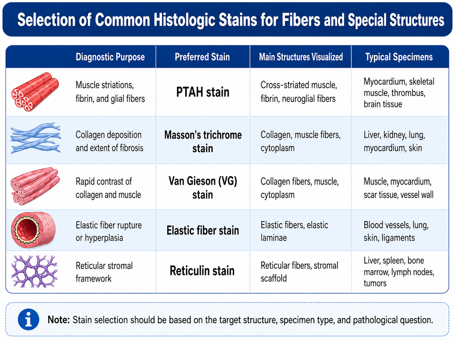
2 PTAH Staining System
2.1 Staining positioning
(1) Striated muscle structure
PTAH, or phosphotungstic acid hematoxylin staining, can be used to display striations and myofibrillar structures in striated muscle. In myocardial and skeletal muscle samples, clear striations usually indicate good preservation of muscle fiber structure. If striations disappear, break, or stain unevenly, HE staining should be used to evaluate muscle fiber necrosis, degeneration, edema, and section orientation.
(2) Fibrin deposition
PTAH can stain fibrin blue or blue-purple, making it suitable for observing thrombi, fibrinous inflammation, exudates around necrotic foci, and intravascular deposits. Fibrin interpretation should be based on tissue location and morphology. When necessary, it should be combined with MSB staining or fibrin-related immunohistochemistry.
(3) Neuroglial fibers
In central nervous system tissue, PTAH can display neuroglial fibers and glial reaction-related structures. This method is suitable for morphological observation but cannot distinguish glial cell subtypes. If astrocytes, microglia, or oligodendrocytes need to be identified, immunomarkers such as GFAP, IBA1, and OLIG2 should be used.
2.2 Method characteristics
(1) Phosphotungstic acid-hematoxylin complex staining
In the PTAH system, phosphotungstic acid participates in both mordanting and differentiation and also affects dye retention by different protein structures. Target structures are usually blue to blue-purple, while the background may appear reddish-brown, brownish-yellow, or pale. The final result is strongly affected by stain maturation, oxidation state, and differentiation conditions.
(2) Sensitivity to fixation conditions
PTAH is sensitive to fixation methods. Insufficient fixation may lead to loose tissue structure and increased background. When fixation conditions vary greatly, the staining intensity of striated muscle, fibrin, and neuroglial fibers may be unstable. Positive controls should be included for retrospective paraffin samples.
(3) Suitable for structural indication, not single-target identification
PTAH can highlight certain proteinaceous structures, but it is not a molecular-specific assay. Blue staining does not necessarily indicate fibrin; it may also represent striated muscle or neuroglial fibers. Interpretation should be based on sample type and lesion location.
3 Masson Trichrome Staining System
3.1 Staining positioning
(1) Collagen fiber deposition
Masson trichrome staining is mainly used to distinguish collagen fibers, muscle fibers, and cytoplasm. Collagen is usually stained blue or green, muscle fibers and cytoplasm are stained red, and nuclei appear dark after nuclear staining. This system is suitable for analyzing liver fibrosis, pulmonary fibrosis, renal interstitial fibrosis, myocardial fibrosis, and skin scars.
(2) Evaluation of tissue remodeling
Masson staining not only displays collagen but also helps observe collagen distribution, deposition range, and the degree of tissue structural remodeling. For example, the scar area after myocardial infarction, destruction of hepatic lobular architecture, renal tubulointerstitial fibrosis, and thickening of alveolar septa can all be clearly evaluated using Masson staining.
(3) Animal models and pharmacodynamic evaluation
In fibrosis models, drug intervention models, and tissue injury repair experiments, Masson staining is commonly used for collagen area quantification. During image analysis, section thickness, staining conditions, imaging parameters, and threshold settings should be standardized to avoid batch effects affecting quantitative results.
3.2 Method characteristics
(1) Strong multicolor differentiation capability
Masson staining uses multiple staining and differentiation steps to create color contrast among collagen, muscle fibers, cytoplasm, and nuclei. Compared with simple collagen staining, it is more suitable for observing tissue structural relationships.
(2) Results are strongly affected by differentiation
Insufficient differentiation can blur the boundary between red and blue or red and green, reducing the contrast between collagen and muscle fibers. Over-differentiation may weaken collagen staining. Differentiation time and staining intensity should be optimized according to tissue type.
(3) Not suitable for displaying fine reticular fibers
Masson staining can display relatively coarse collagen deposition and fibrotic areas, but it is less sensitive than reticular fiber silver staining for fine reticular scaffolds. For analysis of hepatic plate scaffolds, bone marrow reticular structure, or lymphoid tissue framework, reticular fiber staining should be prioritized.
4 VG Staining System
4.1 Staining positioning
(1) Contrast between collagen and muscle
VG staining usually refers to Van Gieson staining. It commonly uses acid fuchsin and picric acid as the core components, staining collagen fibers red and muscle, cytoplasm, and part of the background yellow. Its advantage is a relatively concise system, suitable for rapid differentiation between collagen fibers and muscular tissue.
(2) Observation of scars and fibrosis
VG staining is suitable for observing scars, myocardial fibrosis, vascular wall collagen deposition, and connective tissue proliferation. Compared with Masson staining, VG has fewer color layers, but the contrast between collagen and muscle is direct, making it suitable for structural interpretation and rapid screening.
(3) Vascular wall and muscular tissue
In vascular, muscle, and myocardial samples, VG staining can highlight the difference between collagen and muscular components. If elastic laminae also need to be observed, elastic fiber staining should be added; VG should not be used to assess elastic fiber integrity.
4.2 Method characteristics
(1) Relatively simple operation
The VG workflow is usually simpler than Masson staining and is suitable for batch samples and rapid structural comparison. When collagen is clearly stained red, scar and collagen deposition areas can be intuitively displayed.
(2) Less information layering than Masson
VG mainly addresses the question of how to distinguish collagen from muscle/cytoplasm. It is less suitable than Masson for multilayered analysis of complex tissue remodeling. If comprehensive evaluation of collagen quantification, inflammatory background, and the relationship with muscle fibers is required, Masson usually provides richer information.
(3) Affected by picric acid background
In VG staining, picric acid stains muscle and background yellow. If differentiation, dehydration, or mounting conditions are unstable, the contrast between the yellow background and red collagen may be weakened. Color stability after long-term section storage also needs attention.
5 Elastic Fiber Staining Systems
5.1 Staining positioning
(1) Vascular elastic laminae
Elastic fiber staining is most commonly used to observe the internal and external elastic laminae of blood vessels. In arteriosclerosis, vasculitis, aneurysm, vascular remodeling, and vascular wall injury, rupture, thickening, curling, or disappearance of elastic laminae has important morphological significance.
(2) Elastic tissue in lung and skin
Elastic fibers in alveolar septa, dermal elastic fibers, and ligament elastic structures can all be observed using elastic fiber staining. This system has high application value in studies of chronic lung disease, skin aging, elastic fiber abnormalities, and connective tissue diseases.
(3) Tumor and tissue invasion boundaries
In some tumor tissues, elastic fiber staining can help identify vascular wall structures, serosal elastic layers, or tissue invasion boundaries. Interpretation should be combined with HE and immunohistochemistry results to avoid interpreting elastic fiber rupture alone as evidence of invasion.
5.2 Differences among common methods
(1) Verhoeff method
The Verhoeff method is commonly used to display coarse elastic fibers and vascular elastic laminae. Elastic fibers usually appear black or dark after staining. This method clearly demonstrates elastic layers in vascular walls and is suitable for analyzing arteries and large vessel structures.
(2) Weigert method
Weigert elastic fiber staining is suitable for displaying elastic fiber networks. Background control and differentiation conditions affect the result. When used for lung, skin, and vascular samples, differentiation strength should be adjusted according to elastic fiber thickness.
(3) Orcein method
The orcein method can be used to demonstrate elastic fibers and is commonly applied to skin, blood vessels, and some tissue sections. Its color tone is related to the specific formulation and counterstaining method. It is suitable for routine elastic fiber observation, but its performance for weak or fine elastic fibers should be validated.
(4) Victoria blue method
Victoria blue can display elastic fibers and is commonly used for analyzing blood vessels, lung, and tissue elastic structures. Compared with other elastic staining methods, its background and color tone are more dependent on stain condition, differentiation, and counterstaining combinations.
Table 1 Comparison of Elastic Fiber Staining Methods
Method | Main Target | Suitable Samples | Advantages | Notes |
Verhoeff method | Coarse elastic fibers, vascular elastic laminae | Arteries, vascular walls, aneurysms | Clear elastic lamina outline | Over-differentiation may weaken elastic fiber staining |
Weigert method | Elastic fiber networks | Lung, skin, blood vessels | Suitable for observing fiber network distribution | Background control and differentiation are critical |
Orcein method | Elastic fibers and elastic tissue | Skin, blood vessels, connective tissue | Suitable for routine elastic fiber observation | Tissue background should be interpreted with counterstaining |
Victoria blue method | Elastic fibers and elastic structures | Blood vessels, lung, selected tissue sections | Direct color display, suitable for structural observation | Stain condition and differentiation strongly affect results |
6 Reticular Fiber Staining System
6.1 Staining positioning
(1) Display of tissue scaffold
Reticular fibers are mainly composed of type III collagen-related fibers and form fine, branching scaffolds. Reticular fiber staining usually uses silver impregnation, staining reticular fibers black or dark, and is suitable for observing hepatic lobular scaffolds, splenic cords, lymph node frameworks, bone marrow reticulum, and glandular stromal structures.
(2) Structural assessment of tumor tissue
In tumor pathology, reticular fiber staining can help observe tumor cell nests, stromal scaffolds, and tissue architectural destruction. For example, the distribution pattern of reticular fibers has reference value in hepatocellular carcinoma versus cirrhotic background, bone marrow fibrosis, and destruction of lymphoid tissue architecture.
(3) Bone marrow and hematopoietic tissue
Bone marrow reticular fiber staining is commonly used for evaluating bone marrow fibrosis. Increased, thickened, and interwoven reticular fibers can serve as important morphological indicators of bone marrow microenvironment remodeling. Results should be interpreted using a unified grading system rather than judged only by color intensity.
6.2 Method characteristics
(1) Silver impregnation is sensitive but technically demanding
Reticular fiber staining usually relies on silver salt deposition to display fine fiber structures. Silver impregnation is highly sensitive to slide cleanliness, oxidation, reduction, temperature, and time. Unstable operation may easily cause background deposition, discontinuous fibers, or whole-section blackening.
(2) Suitable for fine fibers, not equivalent to Masson staining
Masson staining displays collagen deposition and fibrosis range, whereas reticular fiber staining displays finer scaffold structures. Both are collagen-related, but the observed targets are different. For scaffold structure analysis in liver, bone marrow, spleen, and lymph nodes, reticular fiber staining is more targeted.
(3) Interpretation depends on tissue architecture
The significance of reticular fibers depends on tissue type. In liver tissue, attention is paid to hepatic plate scaffolds and fibrous septa. In bone marrow, fibrosis degree is assessed. In lymph nodes, scaffold integrity and architectural destruction are evaluated. In tumors, the scaffold pattern around cell nests is important.
7 Method Combinations in Different Research Scenarios
7.1 Fibrosis research
(1) Collagen deposition as the main focus
In liver fibrosis, pulmonary fibrosis, renal interstitial fibrosis, and myocardial fibrosis research, Masson staining is a common first choice. If rapid confirmation of collagen versus muscle background is required, VG staining can also be used as an auxiliary method.
(2) Scaffold structure as the main focus
If hepatic plate architecture, bone marrow fibrosis, or lymphoid tissue scaffold is being studied, reticular fiber staining is more suitable than Masson. Masson can display fibrosis range but cannot fully replace the fine scaffold information provided by reticular fiber silver staining.
7.2 Vascular and elastic tissue research
(1) Vascular wall structure
Vascular lesion studies should prioritize elastic fiber staining to observe the internal elastic lamina, external elastic lamina, elastic fiber rupture, and vascular wall remodeling. If collagen deposition is also a concern, Masson or VG staining can be combined.
(2) Elastic tissue in lung and skin
For pulmonary elastic fiber remodeling, dermal elastic fiber changes, and connective tissue disease research, elastic fiber staining is more targeted than Masson and VG. If collagen proliferation also needs to be evaluated, Masson staining can be combined.
7.3 Muscle and thrombus research
(1) Striated muscle structure
PTAH is preferred for displaying striations in myocardium and skeletal muscle. If myocardial fibrosis is being studied, Masson or VG is more suitable. If both muscle fiber structural disruption and fibrosis are being analyzed, PTAH should be combined with Masson.
(2) Thrombus and fibrin
PTAH can be used to display fibrin-like structures, but when thrombus composition is complex, MSB or immunohistochemistry usually provides more information. PTAH is suitable for auxiliary morphological confirmation and should not be used alone for thrombus component stratification.
Table 2 Typical Experimental Purposes and Special Staining Combinations
Experimental Purpose | Recommended Staining Combination | Main Interpretation Content |
Liver fibrosis evaluation | Masson + reticular fiber staining | Collagen deposition range and hepatic plate scaffold changes |
Myocardial injury and fibrosis | PTAH + Masson/VG | Preservation of muscle striations and formation of collagen scar |
Vascular remodeling | Elastic fiber staining + Masson/VG | Elastic lamina rupture and collagen deposition |
Bone marrow fibrosis | Reticular fiber staining | Bone marrow reticular fiber proliferation and grading |
Thrombus structure analysis | PTAH + MSB/IHC | Fibrin distribution and thrombus component confirmation |
Skin elastic tissue changes | Elastic fiber staining + Masson | Elastic fiber changes and collagen remodeling |
8 Method Boundaries and Common Failure Causes
8.1 Method boundaries
(1) PTAH cannot replace fibrin immunohistochemistry
PTAH can display fibrin-like structures, but it does not have antibody-based molecular specificity. If the research focus is fibrin or fibrinogen localization, immunohistochemistry should be combined.
(2) Masson and VG cannot replace reticular fiber staining
Masson and VG are suitable for contrasting collagen with muscle background, but they cannot fully display fine reticular scaffolds. Silver impregnation should be used for reticular fiber structural analysis.
(3) Elastic fiber staining cannot be replaced by collagen staining
Elastic fibers and collagen fibers differ in structure, distribution, and pathological significance. Elastic lamina rupture in vessels, abnormal pulmonary elastic structures, and dermal elastic fiber changes should be examined using elastic fiber staining rather than relying only on Masson or VG.
8.2 Common failure causes
Table 3 Common Problems and Optimization Directions in Classical Special Staining
Problem | Possible Cause | Staining Involved | Optimization Direction |
Weak blue staining of PTAH target structures | Insufficient stain maturation, over-differentiation, unsuitable fixation | PTAH | Set positive controls, adjust differentiation time, check stain condition |
Unclear boundary between collagen and muscle in Masson staining | Insufficient differentiation, inappropriate staining time | Masson | Optimize differentiation steps and standardize section thickness |
Weak red collagen staining in VG | Decreased stain potency, excessive dehydration, or improper differentiation | VG | Check acid fuchsin solution and shorten excessive processing steps |
Elastic fibers appear discontinuous or too pale | Over-differentiation, aged stain, poor fixation | Elastic fiber staining | Shorten differentiation, renew stain, use positive vascular tissue control |
Whole reticular fiber section turns black | Excessive silver impregnation, slide contamination, excessive reduction | Reticular fiber staining | Control silver staining time, use clean slides, optimize reduction step |
Obvious batch-to-batch variation | Inconsistent stain batches, temperature, time, or differentiation | All special stains | Standardize workflow parameters and include positive controls in each batch |
9 Product Selection for Classical Histological Special Staining
Table 4 Basic Chemical Reagents and Key Dyes for Classical Histological Special Staining
Method System | Product/Reagent Name | CAS No. | Product Category | Application Positioning |
PTAH staining | Hematoxylin | Special staining dye | Core dye in PTAH system; also used in HE, Weigert iron hematoxylin, and related staining systems | |
PTAH staining | Phosphotungstic acid hydrate | Mordant/differentiation-related reagent | Key component of PTAH system; participates in phosphotungstic acid-hematoxylin complex staining and selective structural retention | |
Masson trichrome staining | Acid Red 66 / Biebrich Scarlet | Acid red dye | Used in Masson-type systems to stain cytoplasm, muscle fibers, and erythrocytes red | |
Masson trichrome staining | Acid fuchsin | Acid dye | Used for collagen/muscle background counterstaining and as a red staining component in Masson-related systems | |
Masson trichrome staining | Aniline blue | Collagen staining dye | Used to stain collagen fibers blue; suitable for fibrosis and tissue remodeling observation | |
Masson trichrome staining | Light Green SF Yellowish | Collagen staining dye | Used for collagen fiber green staining in light green-type Masson systems | |
Masson trichrome staining | Fast Green FCF | Acid dye | Used in collagen or background structure staining system development | |
VG staining | Acid fuchsin | Acid dye | Used in VG system to stain collagen fibers red | |
Elastic fiber staining | Orcein | Elastic fiber dye | Used in orcein elastic fiber staining | |
Elastic fiber staining | Basic fuchsin | Basic dye | Used in resorcin-fuchsin-type elastic fiber staining systems | |
Elastic fiber staining | Victoria Blue B | Elastic fiber dye | Used in Victoria blue method for demonstrating elastic fibers and elastic structures | |
Reticular fiber staining | Silver nitrate | Silver impregnation reagent | Core reagent for reticular fiber silver staining, used to visualize fine reticular scaffolds | |
Reticular fiber staining | Chloroauric acid trihydrate | Toning reagent | Used for post-silver staining toning to improve reticular fiber contrast and stability | |
Reticular fiber staining | Sodium thiosulfate | Fixing reagent | Used after silver staining to fix the reaction and reduce nonspecific silver deposition | |
Reticular fiber staining | Nuclear Fast Red | Counterstain | Used for nuclear or background counterstaining after reticular fiber silver staining |
Table 5 Selection of Ready-to-Use Products and Method Systems for Classical Histological Special Staining
Method System | Cat. No. | Product Name | Grade/Specification | Product Category | Application Positioning |
PTAH staining | Mallory PTAH Stain Solution (Spontaneous Oxidation Method) | BioReagent,Biological dye grade,for microscopy | PTAH staining solution | Used to display striated muscle striations, fibrin, neuroglial fibers, and related structures; suitable for natural oxidation PTAH workflow | |
PTAH staining | Mallory Phosphotungstic Acid Hematoxylin Staining Solution (PTAH Natural Oxidation Method) | BioReagent,Biological Stain,for microscopy | PTAH staining solution | Used for phosphotungstic acid hematoxylin natural oxidation staining; suitable for observing muscle fibers, fibrin, and glial fibers | |
PTAH staining | Mallory's Phosphotungstic Acid Hematoxylin Staining Kit (PTAH Chemical Oxidation) | BioReagent, Biological Stain, for microscopy | PTAH staining kit | Used for PTAH chemical oxidation staining; suitable for standardized display of striated muscle, fibrin, and glial fibers | |
Masson trichrome staining | Masson's Trichrome Staining Kit | BioReagent, Biological Stain, for microscopy | Masson staining kit | Used for multicolor differentiation of collagen fibers, muscle fibers, and cytoplasm; suitable for fibrosis and tissue remodeling evaluation | |
Masson trichrome staining | Masson Trichrome Staining Kit (Fast Green Method) | BioReagent, Biological Stain, for microscopy | Masson light green method kit | Used for light green-type Masson staining; suitable for collagen fiber green staining and fibrosis range analysis | |
Masson trichrome staining | Modified Masson Trichrome Staining Solution | BioReagent, Biological Stain, for microscopy | Modified Masson staining solution | Used for observing collagen deposition, muscle fibers, and tissue remodeling; suitable for routine histological special staining workflows | |
VG staining | Van Gieson Staining Solution | BioReagent, Biological Stain, for microscopy | VG staining solution | Used for rapid contrast between collagen fibers and muscle/cytoplasmic background | |
VG staining | Van Gieson Staining Kit | BioReagent, Biological Stain, for microscopy | VG staining kit | Used to stain collagen red and muscle/cytoplasmic background yellow; suitable for observing scars, myocardium, and vascular wall collagen | |
VG staining | Modified Van Gieson Staining Solution | BioReagent, Biological Stain, for microscopy | Modified VG staining solution | Used for rapid differentiation between collagen and muscular tissue; suitable for routine fibrosis and connective tissue proliferation observation | |
VG staining / nuclear stain support | Weigert's Hematoxylin Staining Kit | BioReagent, Biological Stain, for microscopy | Iron hematoxylin nuclear staining kit | Commonly used as acid-resistant nuclear staining before VG, elastic fiber, and other acidic staining systems | |
Elastic fiber staining | Verhoeff Elastic Fiber Staining Kit | BioReagent, Biological Stain, for microscopy | Verhoeff elastic fiber staining kit | Used to observe vascular elastic laminae, coarse elastic fibers, and arterial wall structures | |
Elastic fiber staining | Verhöeff Elastic Fiber Staining Solution (Eosin Counterstain) | BioReagent,Biological Stain,for microscopy | Verhoeff elastic fiber staining solution | Used for Verhoeff elastic fiber display, with eosin counterstaining to enhance tissue background contrast | |
Elastic fiber staining | Verhöeff Elastic Fiber Staining Solution (Orange G Counterstain) | BioReagent,Biological Stain,for microscopy | Verhoeff elastic fiber staining solution | Used for Verhoeff elastic fiber display, with orange counterstaining to distinguish tissue background | |
Elastic fiber staining | Weigert Elastic Fiber Staining Kit | BioReagent, Biological Stain, for microscopy | Weigert elastic fiber staining kit | Used to display elastic fiber networks in blood vessels, lung, skin, and related tissues | |
Elastic fiber staining | Lichen Red Elastic Fiber Staining Solution | BioReagent,Biological Stain,for microscopy | Orcein elastic fiber staining solution | Used to display elastic fibers in skin, blood vessels, and connective tissue | |
Elastic fiber staining | Victoria Blue Elastic Fiber Staining Kit | BioReagent, Biological Stain, for microscopy | Victoria blue elastic fiber staining kit | Used to observe elastic fibers and elastic structures in blood vessels, lung, and tissues | |
Reticular fiber staining | Reticular Fibre Staining Solution (Gomori) | BioReagent, Biological Stain, for microscopy | Reticular fiber silver staining solution | Used to display reticular fiber scaffolds in liver, spleen, bone marrow, lymph nodes, and tumor stroma | |
Reticular fiber staining | Reticular Fibre Staining Solution (Gordon-Sweets) | BioReagent, for microscopy, Biological Stain | Reticular fiber silver staining solution | Used to display fine reticular fibers, tissue scaffolds, and bone marrow fibrosis-related structures |
10 Common Questions
10.1 Should Masson or VG be selected for fibrosis research?
Masson is more suitable for evaluating fibrosis range and tissue remodeling. It provides richer color layering and is commonly used for image quantification. VG is more suitable for rapid contrast between collagen and muscle background and has a relatively simple workflow. If collagen area quantification is required, Masson is generally more commonly used.
10.2 Can PTAH be used for collagen fiber staining?
PTAH is not recommended as the first choice for collagen fiber staining. It is more suitable for displaying striated muscle striations, fibrin, and glial fibers. Collagen deposition and fibrosis evaluation should prioritize Masson or VG staining.
10.3 What is the difference between elastic fiber staining and Masson staining?
Elastic fiber staining displays elastic fibers and elastic laminae and is suitable for analyzing elastic structures in blood vessels, lung, and skin. Masson staining displays collagen fibers and fibrosis range. The two methods target different structures and cannot replace each other.
10.4 Can both reticular fiber staining and Masson staining be used to observe fibrosis?
They have different emphases. Masson displays prominent collagen deposition and fibrosis range, while reticular fiber staining displays fine scaffold structures. For scaffold analysis in liver, bone marrow, spleen, and lymph nodes, reticular fiber staining is more valuable.
10.5 Which special staining should be selected for vascular lesions?
If the focus is rupture, curling, or disappearance of elastic laminae, elastic fiber staining should be selected. If the focus is collagen deposition and fibrosis in the vascular wall, Masson or VG can be combined. If the focus is thrombus fibrin, PTAH or MSB staining can be added.
10.6 Why are positive controls necessary for special staining?
Special staining is highly dependent on stain condition, fixation method, differentiation, and operation time. Positive controls can confirm whether the staining system is valid and prevent technical failure from being misinterpreted as negativity or reduction of the target structure.
The selection of classical special staining should be based on the target structure. PTAH is used for striated muscle, fibrin, and glial fibers. Masson and VG are used for collagen and fibrosis. Elastic fiber staining is used for vascular and elastic structures. Reticular fiber staining is used for tissue scaffolds. Defining method boundaries and including positive controls are essential for obtaining reliable results.
For more related articles, please see below:
[1] Principles and methods of smear staining, microbiological staining, and fundamental dye systems