MHC class I Mouse mAb (AF647) - Anticorpo primario, specific to MHC class I

Cat. No.: Ab189656
Disponibile su ordine
QUALITÀ & PUREZZA ExactAb™ ? ExactAb™ — Aladdin's antibody line emphasizing precision and validated quality. Use for reliable, specificity-validated antibody applications. Validated ? Validated grade — performance confirmed in defined applications before release. Use when you want documented assurance the reagent works as stated. Ex:650nm ? Fluorescence excitation maximum at 650 nm — the wavelength of light that best excites this fluorophore. Match it to your instrument's laser or filter set for the strongest signal. Em:668nm ? Fluorescence emission maximum at 668 nm — the wavelength this fluorophore emits after excitation. Use it to pick the right detection filter and avoid channel cross-talk. 0.5 mg/mL
Application
Flow Cytometry
Host species
Mouse
Reactive species
Mouse
Isotype
Mouse IgG2a
Clone Number
34-1-2S
Conjugation
AF647
★
Dimensione
Germania (EU)
USA*
Prezzo
Qtà
10μg
Ab189656-10μg
—
3 Disponibile
121,40€
25μg
Ab189656-25μg
—
2 Disponibile
225,53€
100μg
Ab189656-100μg
—
1 Disponibile
537,91€
Enter a quantity for the sizes you want to add.
🧪
Perché questa qualità

ExactAb™, Validated, Ex:650nm, Em:668nm, 0.5 mg/mL Em:668nm,Ex:650nm,ExactAb™,Validated per flussi di lavoro cromatografici e analitici sensibili che richiedono un'interferenza minima della linea di base.

🌡

Stoccaggio e spedizione

Store at 2-8°C,Protected from light,Do not freeze Spedizione Wet ice,Do not freeze. Consultare il COA specifico del lotto per le specifiche esatte.

📋

Documenti di qualità

SDS, COA, scheda tecnica e scheda delle specifiche disponibili per il download. COA specifico del lotto accessibile tramite ricerca per numero di lotto.

📚

Prova documentale

Citato in 0 pubblicazioni peer-reviewed su cromatografia, sintesi organica e reazioni di accoppiamento incrociato.

Panoramica

Mouse anti Mouse MHC class I Antibody, Monoclonal (34-1-2S), could be used for Flow, in vivo activation of APCs and so on.

Specifiche

Product Name
MHC class I Mouse mAb (AF647) - Anticorpo primario, specific to MHC class I
Sinonimi
H-2D | H2-D | H2-K1 | K-f | H-2K | H2-K | H2-D1 | H-2K(d) | A-10 alpha chain | FLJ26655 | HLA class I histocompatibility antigen, A-1 alpha chain | HLA class I histocompatibility antigen, A-28 alpha chain | HLA class I histocompatibility antigen, A-9 alph
Specifiche e purezza
ExactAb™, Validated, Ex:650nm, Em:668nm, 0.5 mg/mL
Specie ospite
Mouse
Specificità
MHC class I
Immunogeno
BDF mouse spleen cells
Controllo positivo
Flow: C57BL/6 mouse splenocytes.
Coniugazione
AF647
Eccitazione (nm)
Red Laser(633nm)
Emissione (nm)
668nm
Ex/Em(nm)
Ex:650nm;Em:668nm
Grado
Em:668nm, Ex:650nm, ExactAb™, Validated
Tipo di catena leggera
kappa
Metodo di purificazione
Protein G purified
Concentrazione
0.5 mg/mL
Product Line
Prodotti biologici
Stoccaggio e spedizione
Condizioni di conservazione di stoccaggio
Store at 2-8°C,Protected from light,Do not freeze
Spedito in
Wet ice,Do not freeze
Stabilità e conservazione
The antibody solution should be stored undiluted between 2°C and 8°C, and protected from prolonged exposure to light. Do not freeze.
Immagini

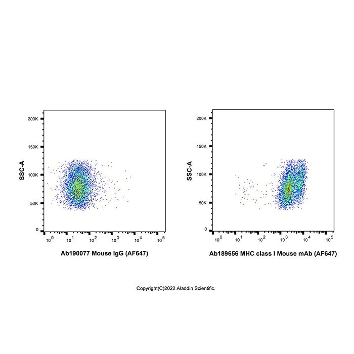
MHC class I Mouse mAb (AF647) (Ab189656) - Flow Cytometry
Flow cytometry analysis of MHC class I expression by C57BL/6 mouse splenocytes (right panel) compared to an isotype control (left panel). Cells were stained with MHC class I Mouse mAb (AF647) (Ab189656) at 0.2μg/test for 1 hour at 4°C. Left panel - Mouse IgG (AF647) (Ab190077). 

MHC class I Mouse mAb (AF647) (Ab189656) - Flow Cytometry
Flow cytometry analysis of MHC class I expression by C57BL/6 mouse splenocytes (red) compared to an isotype control (blue). Cells were stained with MHC class I Mouse mAb (AF647) (Ab189656) at 0.2μg/test for 1 hour at 4°C. Blue - Mouse IgG (AF647) (Ab190077). Black - Unlabelled control, cells without incubation with primary antibody.

Applicazione
Applicazione
Dilution info
Flow Cytometry

≤0.3 μg/test, the suggested use of this reagent is ≤0.3 μg per million cells in 100 µL staining volume

Documentazione

📋 Scheda di dati di sicurezza (SDS)

Documento completo su pericoli, manipolazione, stoccaggio e conformità normativa.

<<<<<<< HEAD Scarica SDS → ======= Scarica SDS → >>>>>>> release248-p3

✅ Certificato di analisi (COA)

Dati di qualità specifici del lotto. Inserisci il numero di lotto per recuperare il COA esatto.

Cerca COA →

📊 Scheda tecnica

Riepilogo di riferimento rapido delle specifiche e applicazioni del prodotto.

Visualizza scheda tecnica →

🔬 Scheda delle specifiche

Attributi di qualità completi e criteri di accettazione per questa qualità.

Visualizza scheda delle specifiche →

Dati avanzati

Obiettivi associati (umani)
HLA-A Tbio HLA class I histocompatibility antigen, A-31 alpha chain (0 Activities)
Activity TypeActivity Value -log(M)Mechanism of ActionActivity ReferencePublications (PubMed IDs)
Certificati (CoA, COO, BSE/TSE e tabella di analisi)
C of A & Other Certificates(BSE/TSE, COO):
Analytical Chart:

Find and download the COA for your product by matching the lot number on the packaging.

3 results found

Lot NumberCertificate TypeDataOggetto
ZJ24F1113782ZJ24F1113782Certificate of AnalysisJun 15, 2026 Ab189656
ZJ24F1113783ZJ24F1113783Certificate of AnalysisJun 15, 2026 Ab189656
ZJ24F1113784ZJ24F1113784Certificate of AnalysisJun 15, 2026 Ab189656
Domande frequenti e articoli
Calcolatori di soluzioni
Recensioni

Recensioni dei clienti

Application Protocols

Manufacturer-tested/validated applications and recommended conditions: Not specified for this item; refer to CoA/Spec Sheet.

General procedural outlines (for planning; not item specifications):

  • Flow cytometry (surface staining): Wash cells with PBS + protein carrier. Block Fc receptors if appropriate. Incubate with the AF647-conjugated antibody in the dark. Wash and analyze on a cytometer equipped with a 633–640 nm laser and far‑red detector (e.g., 660/20 nm). Include viability dye and appropriate controls.
  • Immunofluorescence (cells or sections): Fix (e.g., paraformaldehyde), block with serum/BSA, incubate with the antibody in blocking buffer, wash, and mount with antifade media. Image using far‑red channel settings (Ex ~633–640 nm; Em ~660–700 nm).
  • Western blot (optional approach): After transfer and blocking, incubate membrane with the AF647-conjugated antibody, wash, and image on a near‑IR imager. Note that not all clones recognize denatured epitopes; verify empirically.

Critical notes:

  • Optimize antibody amount, incubation time, and temperature empirically for each assay and sample type.
  • Maintain light protection throughout to preserve AF647 fluorescence.
  • Follow any lot-specific instructions and storage conditions provided with the product.
Biological Roles

Focus: MHC class I biology (general background; not product performance claims).

  • Function: MHC class I molecules present endogenous peptide antigens to CD8+ T cells, enabling immune surveillance of intracellular pathogens and malignantly transformed cells. They are expressed on nearly all nucleated vertebrate cells (literature).
  • Composition: A polymorphic heavy chain (α1–α3 domains) noncovalently associated with β2‑microglobulin and bound peptide. Proper assembly in the endoplasmic reticulum depends on the peptide loading complex (TAP, tapasin, calreticulin, ERp57) (literature).
  • Regulation: Expression is modulated by cytokines (e.g., IFN‑γ upregulates MHC I), cellular stress, and viral immune evasion strategies. Aberrant expression levels can influence cytotoxic T‑cell responses and NK cell activity (literature).
  • Localization: Predominantly at the plasma membrane; internalization and recycling occur, and peptide editing affects surface stability (literature).
  • Research applications enabled by this antibody (general): Quantifying basal and cytokine-induced MHC I expression, assessing antigen presentation competency, monitoring immune modulation in cell lines and primary cells, and validating genome editing/knockdown effects on antigen processing pathways.

Item-specific target epitope, isotype, and species reactivity: Not specified for this item; refer to CoA/Spec Sheet.

Buffer Applications

Applicability:

  • While not a buffer component, this AF647-conjugated antibody is typically used in standard biological buffers.

General buffer recommendations (literature/best practice):

  • Staining/wash buffer: PBS (10–20 mM phosphate, 150 mM NaCl, pH 7.2–7.4) with 0.5–1% BSA or 1–5% serum to block nonspecific binding; optional 0.05–0.1% Tween 20 for wash steps in IF.
  • Flow cytometry: PBS + 1–2% FBS or BSA; include EDTA (1–5 mM) to reduce cell clumping if needed. Keep samples on ice or at 4°C to minimize capping/internalization for live-cell staining.
  • Fixation/permeabilization: For surface MHC I, fixation with 1–4% paraformaldehyde is often sufficient; permeabilization is typically unnecessary unless co-staining intracellular markers. Verify AF647 compatibility.
  • Storage buffer: Maintain antibody in supplier-provided buffer. If aliquoting, use low-protein-binding tubes and avoid repeated freeze–thaw (this item is specified Do not freeze).

Item-specific buffer composition and recommended dilutions: Not specified for this item; refer to CoA/Spec Sheet.

Green Alternatives

Applicability:

  • As a fluorescent antibody reagent, “green chemistry” considerations focus on minimizing hazardous preservatives, reducing waste, and selecting imaging/flow protocols that avoid toxic fixatives and solvents.

General guidance (literature/best practice):

  • Buffer systems: Use PBS/TBS without toxic biocides when biosafety permits; adopt sterile technique and small aliquots to minimize contamination in the absence of azide.
  • Waste minimization: Plan staining volumes carefully (e.g., microvolumes in flow cytometry) to reduce reagent use and hazardous waste generation.
  • Fixation choices: Prefer neutral buffered formalin alternatives or low-percentage paraformaldehyde with proper ventilation; avoid methanol fixation for AF647 if it diminishes fluorescence.
  • Imaging media: Choose low-toxicity, glycerol-based antifade mountants that are compatible with AF647 and avoid aromatic amine scavengers that can generate hazardous waste.
  • Energy and logistics: The product is shipped on wet ice and stored at 2–8°C (item-specific); consolidate orders and coordinate experiments to minimize repeated cold-chain shipments.

Comparison note (general):

  • When panel design allows, far‑red fluorophores like AF647 reduce the need for high laser powers and can improve signal-to-background on autofluorescent samples, potentially lowering acquisition times and associated energy use, though benefits are context dependent.
Pharmaceutical Uses

Scope:

  • This product is designated for research use only. It is not intended for human or animal therapeutic or diagnostic applications.

General context (non-clinical, manufacturing/research tools):

  • Process analytics: Fluorescent antibodies are used in development laboratories for cell line characterization, monitoring MHC I expression during cell culture optimization, and assessing immunogenicity risks in bioprocess development (research setting).
  • Companion tools: In preclinical research, AF647-conjugated antibodies serve as analytical reagents for flow cytometry, imaging, and blotting—supporting target verification and mechanism studies.

Regulatory status:

  • No pharmacopeial monograph or GMP grade is indicated. Any use in regulated workflows would require appropriate qualification/validation and vendor documentation.

Item-specific claims on clinical/diagnostic use: None; For research use only (as provided).

Physical Properties

Item-specific properties:

  • Physical state/appearance: Not specified for this item; refer to CoA/Spec Sheet.
  • Concentration, buffer formulation, and protein content: Not specified for this item; refer to CoA/Spec Sheet.
  • Excitation/Emission (AF647): Ex 650 nm, Em 668 nm (provided).

Literature/general values for context (not item specifications):

  • Antibody approximate MW: ~150 kDa for an intact IgG (without accounting for dye load).
  • AF647 photophysics: Bright far‑red fluorophore with typical excitation peak ~650 nm and emission peak ~668 nm; high photostability relative to many cyanine dyes.
  • Solubility: IgG antibodies are soluble in aqueous buffers (e.g., PBS) at neutral pH; stability is enhanced with protein stabilizers (e.g., BSA) and sugars (e.g., trehalose) (literature). Specific buffer excipients for this item are not specified.
  • pI/logP/pKa: Not applicable as such for intact antibodies; the protein exhibits a distribution of pKa values across ionizable residues (literature). Not specified for this item.
  • Refractive index/density: Not typically defined for antibody solutions beyond the solvent; not specified for this item.

Practical notes (general):

  • AF647 detection aligns with common far‑red channels (e.g., 633–640 nm laser excitation) and emission detection around 660–680 nm.
  • Avoid high-intensity light exposure during handling to minimize photobleaching; store protected from light (item-specific storage guidance provided under Storage & Reconstitution).
Quality and Grades

Item-specific quality designation:

  • Listed grade: ExactAb™, Validated; AF647 conjugate (Ex 650 nm / Em 668 nm).

Interpretation and implications (general guidance):

  • “Validated” typically indicates the product has undergone internal quality control to confirm identity, integrity, and performance characteristics in one or more assay formats. However, the specific validation assays, acceptance criteria, and lot-release tests for this item are not specified; refer to the product CoA/Spec Sheet for details.
  • ExactAb™: This appears to be a supplier-specific program/mark. Without a formal definition provided in the Product Data, users should consult the vendor documentation for the scope (e.g., clonality confirmation, conjugation QC, residual free dye testing, aggregate assessment). Not specified for this item otherwise.

Conjugate-specific considerations (general):

  • AF647-labeled antibodies are commonly assessed for degree of labeling (DOL), free dye content, and functional binding post-conjugation (e.g., by antigen-positive cell staining or ELISA). Actual DOL and test results are not specified for this item.
  • Fluorochrome choice affects panel design: AF647 occupies the far‑red channel; verify tandem dye conflicts and compensation controls when multiplexing.

What to check on receipt (best practice):

  • Confirm lot number and any provided QC metrics (protein concentration, purity, endotoxin level, preservative, DOL) on the CoA/Spec Sheet—these are not specified in the Product Data here.
  • Inspect for shipping integrity (remained cold, not frozen) and fluorescence performance controls as applicable.
Reaction and Applications

Scope:

  • As an AF647-conjugated monoclonal antibody, this reagent is used for detection/quantification of MHC class I by fluorescence-based methods rather than for chemical synthesis.

Common research applications (literature/general):

  • Flow cytometry: Surface staining of live or fixed cells to quantify MHC I expression; AF647 channel minimizes spectral overlap with FITC/PE and suits 633–640 nm laser excitation.
  • Fluorescence microscopy (IF/IC): Visualization of MHC I localization on cell membranes; far‑red emission reduces cellular autofluorescence.
  • High-content imaging and imaging flow cytometry: Multiplexing with other fluorophores; AF647’s brightness aids detection of low-abundance targets.
  • Immunoblotting: Less common for fluor-conjugates, but direct detection on near‑infrared imagers is feasible when antigen is properly denatured and epitope preserved.
  • Immunoprecipitation is typically performed with unconjugated antibodies; AF647 conjugation may sterically affect binding in capture formats (general caution).

Practical tips (general):

  • Titrate per lot and application to achieve optimal signal-to-noise; do not rely on fixed dilutions without optimization.
  • Include isotype controls and fluorescence-minus-one (FMO) controls for panel design.
  • Protect from light throughout staining and acquisition to preserve AF647 signal.

Item-specific application claims from manufacturer: Not specified for this item; refer to CoA/Spec Sheet.

Reaction Conditions

Not applicable as a substrate for chemical reactions. However, practical conditions for analytical use are relevant.

General conditions for common applications (literature guidance; not item specifications):

  • Flow cytometry surface staining: Perform at 2–8°C or room temperature for 15–30 minutes in the dark; wash with PBS-based buffer. Optimize antibody amount per 10^5–10^6 cells empirically. Avoid prolonged incubations that may increase background.
  • Immunofluorescence (fixed cells/tissue): Block with serum/protein buffer; incubate with the AF647-conjugated antibody under subdued light. Rinse thoroughly; mount with antifade media compatible with far‑red dyes. Minimize exposure to blue/green light sources.
  • Cytokine induction studies: For assessing MHC I regulation, treat cells with IFN‑γ (concentration/time per literature) and stain post-treatment to quantify upregulation; include untreated controls.

Controls and compensation (general):

  • Include isotype controls and FMO controls for panel design.
  • Set compensation using single-stained controls collected on the same instrument settings; AF647 typically requires compensation into adjacent far‑red/near‑IR channels.

Item-specific recommended dilutions, incubation times, and buffers: Not specified for this item; refer to CoA/Spec Sheet.

Safety and Handling

GHS/SDS information:

  • Signal word, hazard statements, GHS classification, pictograms: Not specified for this item; refer to SDS for authoritative safety information.

General laboratory precautions (good practice):

  • Personal protective equipment: Lab coat, gloves (nitrile recommended), and safety glasses when handling antibody solutions and any accompanying buffers or preservatives.
  • Engineering controls: Handle in a clean area; use biological safety cabinet if working with potentially biohazardous samples.
  • Incompatibilities: Protect from strong oxidizers and reactive chemicals. Protect from light to preserve AF647 fluorescence. Do not freeze this item per product instructions.
  • Hygiene: Avoid ingestion and contact with skin/eyes. Wash hands thoroughly after use. Do not pipette by mouth.

First-aid overview (general):

  • Skin/eye contact: Rinse with water for several minutes; remove contact lenses if present and easy to do. Seek medical attention if irritation persists.
  • Inhalation/ingestion: Move to fresh air; rinse mouth. Seek medical attention if symptoms occur.

Special risks/handling for fluorescent antibodies (general):

  • Photobleaching: Work under subdued light and store vials wrapped in foil or in amber containers.
  • Preservatives: Some antibody formulations contain sodium azide or other biocides; the presence/absence is not specified for this item. If azide is present, avoid contact with acids and copper-containing plumbing to prevent formation of explosive metal azides; consult SDS.
  • Biological use note: For research use only (as specified). Not for human or animal therapeutic or diagnostic use.
Solvent Selection

Applicability:

  • This product is a protein reagent intended for use in aqueous biological buffers. Organic solvent selection is generally not applicable.

General buffer/solvent guidance (literature/best practice):

  • Primary diluent: Phosphate-buffered saline (PBS, pH ~7.2–7.4) or Tris-buffered saline (TBS) are standard for antibodies. For flow cytometry and staining, PBS with 0.5–1% BSA or serum and 0.05–0.1% sodium azide (if compatible with downstream use) can reduce nonspecific binding and microbial growth.
  • Detergents: For immunofluorescence and some wash steps, 0.05–0.1% Tween 20 in PBS/TBS may reduce background. Avoid high detergent levels that can destabilize antibody-antigen interactions.
  • Additives: Carrier proteins (e.g., 0.1–1% BSA) and stabilizers (e.g., trehalose, glycerol) can enhance storage stability. Presence/absence for this item is not specified.
  • Organic co-solvents: Avoid DMSO or alcohols unless strictly necessary; they can quench fluorescence and denature antibodies. If mounting media contain antifade reagents, ensure AF647 compatibility.

Comparison note (general):

  • AF647 performs well in standard aqueous buffers and common mounting media formulated for far‑red dyes. If using clearing solvents or high refractive index media, verify AF647 stability with manufacturer data or small-scale tests.
Storage and Reconstitution

Item-specific storage and shipping conditions (from Product Data):

  • Storage: Store at 2–8°C.
  • Light sensitivity: Protect from light.
  • Do not freeze: Freezing is contraindicated for this item.
  • Shipping: Shipped on wet ice; do not freeze during transit.

Reconstitution and formulation:

  • Physical form, buffer composition, and whether reconstitution is required: Not specified for this item; refer to CoA/Spec Sheet. If provided as a liquid, gentle mixing by inversion is recommended; avoid vortexing to minimize aggregation and foaming.

Handling best practices (general):

  • Upon receipt, verify the vial remained cold and protected from light. Store immediately at 2–8°C in the original light-protective container.
  • Avoid repeated vial openings; if frequent use is expected and the formulation permits, prepare working aliquots under sterile conditions. Because this product must not be frozen, aliquotting is only applicable if aliquots are also stored at 2–8°C and used promptly within their stability window per the CoA.
  • Prevent microbial contamination by using sterile tips and buffers. If the formulation lacks preservatives, use small aliquots and practice aseptic technique.
  • Do not expose to high temperatures; minimize time at room temperature during experimental setup.

Stability/shelf life: Not specified for this item; refer to CoA/Spec Sheet for lot-specific expiry and stability guidance.

Structure and Identity

Brief: MHC class I Mouse mAb (AF647) is a fluorescently labeled mouse monoclonal antibody directed against major histocompatibility complex (MHC) class I, conjugated to the far‑red dye Alexa Fluor 647 (AF647).

Item-specific facts (from Product Data):

  • SKU: Ab189656
  • Product designation: MHC class I Mouse mAb (AF647)
  • Conjugate: AF647; excitation 650 nm, emission 668 nm
  • Grade: ExactAb™, Validated
  • Structural identifiers (SMILES, InChIKey, CAS, CID): Not specified for this item; refer to CoA/Spec Sheet.
  • Molecular formula and molecular weight: Not specified for this item; refer to CoA/Spec Sheet.

General structural description (literature/general knowledge):

  • Antibody class: Typically an IgG monoclonal antibody of mouse origin, composed of two heavy and two light chains linked by disulfide bonds; Y‑shaped heterotetramer with Fc and two Fab fragments.
  • Approximate size: Intact IgG is ~150 kDa; hydrodynamic diameter ~10–15 nm (literature).
  • Conjugation: AF647 is commonly attached via amide linkages formed between dye NHS esters and antibody lysine residues (literature). Degree of labeling (DOL) is a key parameter, but is not specified for this item.
  • Target: MHC class I heavy chain/complex on nucleated cells; exact epitope/clone not specified for this item.

2D/functional features in words (general):

  • Two antigen-binding sites (paratopes) in the Fab regions recognizing an epitope on MHC I.
  • A far‑red fluorophore (AF647) covalently linked on surface-exposed lysines enabling detection in the 650/668 nm channel, with minimal spectral overlap into green/yellow channels (literature).
Synthetic Utility

Not a chemical building block; synthetic utility in classical organic synthesis does not apply.

Related conjugation chemistry (general background):

  • AF647 coupling: Commonly achieved via N‑hydroxysuccinimide (NHS) ester reaction with solvent-exposed lysine ε‑amines on the antibody at pH ~8.3, producing stable amide bonds. Alternative site-specific strategies include enzymatic glycan remodeling or cysteine-selective maleimide chemistry on reduced interchain disulfides.
  • Degree of labeling (DOL): Typically optimized to balance brightness and antigen binding; excessive labeling can reduce affinity or increase aggregation. DOL for this item is not specified.
  • Purification: Post-conjugation removal of free dye by desalting, spin columns, or size-exclusion chromatography is standard practice.

Use in assay assembly (general):

  • As a directly labeled detection reagent, it removes the need for secondary antibodies, simplifying workflows and reducing variability in multiplex applications.

Item-specific conjugation details (chemistry, DOL, residual free dye): Not specified for this item; refer to CoA/Spec Sheet.

Target Specificity

Item-specific details required for rigorous targeting information are not provided in the Product Data. Accordingly:

  • Antigen/epitope: Not specified for this item; refer to CoA/Spec Sheet.
  • Clone name/ID: Not specified for this item; refer to CoA/Spec Sheet.
  • Isotype/subclass: Not specified for this item; refer to CoA/Spec Sheet.
  • Species reactivity/cross-reactivity: Not specified for this item; refer to CoA/Spec Sheet.
  • Conjugate: AF647 (Ex 650 nm / Em 668 nm) — provided.

General background (for context only):

  • “MHC class I” commonly refers to the heavy chain isoforms forming antigen-presenting complexes with β2‑microglobulin on most nucleated cells. Specific clones may recognize pan‑MHC I or allele-restricted determinants. Without clone/epitope information, users should verify compatibility with their species/allele of interest.

Domande frequenti

What does ExactAb? mean?
ExactAb? indicates the product has been validated for use in rigorous antibody validation workflows. Validation may include positive/negative control samples, signal-to-noise testing, and protocol-specific QC. Lot-specific test data is published on the Certificate of Analysis where available.
What does Validated mean?
Validated indicates the product has been validated for use in rigorous antibody validation workflows. Validation may include positive/negative control samples, signal-to-noise testing, and protocol-specific QC. Lot-specific test data is published on the Certificate of Analysis where available.
How should this product be stored?
Store at 2–8 °C, protected from light. Do not freeze. Freezing may cause irreversible damage to this material.
How is this product shipped?
This product ships chilled on wet ice. Unpack on arrival and transfer it to the storage condition stated above.
What documentation is provided?
Available product documentation, including Certificates of Analysis (COA), Safety Data Sheets (SDS), and specification sheets, is shown in the product document area. Document availability and access follow the current site policy.
Which species reactivity has this antibody been validated against?
Validated reactivity is listed on the PDP and on each lot's datasheet. We validate against the most commonly tested species for the target (MHC class I Mouse mAb (AF647)); cross-reactivity in untested species may exist but is not guaranteed. Contact technical support for specific species inquiries.

Hai bisogno di aiuto per scegliere la qualità?

La nostra guida alla selezione delle qualità copre purezza, stato dello stabilizzatore e idoneità all'applicazione per tutte le varianti del nostro catalogo.

Visualizza la guida alle qualità Validated → Visualizza la guida alle qualità ExactAb™ → Visualizza la guida alle qualità Ex:650nm → Visualizza la guida alle qualità Em:668nm →

Prodotti correlati

Goat Anti-Mouse IgG H&L Antibody - Anticorpo secondario, for ELISA,LF,GICA various applications, from Mouse, generated in Goat
Goat Anti-Mouse IgG H&L (HRP) - Anticorpo secondario, for WB,ELISA,IHC-P various applications, from Mouse, generated in Goat, IgG
Mouse IgG - Anticorpo di controllo isotipo, generated in Rat, >95%, IgG
Rabbit Anti-Mouse IgG H&L (HRP) - Anticorpo secondario, IgG
BALB/c Mouse IgG - Anticorpo di controllo isotipo, IgG
Rabbit Anti-Mouse IgG antibody - Anticorpo secondario
Goat Anti-Mouse IgG H&L (FITC) - Anticorpo secondario
Goat Anti-Mouse IgG H&L (TRITC) - Anticorpo secondario
Goat Anti-Mouse IgG H&L Antibody - Anticorpo secondario, Goat IgG
Goat Anti-Mouse IgG H&L (Cy5) - Anticorpo secondario
Goat Anti-Mouse IgG H&L (HRP) - Anticorpo secondario
Goat Anti-Mouse IgG H&L (Biotin) - Anticorpo secondario
Rabbit Anti-Mouse IgG (HRP) - Anticorpo secondario
Rabbit Anti-Mouse IgG (Biotin) - Anticorpo secondario
Goat Anti-Mouse IgG H&L (HRP) - Anticorpo secondario, Goat IgG
Goat Anti-Mouse IgG H&L (TRITC) - Anticorpo secondario, Goat IgG
Goat Anti-Mouse IgG H&L (Biotin) - Anticorpo secondario, Goat IgG
Goat Anti-Mouse IgG H&L (FITC) - Anticorpo secondario, Goat IgG
Goat Anti-Mouse IgG H&L (Cy5) - Anticorpo secondario, Goat IgG
Goat Anti-Mouse IgG H&L (PE) - Anticorpo secondario, Goat IgG
Goat Anti-Mouse IgG H&L (APC) - Anticorpo secondario, Goat IgG
Mouse IgG (Biotin) - Anticorpo di controllo isotipo, Mouse IgG
Mouse IgG (PE) - Anticorpo di controllo isotipo, Mouse IgG
Mouse IgG (APC) - Anticorpo di controllo isotipo, Mouse IgG
Mouse IgG (FITC) - Anticorpo di controllo isotipo, Mouse IgG
Mouse IgG2a-Fc - Anticorpo di controllo isotipo, Mouse IgG2a
Capra Anti-Mouse IgG H&L (Fosfatasi alcalina) - Anticorpo secondario, Goat IgG
MAb di topo di classe MHC I - Anticorpo primario, specific to MHC class I, Mouse IgG2a
Goat Anti-Mouse IgG H&L (AF405) - Anticorpo secondario, Goat IgG
Goat Anti-Mouse IgG H&L (AF647) - Anticorpo secondario, Goat IgG
Mouse IgG2b-kappa, anti-Spike RBD - Anticorpo di controllo isotipo, specific to Spike RBD, Mouse IgG2b
Mouse IgG2a-kappa, anti-Spike RBD - Anticorpo di controllo isotipo, specific to Spike RBD, Mouse IgG2a
Mouse IgG3-kappa, anti-HEL - Anticorpo di controllo isotipo, specific to HEL, Mouse IgG3
Mouse IgG2c-kappa, anti-HEL - Anticorpo di controllo isotipo, specific to HEL, Mouse IgG2c
Mouse lgG2A(D265A)-kappa, anti-HEL - Anticorpo di controllo isotipo, specific to HEL, Mouse IgG2a(D265A)
Mouse IgG1(D265A)-kappa, anti-HEL - Anticorpo di controllo isotipo, specific to HEL, Mouse IgG1(D265A)
Mouse IgG (AF488) - Anticorpo di controllo isotipo, IgG
Mouse IgG (PerCP) - Anticorpo di controllo isotipo, IgG
Mouse IgG (AF405) - Anticorpo di controllo isotipo, IgG
Mouse IgG (AF647) - Anticorpo di controllo isotipo, IgG
Goat Anti-Mouse IgG H&L (AF555) - Anticorpo secondario, specific to Mouse IgG, Goat IgG
Mouse IgG (AF700) - Anticorpo di controllo isotipo, IgG
Goat Anti-Mouse IgG H&L (AF700) - Anticorpo secondario, specific to Mouse IgG, Goat IgG
Mouse IgG (PE-Cy5) - Anticorpo di controllo isotipo
Mouse IgG (PE-Cy7) - Anticorpo di controllo isotipo
Mouse IgG2b-kappa, anti-E8L - Anticorpo di controllo isotipo, specific to E8L
Mouse IgG2a-kappa, anti-A29L - Anticorpo di controllo isotipo, specific to A29L
MHC class I Mouse mAb (APC) - Anticorpo primario, specific to MHC class I
MHC class I Mouse mAb (Biotin) - Anticorpo primario, specific to MHC class I
MHC class I Mouse mAb (FITC) - Anticorpo primario, specific to MHC class I
Mouse IgG2c-kappa (AF647), anti-HEL - Anticorpo di controllo isotipo, specific to HEL
Mouse IgG2c-kappa (AF700), anti-HEL - Anticorpo di controllo isotipo, specific to HEL
Mouse IgG2c-kappa (AF405), anti-HEL - Anticorpo di controllo isotipo, specific to HEL
Mouse IgG2c-kappa (Biotin), anti-HEL - Anticorpo di controllo isotipo, specific to HEL
Mouse IgG2a-kappa (APC), anti-Spike RBD - Anticorpo di controllo isotipo, specific to Spike RBD
Mouse IgG2a-kappa (FITC), anti-Spike RBD - Anticorpo di controllo isotipo, specific to Spike RBD
Mouse IgG2a-kappa (AF700), anti-Spike RBD - Anticorpo di controllo isotipo, specific to Spike RBD
Mouse IgG2a-kappa (AF647), anti-Spike RBD - Anticorpo di controllo isotipo, specific to Spike RBD
Mouse IgG2a-kappa (AF555), anti-Spike RBD - Anticorpo di controllo isotipo, specific to Spike RBD
Mouse IgG2a-kappa (AF405), anti-Spike RBD - Anticorpo di controllo isotipo, specific to Spike RBD
Mouse IgG2c-kappa (FITC), anti-HEL - Anticorpo di controllo isotipo, specific to HEL
Mouse IgG2c-kappa (AF555), anti-HEL - Anticorpo di controllo isotipo, specific to HEL
Mouse IgG2c-kappa (APC), anti-HEL - Anticorpo di controllo isotipo, specific to HEL
Mouse IgG2c-kappa (PE), anti-HEL - Anticorpo di controllo isotipo, specific to HEL
Mouse IgG2a-kappa (Biotin), anti-Spike RBD - Anticorpo di controllo isotipo, specific to Spike RBD
Mouse IgG2a-Fc (Biotin) - Anticorpo di controllo isotipo
Mouse IgG2a-Fc (FITC) - Anticorpo di controllo isotipo
Mouse IgG (AF555) - Anticorpo di controllo isotipo
Mouse IgG2b-kappa (Biotin), anti-Spike RBD - Anticorpo di controllo isotipo, specific to Spike RBD
Mouse IgG2b-kappa (FITC), anti-Spike RBD - Anticorpo di controllo isotipo, specific to Spike RBD
Mouse IgG2b-kappa (APC), anti-Spike RBD - Anticorpo di controllo isotipo, specific to Spike RBD
Mouse IgG2b-kappa (APC-Cy7), anti-Spike RBD - Anticorpo di controllo isotipo, specific to Spike RBD
Goat Anti-Mouse IgG H&L (PE-Cy5) - Anticorpo secondario, specific to Mouse IgG
Centrose patent anti-dysadherin (anti-Dysadherin) - Anticorpo primario, specific to FXYD5
Mouse IgG2a-kappa (AF405), anti-A29L - Anticorpo di controllo isotipo, specific to A29L
Mouse IgG2a-kappa (AF488), anti-A29L - Anticorpo di controllo isotipo, specific to A29L
Mouse IgG2a-kappa (AF647), anti-A29L - Anticorpo di controllo isotipo, specific to A29L
Mouse IgG2a-kappa (AF700), anti-A29L - Anticorpo di controllo isotipo, specific to A29L
Mouse IgG2a-kappa (Biotin), anti-A29L - Anticorpo di controllo isotipo, specific to A29L
Mouse IgG2a-kappa (FITC), anti-A29L - Anticorpo di controllo isotipo, specific to A29L
Mouse IgG3-kappa (Biotin), anti-HEL - Anticorpo di controllo isotipo, specific to HEL
Mouse IgG3-kappa (FITC), anti-HEL - Anticorpo di controllo isotipo, specific to HEL
Mouse IgG3-kappa (PE), anti-HEL - Anticorpo di controllo isotipo, specific to HEL
Mouse IgG3-kappa (AF488), anti-HEL - Anticorpo di controllo isotipo, specific to HEL
Mouse IgG3-kappa (APC), anti-HEL - Anticorpo di controllo isotipo, specific to HEL
Mouse IgG3-kappa (AF647), anti-HEL - Anticorpo di controllo isotipo, specific to HEL
Mouse IgG2b-kappa (AF555), anti-Spike RBD - Anticorpo di controllo isotipo, specific to Spike RBD
Goat Anti-Mouse IgG H&L (AF488) - Anticorpo secondario
Goat Anti-Mouse IgG H&L (PE-Cy7) - Anticorpo secondario
Mouse IgG (PE-Cy5.5) - Anticorpo di controllo isotipo
Goat Anti-Mouse IgG H&L (AF750) - Anticorpo secondario
Mouse IgG2a-kappa (AF555), anti-A29L - Anticorpo di controllo isotipo, specific to A29L
Mouse IgG2a-kappa (PE-Cy5.5), anti-A29L - Anticorpo di controllo isotipo, specific to A29L
Goat Anti-Mouse IgG H&L (AF546) - Anticorpo secondario
Goat Anti-Mouse IgG H&L (Oregon Green 488) - Anticorpo secondario
Goat Anti-Mouse IgG H&L (Cy3) - Anticorpo secondario
Mouse IgG2a-kappa (APC), anti-A29L - Anticorpo di controllo isotipo, specific to A29L
Mouse IgG3-kappa (AF555), anti-HEL - Anticorpo di controllo isotipo, specific to HEL
Mouse IgG (PE-AF647) - Anticorpo di controllo isotipo

Desidera che le inviamo un messaggio quando avremo sconti disponibili?

Ricordamelo più tardi

Grazie! Controlli la sua casella di posta per confermare.

Ops! Le notifiche sono disabilitate.