
What Determines Collagen Function: From Triple-Helix Formation to Hierarchical Assembly, Crosslink Maturation, and Dynamic Remodeling
What Determines Collagen Function: From Triple-Helix Formation to Hierarchical Assembly, Crosslink Maturation, and Dynamic Remodeling
Introduction
Collagen is often described as a “supporting component” of the skin, bone, cartilage, and tendons. Although this description reflects the structural role of collagen, it can easily lead to an oversimplified assumption: the higher the collagen content, the better the condition of the tissue.
In reality, collagen content indicates only how much collagen is present in a tissue; it does not, by itself, reveal whether that collagen has formed normally functioning structures. Inappropriate collagen types, abnormal triple-helix folding, impaired fibril assembly, disordered fiber alignment, abnormal degrees of crosslinking, and imbalances between synthesis and degradation can all weaken tissue function. In scars and organ fibrosis, collagen deposition may increase substantially, while normal tissue architecture and mechanical properties are nevertheless disrupted.[12,13]
The development of collagen function involves a continuous structural hierarchy:
Amino acid sequence and post-translational modifications → formation of a triple helix by three α chains → procollagen processing and extracellular assembly → formation of fibrils, fibers, or network structures → spatial organization and intermolecular crosslinking → tissue mechanics and cellular regulation → synthesis, degradation, and continuous remodeling
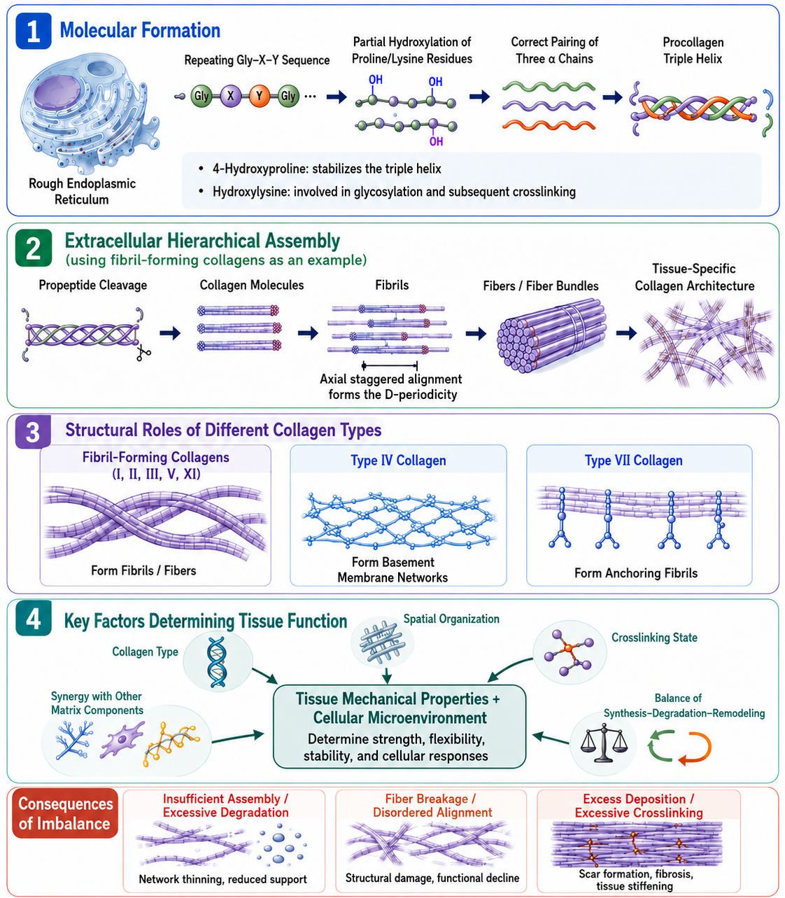
Collagen function is established through the combined contribution of multiple structural levels. Gly–X–Y repeat sequences, hydroxylation, and α-chain pairing generate a stable procollagen triple helix. Following propeptide cleavage, different collagen types further assemble into fibrils, basement membrane networks, or anchoring fibrils. Collagen type, spatial organization, crosslinking state, other matrix components, and the balance among synthesis, degradation, and remodeling collectively determine tissue mechanical properties and the cellular microenvironment. A schematic illustration of collagen from triple-helix formation and extracellular hierarchical assembly to tissue function and dynamic remodeling is shown below.
1 What Is Collagen?
1.1 Collagen Is a Protein Family
Collagens are a group of extracellular matrix proteins characterized by collagen triple-helical domains. Different collagens are encoded by different genes and vary in their α-chain composition, non-collagenous domains, tissue distribution, and higher-order assembly patterns.[1]
According to their higher-order modes of assembly, collagens can be broadly classified as follows:
Structural category | Representative types | Major higher-order structures | Representative functions |
Fibril-forming collagens | Types I, II, III, V, and XI | Fibrils, fibers, and fiber bundles | Build the load-bearing framework of tissues such as skin, tendon, bone, and cartilage |
Network-forming collagens | Type IV | Basement membrane collagen networks | Form the principal structural scaffold of basement membranes |
Anchoring collagen | Type VII | Anchoring fibrils | Connect basement membranes to the underlying connective tissue |
Fibril-associated collagens | Types IX, XII, and XIV, among others | Associated with the surfaces of collagen fibers | Regulate interactions between fibers and surrounding matrix components |
Transmembrane collagens | Types XIII and XVII, among others | Transmembrane structures | Participate in connections between cells and the extracellular matrix |
This classification reflects the distinct structural tasks performed by different collagens. Type I collagen is suited to forming load-bearing fibrils, type IV collagen primarily forms basement membrane networks, and type VII collagen forms anchoring fibrils. Although they all contain triple-helical domains, they cannot substitute for one another.[1,6–8]
1.2 Collagen Is an Important Component of the Extracellular Matrix
The extracellular matrix (ECM) is an extracellular network composed of collagen, elastin, fibronectin, laminin, proteoglycans, glycosaminoglycans, and other components.
Collagen primarily provides tensile resistance and structural organization, but the overall properties of a tissue also depend on other components. In the skin, collagen fibers, elastic fibers, proteoglycans, and water collectively influence the strength and flexibility of the dermis. In cartilage, the type II collagen network restrains excessive swelling of the water-rich proteoglycan system and works together with it to bear complex loads. In bone, type I collagen forms the organic matrix framework, whereas the mineral phase provides relatively high stiffness and compressive strength.
Tissues with similar collagen contents may nevertheless exhibit completely different mechanical properties because of differences in collagen type, fiber orientation, crosslinking state, and other matrix components.
1.3 Collagen Is Also Part of the Cellular Microenvironment
Cells can recognize collagen structures through collagen-binding receptors and sense collagen fiber density, orientation, ligand accessibility, and local mechanical conditions. Changes in the collagen environment can affect cell adhesion, morphology, cytoskeletal organization, migration, and matrix synthesis.
Cells can also remodel the collagen network in return. Fibroblasts and other cells can secrete collagen, exert traction on fibers, alter fiber orientation, and cleave the surrounding matrix through proteases. Collagen is therefore not a passive material that remains unchanged after formation, but rather a tissue environment that is continuously constructed and remodeled by cells.[11,14]
2 How Is the Triple Helix Formed?
2.1 Gly–X–Y Repeat Sequences Are the Structural Basis of Collagen
Collagen triple-helical domains are generally composed of continuous Gly–X–Y tripeptide repeat sequences. Gly represents glycine, while the X and Y positions can contain various amino acids, among which proline and 4-hydroxyproline are particularly common.
The side chain of glycine consists of only one hydrogen atom and is therefore very small, allowing it to occupy the tightly packed center of the triple helix. If glycine at this position is replaced by a bulkier amino acid, the close packing of the three chains may be disrupted. Proline restricts the conformation of the polypeptide backbone, while 4-hydroxyproline helps preorganize chain conformation and enhances the thermal stability of the triple helix through stereoelectronic effects and other mechanisms.[2,17]
Different Gly–X–Y tripeptides do not contribute equally to stability, and the collagen triple helix may therefore exhibit local differences in stability along the length of the molecule. The triple-helical domains of non-fibril-forming collagens may also contain interruptions in the Gly–X–Y repeats. These interruptions alter local flexibility and contribute to the formation of higher-order structures and functions.
The “α” in a collagen “α chain” is part of the nomenclature for collagen polypeptide chains and does not refer to the α helix found in the secondary structure of ordinary proteins. A single collagen chain mainly adopts an extended conformation, and three chains then wind around one another to form the characteristic right-handed collagen triple helix.
2.2 Hydroxylation Supports Collagen Folding and Maturation
After procollagen α chains enter the endoplasmic reticulum, some proline and lysine residues undergo hydroxylation. After procollagen α chains enter the endoplasmic reticulum, some proline and lysine residues undergo hydroxylation. Collagen prolyl hydroxylases, particularly collagen prolyl 4-hydroxylase, and lysyl hydroxylases are Fe²⁺- and 2-oxoglutarate-dependent dioxygenases.[3]
Hydroxylation of proline primarily produces hydroxyproline, whereas hydroxylation of lysine primarily produces hydroxylysine. These reactions require Fe²⁺. Ascorbic acid helps maintain the iron ion in the active site of the hydroxylases in its reduced state, allowing the enzymes to sustain effective catalysis.[3]
4-Hydroxyproline plays an important role in stabilizing the triple helix at human body temperature. Hydroxylysine has somewhat different functions: some hydroxylysine residues can undergo further glycosylation, while certain sites also participate in intermolecular collagen crosslinking. Hydroxylation therefore affects both collagen molecular folding and extracellular fiber maturation.
2.3 The Three α Chains Must Be Correctly Selected and Paired
Typical fibril-forming collagens are synthesized as procollagens. Procollagen contains a central triple-helical domain and N- and C-terminal propeptides at both ends.
The C-terminal propeptides participate in α-chain recognition and trimer formation. After three appropriate α chains have paired, triple-helix folding generally proceeds from the C terminus toward the N terminus. For example, a common type I collagen molecule consists of two α1(I) chains and one α2(I) chain.
The synthesis of α chains does not necessarily mean that normal collagen will be formed. Incorrect chain selection, insufficient hydroxylation, abnormal molecular chaperone function, or altered folding kinetics may affect triple-helix stability, secretion efficiency, and subsequent assembly.[3]
The N- and C-terminal propeptides also increase procollagen solubility and prevent premature fibril formation within the cell. Thus, procollagen has already formed a triple helix inside the cell but has not yet entered the mature fibrillar state.
3 How Do Collagen Molecules Assemble into Higher-Order Structures?
3.1 Propeptide Processing Initiates the Assembly of Fibril-Forming Collagens
After procollagen is secreted, the N- and C-terminal propeptides are generally removed by the corresponding proteases. Once the propeptides have been cleaved, collagen molecules become less soluble and can align with a specific axial stagger, forming periodic fibrillar structures containing gap and overlap regions.
Fibril formation does not occur merely through the random precipitation of collagen molecules in the extracellular space. In situ studies of tendon fibroblasts have shown that some processing of type I procollagen can begin in the later stages of the secretory pathway, suggesting that procollagen processing, secretion, and extracellular assembly may be spatially and temporally coordinated.[4]
3.2 Small Amounts of Type V Collagen Can Regulate Type I Collagen Fibril Formation
Collagen fibrils in tissues such as skin, tendon, and cornea are generally not composed of a single collagen type. Type I collagen is often the major quantitative component, whereas type V collagen participates as a less abundant regulatory component in the formation of heterotypic fibrils.
Mouse genetic studies have shown that Col5a1 deficiency severely impairs the initiation of type I collagen fibril assembly, indicating that type V collagen participates in fibril nucleation and diameter regulation. The low abundance of type V collagen does not imply that its structural role is weak.[5]
Cartilage fibrils are also composed of multiple collagen types. Type II collagen is the principal component, whereas types IX and XI collagen participate in surface interactions and regulation of fibril diameter.
3.3 Type IV Collagen Forms Basement Membrane Networks
Type IV collagen differs from typical fibril-forming collagens. Its triple-helical domain contains multiple interruptions in the Gly–X–Y repeats, giving the molecule greater flexibility, while domains at both ends of the molecule participate in specific interactions.
The N-terminal 7S region of type IV collagen molecules can form multimolecular junctions, whereas the C-terminal non-collagenous domain 1 (NC1) participates in α-chain selection and connects two type IV collagen protomers. Interactions at both ends collectively construct the type IV collagen network in basement membranes.[6,7]
The type IV collagen network works together with laminin, nidogen, basement membrane proteoglycans, and other components to maintain basement membrane structure. Its principal role is not to form thick fiber bundles with typical periodic banding, but to establish a continuous and relatively flexible tissue interface.
3.4 Type VII Collagen Forms Anchoring Fibrils
Type VII collagen molecules can form antiparallel dimers stabilized by disulfide bonds and then laterally aggregate into anchoring fibrils. Anchoring fibrils are located beneath the basement membrane and connect it to the connective tissue of the papillary layer, helping maintain mechanical continuity between the epidermis and dermis.[8]
When the structure or assembly of type VII collagen is abnormal, the connection between the epidermis and underlying connective tissue may become fragile even if other basement membrane components remain present.
4 How Do Spatial Organization and Crosslinking Establish Tissue Function?
4.1 Fiber Alignment Determines How Loads Are Transmitted
The mechanical properties of a single collagen fiber cannot fully represent those of an entire tissue. Fiber orientation, diameter, density, curvature, branching, and interlamellar connections collectively determine how loads are transmitted.
In tendons, collagen fibers are primarily aligned along the direction of force, which facilitates the transmission of directional tensile forces generated by muscles. In the dermis, collagen fibers interweave in multiple directions, allowing the skin to accommodate tension and deformation from different directions. In articular cartilage, collagen fibers adopt different orientations at different depths and work together with proteoglycans and water to form a layered load-bearing structure.
When a collagen network is stretched, initially curved or wavy fibers may first rotate and straighten, after which increasing numbers of fibers become load-bearing. The resulting mechanical response is generally not a fixed linear relationship. Reconstituted type I collagen networks in vitro can exhibit strain stiffening over a certain strain range, meaning that progressively greater force is required to continue deforming the network as deformation increases.[9]
This type of mechanical behavior arises from the combined action of multiple structural levels. The triple helix provides a stable molecular unit, fibrillar connections transmit loads at the molecular scale, and fiber alignment and the three-dimensional network determine load distribution at the tissue scale.
4.2 Lysyl Oxidase-Related Crosslinking Stabilizes Fibrils
After collagen fibrils have formed, members of the lysyl oxidase (LOX) family can catalyze the oxidative deamination of specific lysine or hydroxylysine residues in the telopeptide regions, producing the reactive aldehydes allysine or hydroxyallysine, respectively. These aldehyde groups then undergo non-enzymatic condensation with adjacent aldehyde or amino groups in neighboring collagen molecules, progressively forming divalent and multivalent intermolecular crosslinks.[10]
An appropriate degree of enzymatic crosslinking increases fibrillar continuity, tensile strength, and long-term stability. The extent of lysine hydroxylation and the pathways of crosslink formation differ among tissues, resulting in differences in the mechanical properties of mature collagen.
4.3 The Degree of Crosslinking Must Be Compatible with Tissue Turnover
When collagen crosslinking is insufficient, fibrils may lack adequate stability. When crosslinking is excessive, collagen fibers may become rigid and less susceptible to proteolytic cleavage.
In addition to LOX-related enzymatic crosslinking, long-lived collagen may also undergo non-enzymatic glycation. Reducing sugars and their derivatives react with amino groups in collagen and, following multiple rearrangement and oxidation steps, can form advanced glycation end products (AGEs). Some AGEs create additional linkages between collagen molecules.
Age-related modifications can alter collagen fiber stiffness, thermal stability, and susceptibility to proteolysis, although the effects vary among tissues and crosslink types. Increased collagen stiffness does not necessarily indicate enhanced tissue function and may instead be accompanied by reduced compliance and restricted turnover.[10]
4.4 Collagen Function Depends on the Entire Extracellular Matrix System
Fibronectin, proteoglycans, glycosaminoglycans, elastin, and water are also present around collagen fibers. Fibronectin can participate in collagen deposition and network organization. Proteoglycans can regulate fibril spacing, diameter, and the hydration environment. Cellular traction forces can straighten, move, and realign collagen fibers.
The same collagen type may form different fiber structures and exhibit different mechanical behaviors in different matrix environments. Collagen type provides the basic capacity for assembly, whereas tissue function is jointly determined by collagen proportions, spatial organization, crosslinking state, other matrix components, and cellular activity.
5 How Is the Collagen Network Continuously Renewed?
5.1 Increased Collagen Synthesis Does Not Necessarily Mean an Increase in Functional Networks
Several steps must occur between increased collagen gene expression and improved tissue function:
Gene transcription → α-chain translation → hydroxylation and glycosylation → triple-helix formation → procollagen secretion → propeptide processing → fibril assembly → spatial organization → crosslink maturation
An intervention that increases the expression of collagen genes such as COL1A1 demonstrates only that one stage of the synthetic process has changed. Whether newly synthesized collagen forms a stable triple helix, is normally secreted, assembles into fibrils, and correctly integrates with the existing network must still be evaluated using protein analysis, structural imaging, and mechanical testing.[3,4]
5.2 Collagen Degradation Is Part of Normal Remodeling
Matrix metalloproteinases (MMPs) participate in the degradation of collagen and other extracellular matrix components. MMP1, MMP8, MMP13, and related enzymes can cleave fibril-forming collagens at specific sites. The resulting fragments are then further degraded by other proteases or taken up and cleared by cells.
Normal collagen degradation serves several functions:
1. Removes damaged or structurally abnormal collagen;
2. Provides space for the assembly of newly synthesized collagen;
3. Adjusts fiber orientation and network porosity;
4. Supports tissue growth and wound repair;
5. Prevents the persistent, disordered accumulation of collagen.
A 2024 mouse skin study showed that, while depositing type I collagen, dermal fibroblasts can use membrane-type matrix metalloproteinase 14 (MMP14) to remodel the collagen fibrils surrounding the cells. Mmp14 deficiency impaired pericellular collagen remodeling and affected fibroblast survival, demonstrating that local proteolysis is not merely a process of tissue destruction but also participates in normal matrix formation.[11]
5.3 Collagen Fibers Continuously Grow and Rearrange under Cellular Activity
A 2025 live-cell imaging study used a fluorescent fusion-labeled type I collagen α2 chain to observe the intracellular transport, secretion, and extracellular fiber formation of collagen. Newly formed fibers were observed to extend along existing fibers and to undergo branching, bundling, looping, and mutual entanglement.[14]
These observations indicate that collagen networks are not formed through the random extracellular precipitation of molecules. The sites of cellular secretion, the orientation of existing fibers, local traction forces, and interactions among fibers all influence network growth.
The study used cell culture and a fluorescent fusion collagen system, allowing dynamic processes to be visualized at the cellular level. However, the observations still require validation against in situ structures in native human tissues.
5.4 Collagen Homeostasis Requires Both Stability and Renewability
Collagen state | Major change | Possible outcome |
Normal turnover | Synthesis, assembly, crosslinking, and degradation are coordinated | The network remains stable while retaining remodeling capacity |
Insufficient synthesis | Newly formed collagen cannot compensate for losses | The network becomes thinner and fiber continuity declines |
Excessive degradation | Mature fibers undergo persistent fragmentation | Tissue support and the cell-adhesion environment are impaired |
Insufficient degradation | Damaged or excess collagen is difficult to remove | Abnormal matrix accumulation |
Excessive crosslinking | Collagen stiffness increases and proteolysis becomes restricted | Tissue stiffening and reduced renewal capacity |
Abnormal assembly | Collagen is present but fails to form a normal network | Collagen content does not correspond to tissue function |
The collagen network must possess sufficient structural stability while also being capable of removing damaged components and incorporating newly synthesized collagen. Insufficient stability makes fibers susceptible to damage, whereas excessive stability may restrict normal remodeling.
6 How Does Collagen Imbalance Affect Tissues?
6.1 Collagen Loss Can Occur at Multiple Stages
A reduction in tissue collagen is not necessarily caused only by decreased collagen gene expression. It may also result from:
1. A reduction in the number or activity of collagen-producing cells;
2. Abnormal α-chain hydroxylation and triple-helix folding;
3. Impaired procollagen secretion or propeptide processing;
4. Reduced efficiency of fibril assembly;
5. Increased collagen degradation.
Reduced collagen staining or collagen content reveals only the final outcome and cannot directly identify the specific impaired stage. Distinguishing among insufficient synthesis, failed assembly, and enhanced degradation helps provide a more accurate understanding of tissue changes.
6.2 Collagen Fiber Fragmentation Can In Turn Affect Fibroblasts
Changes in collagen in aged skin involve not only a reduction in content but also fragmentation of type I collagen fibers, decreased continuity, and alterations in spatial organization. After collagen fibers become fragmented, the mechanical connections between fibroblasts and the intact matrix are reduced, altering cell spreading and tension and potentially further increasing matrix metalloproteinase 1 (MMP1) expression.[12]
Studies of human skin tissue and three-dimensional collagen models support a mutually reinforcing relationship among collagen fragmentation, reduced cellular tension, and increased collagen degradation. However, skin aging is also influenced by ultraviolet radiation, oxidative stress, inflammation, cellular senescence, and changes in other extracellular matrix components and therefore cannot be attributed to a single collagen pathway.
6.3 Excessive Collagen Deposition Can Also Disrupt Tissue Function
Increased collagen deposition during the early stages of wound repair helps restore tissue continuity. If profibrotic signaling persists, collagen synthesis remains greater than degradation, fibers continue to accumulate and undergo extensive crosslinking, and normal tissue architecture may gradually be replaced by dense matrix.
Studies of renal fibrosis have shown that elevated LOX expression and enhanced collagen crosslinking can promote matrix stiffening. In the corresponding experimental models, LOX inhibition reduced collagen crosslinking and alleviated some fibrotic features.[13]
Scarring and fibrosis cannot be evaluated solely on the basis of total collagen content. Collagen-type proportions, fiber orientation, degree of bundling, crosslinking state, matrix stiffness, and collagen clearance capacity must also be examined. Increased collagen deposition is not equivalent to restoration of normal tissue architecture.
7 Collagen Research Is Shifting from Content Assessment to Structural and Dynamic Evaluation
7.1 Second-Harmonic Generation Imaging Can Visualize the Spatial Structure of Collagen
Conventional hydroxyproline assays, histological staining, and immunological detection can assess total collagen or specific collagen types, but provide limited information about fiber orientation, hierarchical structure, and molecular organization.
Second-harmonic generation (SHG) microscopy does not require exogenous fluorescent labeling and is suitable for observing highly ordered fibril-forming collagens. Polarization-resolved SHG can also be used to analyze fiber orientation and certain molecular organization parameters.
A 2025 study used polarization-resolved SHG to compare engineered collagen matrices with different type I/type III collagen ratios, as well as collagen structures in healthy and scarred skin. The results showed that collagen-type proportions and tissue condition were associated with differences in imaging parameters and fiber orientation.[15]
SHG signals primarily reflect the nonlinear optical properties of ordered collagen structures and cannot independently replace collagen-type quantification, crosslink analysis, or tissue mechanical measurements.
7.2 Dynamic Imaging Complements Static Structural Studies
Live-cell collagen imaging can visualize collagen secretion, fiber elongation, and network rearrangement, compensating for the limitation of conventional fixed sections, which reveal only the final structure.[14]
A 2026 study also used time-resolved cryo-electron microscopy (cryo-EM) to observe the assembly of the simplified recombinant collagen model CF-1552 from triple helices into higher-order structures and captured spindle-shaped and axial assembly intermediates. The study proposed that collagen assembly may proceed through intermediate states that are relatively easy to rearrange before gradually transitioning into stable fibers.[16]
The study used an engineered recombinant collagen model lacking post-translational modifications and displaying relatively slow assembly, rather than full-length human collagen in native tissue. The axial staggering and lateral packing of CF-1552 intermediates still differed from those of native collagen, and the CF-1552 model itself did not reproduce the characteristic D-periodic banding of native fibril-forming collagens. The model therefore provides a controllable experimental system for investigating early hierarchical collagen assembly, but whether the proposed intermediates and assembly pathways are broadly applicable to native collagen still requires further verification using full-length collagen, in situ tissues, and other models.[16]
7.3 Collagen Function Requires Multilevel Evaluation
Evaluation level | Major indicators |
Synthesis level | Collagen gene expression and procollagen and mature collagen content |
Molecular level | α-chain composition, hydroxylation, glycosylation, and triple-helix stability |
Assembly level | Propeptide processing, fibril formation, and fiber diameter |
Spatial level | Fiber orientation, density, branching, continuity, and three-dimensional structure |
Chemical maturation level | Enzymatic crosslinking, non-enzymatic glycation, and crosslink types |
Dynamic level | Synthesis rate, degradation rate, and cell-mediated remodeling |
Functional level | Tissue stiffness, strength, toughness, and cellular responses |
8 Classification and Research Applications of Representative Chemicals Related to Collagen Structure Formation, Hydroxylation and Maturation, Crosslinking and Glycation, Dynamic Remodeling, and Analysis
Table 1. Products Related to Core Collagen Materials, Structural Controls, and Sample Preparation
Category | CAS No. | Aladdin Cat. No. | Name | Specification or Purity | Product Features and Applications |
Denatured collagen structural control | 9000-70-8 | Gelatin | Photographic grade, gel strength ~250 g Bloom | Used to study collagen triple-helix denaturation, gel formation, thermal responses, and structural differences between native and denatured collagen. | |
Medium for collagen extraction and dispersion | 64-19-7 | Glacial acetic acid | Guaranteed reagent, ≥99.5% | Used for acid-soluble collagen extraction, preparation of acidic collagen solutions, sample preparation before neutralization-induced fibril formation, and collagen material research. | |
Neutralizing reagent for collagen systems | 1310-73-2 | Sodium hydroxide | BP, low endotoxin, European Pharmacopoeia (Ph. Eur.), NF, ≥98%, pellets | Used for neutralization of acidic collagen solutions, pH adjustment, investigation of collagen fibril formation and gelation conditions, and preparation of cell culture matrices. | |
Enzyme for limited acidic digestion of collagen and peptide preparation | 9001-75-6 | Recombinant pepsin (mass spectrometry grade) | Animal-origin-free, carrier-free, biologically active, mass spectrometry grade (MS), ActiBioPure™, EnzymoPure™, for protein sequencing, ≥10,000 U/g enzyme powder; ≥20,000 U/g protein | Used for limited collagen digestion under acidic conditions, collagen peptide preparation, mass spectrometric analysis, and identification of collagen structural domains. | |
Recombinant type III collagen material | 9007-34-5 | Recombinant humanized type III collagen | Animal-origin-free, carrier-free, recombinant, ≥90% (SDS-PAGE), expressed in Nicotiana benthamiana | Used for structural characterization, cell adhesion, cytocompatibility, extracellular matrix construction, and tissue repair-related research involving recombinant type III collagen materials. | |
Bovine type I collagen material | 9007-34-5 | Bovine collagen I | Moligand™ | Used to study the type I collagen triple helix, fibril formation, gel construction, cell culture matrices, and collagen mechanics. | |
Bovine type I collagen solution | 9007-34-5 | Bovine collagen solution | Moligand™, ≥95%, Type I, sterile filtered, BSE-free, suitable for biomedical research | Used for type I collagen coating, three-dimensional gel formation, fibril assembly, extracellular matrix simulation, and biomaterials research. | |
Porcine type I collagen material | 9007-34-5 | Porcine type I collagen (Col I) | Moligand™, R&D grade | Used to study the structure, fibril-forming properties, material compatibility, and tissue engineering applications of type I collagen from different animal sources. |
Table 2. Products Related to Characteristic Collagen Amino Acids, Hydroxylation, and Triple-Helix Formation
Category | CAS No. | Aladdin Cat. No. | Name | Specification or Purity | Product Features and Applications |
Core amino acid of the triple helix | 56-40-6 | Glycine | UltraBio™, molecular biology grade, ultrapure grade, ≥99% (NT) | Used to study collagen Gly–X–Y repeat sequences, central packing within the triple helix, collagen-mimetic peptides, and structural stability. | |
Collagen-enriched amino acid | 147-85-3 | L-Proline | Animal-origin-free, USP, European Pharmacopoeia (Ph. Eur.), for cell culture, ≥99% | Used to study collagen sequence composition, proline hydroxylation, triple-helical conformation, and collagen synthesis in cell culture systems. | |
Hydroxyproline control and culture component | 51-35-4 | trans-4-Hydroxy-L-proline | Moligand™, for cell culture, ≥98.5% | Used for collagen hydroxyproline quantification and studies of triple-helix stability, collagen metabolism, and cell culture. | |
Amino acid precursor for collagen crosslinking | 56-87-1 | L-Lysine | Moligand™, ≥98%, metals <500 ppm | Used to study collagen lysine hydroxylation, lysyl oxidation, intermolecular crosslinking, and collagen-mimetic systems. | |
Hydroxylysine control for method development | 13204-98-3 | 5-Hydroxy-DL-lysine hydrochloride | ≥97% | Used for establishing chromatographic and mass spectrometric methods for hydroxylysine, peak identification, and research on collagen crosslink precursors. | |
Hydroxylase cofactor | 50-81-7 | L-Ascorbic acid | Anhydrous grade, Moligand™, ACS, ≥99% | Used to support proline and lysine hydroxylation in procollagen, fibroblast collagen synthesis, and extracellular matrix deposition. | |
Dioxygenase cosubstrate | 328-50-7 | K657647 | α-Ketoglutaric acid | Moligand™, for cell culture, ≥99%, endotoxin ≤10 EU/g, total microbial count ≤100 cfu/g | Used to study procollagen hydroxylase reactions and Fe²⁺- and α-ketoglutarate-dependent dioxygenases. |
Source of ferrous ions for hydroxylation reactions | 7782-63-0 | Ferrous sulfate heptahydrate | Suitable for plant cell culture, ≥99% | Used in cofactor systems for collagen hydroxylases, ferrous ion-dependent enzymatic reactions, and studies of collagen maturation conditions. | |
Hydroxylation reaction product control | 110-15-6 | Succinic acid | AR, ≥99.5% | Used for product analysis of α-ketoglutarate-dependent hydroxylation reactions, metabolic controls, and enzymatic reaction systems. | |
Dioxygenase pathway inhibitor | 89464-63-1 | Dimethyloxalylglycine | ≥98% (HPLC) | Used to inhibit 2-oxoglutarate-dependent dioxygenases and to study HIF-related hydroxylation pathways, collagen hydroxylation-related mechanisms, and regulation of collagen synthesis. | |
Inhibitor for procollagen hydroxylation research | 3943-89-3 | Ethyl 3,4-dihydroxybenzoate | ≥98% | Used to inhibit collagen prolyl 4-hydroxylation and to study hydroxyproline formation, procollagen synthesis and secretion, and triple-helix stability. |
Table 3. Products Related to Enzymatic Collagen Crosslinking, Material Crosslinking, and Non-Enzymatic Glycation
Category | CAS No. | Aladdin Cat. No. | Name | Specification or Purity | Product Features and Applications |
Aldehyde-based material crosslinker | 111-30-8 | Glutaraldehyde (50%) | Photographic grade, 50% in H₂O | Used for in vitro crosslinking and fixation of collagen membranes, hydrogels, and scaffolds, and for studies of resistance to enzymatic degradation and mechanical properties. | |
Lysyl oxidase pathway inhibitor | 151-18-8 | 3-Aminopropionitrile (BAPN) | Moligand™, ≥98%, stabilized with 0.1% potassium carbonate | Used to inhibit lysyl oxidase-related collagen crosslinking and to study fibril maturation, tissue stiffness, and fibrotic processes. | |
Naturally derived material crosslinker | 6902-77-8 | Genipin | Moligand™, ≥98% | Used for crosslinking collagen hydrogels, membranes, and tissue-engineering scaffolds, and for studies of material stability, mechanics, and degradation properties. | |
Carbodiimide coupling reagent | 25952-53-8 | 1-Ethyl-3-(3-dimethylaminopropyl)carbodiimide hydrochloride | ≥98% | Used for in vitro coupling between collagen carboxyl and amino groups, collagen scaffold crosslinking, and immobilization of bioactive molecules. | |
Carbodiimide activation additive | 6066-82-6 | N-Hydroxysuccinimide (NHS) | ≥98% | Used with carbodiimides for activation of collagen carboxyl groups, amide bond formation, material crosslinking, and surface functionalization. | |
Rapid glycation model sugar | 50-69-1 | D-(−)-Ribose | For cell culture, ≥99% (HPLC) | Used to study non-enzymatic collagen glycation, advanced glycation end-product formation, fiber stiffening, and susceptibility to degradation. | |
Control for physiological glycation conditions | 50-99-7 | D-(+)-Glucose | Anhydrous grade, UltraBio™, ≥99.5% (HPLC), sum of enantiomers | Used to study slow non-enzymatic collagen glycation, sugar concentration responses, crosslink accumulation, and extracellular matrix aging. | |
Reactive dicarbonyl glycating agent | 107-22-2 | Glyoxal solution | Molecular biology grade, 40% in H₂O (8.8 M) | Used to study collagen carbonyl stress, advanced glycation end-product formation, intermolecular crosslinking, and changes in material properties. | |
Reactive dicarbonyl glycating agent | 78-98-8 | Methylglyoxal | Moligand™, 40% solution | Used to study non-enzymatic collagen glycation, dicarbonyl stress, crosslink formation, and extracellular matrix damage. | |
Analytical control for mature collagen crosslinks | 63800-01-1 | Pyridinoline | ≥99% | Used to establish qualitative and quantitative methods for mature collagen pyridinoline crosslinks and for studies of bone and cartilage collagen metabolism and chromatographic and mass spectrometric analysis. |
Table 4. Products Related to Collagen Degradation, Matrix Remodeling, and Content Analysis
Category | CAS No. | Aladdin Cat. No. | Name | Specification or Purity | Product Features and Applications |
Enzyme for collagen degradation and tissue digestion | 9001-12-1 | Collagenase from Clostridium histolyticum | Sterile filtered, for cell culture, lyophilized powder, 0.5–5.0 FALGPA units/mg solid | Used for collagen fiber degradation, tissue dissociation, cell isolation, assessment of collagen material resistance to enzymatic degradation, and matrix remodeling research. | |
Broad-spectrum matrix metalloproteinase inhibitor | 142880-36-2 | GM 6001, broad-spectrum MMP inhibitor | Moligand™, ≥98% | Used to inhibit multiple matrix metalloproteinases and to study collagen degradation, extracellular matrix remodeling, and mechanisms of fiber protection. | |
Metalloproteinase inhibitor | 66-71-7 | 1,10-Phenanthroline, anhydrous | Moligand™, ≥99% | Used to inhibit metalloproteinase activity through metal-ion chelation and to study collagen degradation and enzyme-dependent mechanisms. | |
Metal-ion chelator | 60-00-4 | Ethylenediaminetetraacetic acid (EDTA) | For cell culture, ≥99% | Used to chelate divalent metal ions, inhibit metal-dependent proteases, terminate enzymatic reactions, and process samples. | |
Pro-metalloproteinase activator | 6283-24-5 | p-Aminophenylmercuric acetate (APMA) | Moligand™, ≥98% | Used for in vitro activation of certain pro-matrix metalloproteinases, enzyme activity assays, and collagen degradation systems. | |
Reagent for acid hydrolysis of collagen | 7647-01-0 | H485680 | Hydrochloric acid, fuming, 37% (controlled precursor chemical) | Guaranteed reagent, suitable for analysis, max. 0.001 ppm Hg | Used for acid hydrolysis of collagen samples, hydroxyproline release, amino acid composition analysis, and collagen content determination. |
Histological dye for collagen | 2610-10-8 | Direct Red 80 | Dye content 25% | Used for histological staining of collagen fibers, observation of fiber distribution, polarization analysis, and assessment of scarring and fibrosis. | |
Hydroxyproline oxidation reagent | 7080-50-4 | Chloramine-T trihydrate | AR, ≥98% (T) | Used in the oxidation step of colorimetric hydroxyproline assays and for analysis of total collagen content in tissues and collagen materials. | |
Colorimetric chromogenic reagent for hydroxyproline | 100-10-7 | p-Dimethylaminobenzaldehyde | ≥98% | Used for chromogenic detection of hydroxyproline following chloramine-T oxidation, colorimetric determination of hydroxyproline in tissues and collagen materials, and indirect estimation of total collagen content. |
Note: The products listed above are representative Aladdin research products. Additional product specifications, grades, and certificates of analysis can be retrieved from the Aladdin website using the product name, CAS number, or catalog number.
References
[1] Reactome. Collagen formation. Homo sapiens. Stable identifier: R-HSA-1474290. Accessed July 28, 2026.
[2] Ramshaw JAM, Shah NK, Brodsky B. Gly-X-Y tripeptide frequencies in collagen: a context for host–guest triple-helical peptides. Journal of Structural Biology. 1998;122(1–2):86–91.
[3] Reactome. Collagen biosynthesis and modifying enzymes. Homo sapiens. Stable identifier: R-HSA-1650814. Accessed July 28, 2026.
[4] Canty-Laird EG, Lu Y, Kadler KE. Stepwise proteolytic activation of type I procollagen to collagen within the secretory pathway of tendon fibroblasts in situ. Biochemical Journal. 2012;441(2):707–717.
[5] Wenstrup RJ, Florer JB, Brunskill EW, Bell SM, Chervoneva I, Birk DE. Type V collagen controls the initiation of collagen fibril assembly. Journal of Biological Chemistry. 2004;279(51):53331–53337.
[6] Timpl R, Wiedemann H, van Delden V, Furthmayr H, Kühn K. A network model for the organization of type IV collagen molecules in basement membranes. European Journal of Biochemistry. 1981;120:203–211.
[7] Sundaramoorthy M, Meiyappan M, Todd P, Hudson BG. Crystal structure of NC1 domains: structural basis for type IV collagen assembly in basement membranes. Journal of Biological Chemistry. 2002;277(34):31142–31153.
[8] Sakai LY, Keene DR, Morris NP, Burgeson RE. Type VII collagen is a major structural component of anchoring fibrils. Journal of Cell Biology. 1986;103(4):1577–1586.
[9] Bakshi S, Vaisakh VM, Sarkar R, Majumdar S. Strain localization and yielding dynamics in disordered collagen networks. Soft Matter. 2021;17(26):6435–6444.
[10] Panwar P, Lamour G, Mackenzie NCW, et al. Changes in structural-mechanical properties and degradability of collagen during aging-associated modifications. Journal of Biological Chemistry. 2015;290(38):23291–23306.
[11] Sabeh F, Li XY, Olson AW, et al. Mmp14-dependent remodeling of the pericellular–dermal collagen interface governs fibroblast survival. Journal of Cell Biology. 2024;223(9):e202312091.
[12] Fisher GJ, Quan T, Purohit T, et al. Collagen fragmentation promotes oxidative stress and elevates matrix metalloproteinase-1 in fibroblasts in aged human skin. American Journal of Pathology. 2009;174(1):101–114.
[13] Zhang X, Zhou W, Niu Y, et al. Lysyl oxidase promotes renal fibrosis via accelerating collagen cross-link driving by β-arrestin/ERK/STAT3 pathway. FASEB Journal. 2022;36(8):e22427.
[14] Kent O, Casey ER, Brown M, et al. New imaging tools reveal live cellular collagen secretion, fibril dynamics and network organisation. Scientific Reports. 2025;15:13764.
[15] Zhou M, Gomes MP, Elgersma A, et al. Multiscale investigation of collagen structure in human skin and gel matrices using polarization resolved second harmonic generation microscopy. Scientific Reports. 2025;15:20025.
[16] Fang B, Zeng D, Duan Z, et al. Real-time visualization of collagen assembly uncovers metastable properties in hierarchical organization. Nature Communications. 2026;17:6382.
[17] Shoulders MD, Raines RT. Collagen structure and stability. Annual Review of Biochemistry. 2009;78:929–958.
For more related articles, see below:
Applicability Comparison of Vitronectin, Laminin and Fibronectin in Cell Culture Coating Systems
Commonly Used Proteases in Research: Fundamentals and Representative Applications
Beneath the Skin: Protein- and Peptide-Based Actives in Skincare and Aesthetic Medicine
Review of Collagenase: Enzymological Features, Type Systems, and Research Applications