GRADE & PURITYExactAb™?ExactAb™ — Aladdin's antibody line emphasizing precision and validated quality. Use for reliable, specificity-validated antibody applications.Validated?Validated grade — performance confirmed in defined applications before release. Use when you want documented assurance the reagent works as stated.0.5 mg/mL
ExactAb™, Validated, 0.5 mg/mL ExactAb™,Validated for sensitive chromatographic and analytical workflows requiring minimal baseline interference.
🌡
Storage & shipping
Store at 2-8°C,Protected from light,Do not freeze Ships Wet ice,Do not freeze Check lot-specific COA for exact specifications.
📋
Quality documents
SDS, COA, datasheet, and spec sheet available for download. Lot-specific COA accessible via lot number lookup.
📚
Literature proof
Cited in 0 peer-reviewed publications across chromatography, organic synthesis, and cross-coupling reactions.
Visão geral
Mouse IgG; Polyclonal, Isotype Control, could be used for Flow Cytometry.
Specifications
Product Name
Mouse IgG (TRITC) - Isotype Control Antibody
Sinónimos
Mouse IgG
Especificações e pureza
ExactAb™, Validated, 0.5 mg/mL
Espécie hospedeira
Mouse
Conjugação
TRITC
Excitação (nm)
Green Laser(532nm)
Emissão (nm)
568nm
Ex/Em(nm)
Ex:553nm;Em:568nm
Grau
ExactAb™, Validated
Método de purificação
Affinity chromatography
Concentração
0.5 mg/mL
Armazenamento e envio
Condições de armazenamento de armazenamento
Store at 2-8°C,Protected from light,Do not freeze
Enviado em
Wet ice,Do not freeze
Estabilidade e armazenamento
The antibody solution should be stored undiluted between 2°C and 8°C, and protected from prolonged exposure to light. Do not freeze.
Imagens
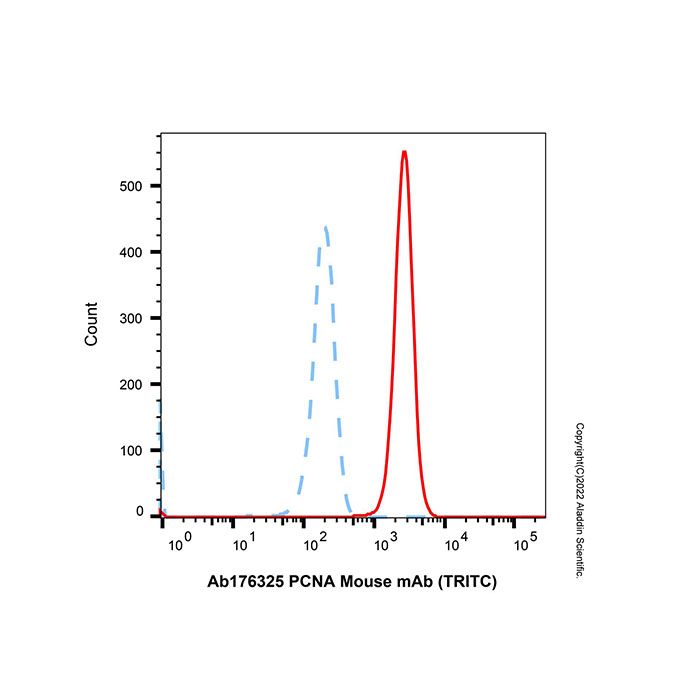
Mouse IgG (TRITC) (Ab225743) - Flow Cytometry Flow cytometry analysis of PCNA expression by Hela cells (Red) compared to an isotype control (Blue). Cells were fixed and permeabilized with 100% methanol and then stained with PCNA Mouse mAb (TRITC) (Ab176325) (Red) at 1/50 dilution for 30 minutes at 4°C in the dark. Blue - Mouse IgG (TRITC) (Ab225743).
Documentation
📋 Safety Data Sheet (SDS)
Comprehensive hazard, handling, storage, and regulatory compliance document.
Determine the necessary mass, volume, or concentration for preparing a solution.
Dilution Calculator
Determine the dilution needed to prepare a stock solution.
Reconstitution Calculator
Revisões
Avaliações dos Clientes
Application Protocols
Item-specific tested applications, recommended dilutions, and positive controls: Not specified for this item; refer to CoA/Spec Sheet.
Important note
In the absence of manufacturer-validated protocols for this SKU, users should follow institutional standard operating procedures and optimize conditions empirically for their sample type and instrumentation. No protocol claims are made for this product.
Biological Roles
Literature/general (for context; not item-specific)
Mouse IgG is a major immunoglobulin class mediating humoral immunity. It comprises two heavy and two light chains forming antigen-binding sites through hypervariable complementarity-determining regions (CDRs).
Effector functions are mediated via the Fc region, including engagement of Fcγ receptors and complement component C1q (subclass dependent). Glycosylation at Fc Asn297 modulates receptor affinity and effector responses.
In research, purified mouse IgG (conjugated or unconjugated) is widely used as:
Primary antibodies against specific antigens (when raised in mouse), or
Isotype controls to benchmark nonspecific binding.
TRITC is a xanthene-based rhodamine dye; when conjugated to proteins, it provides bright orange fluorescence suitable for microscopy and cytometry. Photophysical properties can vary with local environment and degree of labeling.
Item-specific
Target antigen, clone, subclass, and specific biological binding properties are not specified for this item; refer to CoA/Spec Sheet.
Buffer Applications
Applicability
This product is not a buffering agent. However, its performance depends on compatible buffer systems.
General guidance (not item-specific)
Common working buffers: PBS (pH ~7.2–7.6) or TBS with 0.1% BSA to reduce nonspecific binding; optional 0.05–0.1% Tween‑20 for washes.
Blocking solutions: 1–5% BSA, serum, or casein, selected to avoid species cross-reactivity; validate for your assay.
pH and ionic strength: Maintain near-physiological conditions to preserve IgG structure and TRITC fluorescence; avoid low pH elution buffers except during affinity purification steps.
Mounting media for IF: Use antifade media compatible with rhodamine/TRITC emission to enhance stability and signal retention.
Item-specific buffer composition or recommended formulations are not specified for this item; consult CoA/Spec Sheet.
Green Alternatives
Context
As a fluorescent antibody reagent, environmental impact is dominated by buffer preparation, single-use plastics, and imaging workflows rather than solvent-intensive chemistry.
Greener practices (general guidance)
Buffer preparation: Use phosphate or Tris buffers prepared with deionized water; avoid unnecessary organic additives. Prepare concentrates to reduce packaging and shipping footprint.
Waste minimization: Scale staining volumes to the smallest practical amount; reuse buffer reservoirs when contamination risk is controlled.
Illumination efficiency: Use LED light sources and appropriate filters/exposure times to reduce energy usage and extend sample life (less photobleaching → fewer repeats).
Packaging: Consolidate orders and choose refrigerated ground shipping when feasible to reduce cold-chain emissions.
Alternatives and trade-offs (literature/general)
Alternative fluorophores (e.g., rhodamine derivatives with higher photostability, or green-chemistry–manufactured dyes) may reduce the need for repeats due to bleaching; however, changing fluorophores impacts panel design, instrument settings, and compensation.
Glycerol- and sugar-based antifade mountants can improve data quality without hazardous solvents; confirm compatibility with TRITC spectra.
Pharmaceutical Uses
Applicability
Research use only (per Product Data). Not for diagnostic or therapeutic use.
Literature/general (non-clinical)
Fluorescent IgG conjugates can serve as process controls, analytical reagents, and reference materials in bioprocess development, immunoassay method development, and device qualification (e.g., optical system checks) in preclinical research environments.
No pharmacopeial monograph or excipient role is implied for this item. Any GMP or clinical application would require separate qualification and is outside the scope of this product.
Physical Properties
Item-specific (from Product Data)
Appearance: Not specified for this item; refer to CoA/Spec Sheet.
Molecular Formula: Not specified for this item; refer to CoA/Spec Sheet.
Molecular Weight: Not specified for this item; refer to CoA/Spec Sheet.
Literature/general values (for context; not item specifications)
Antibody solution form: Typically supplied in aqueous buffer (e.g., PBS or TBS) with protein concentrations in the 0.1–5 mg/mL range; exact buffer and concentration are not specified for this item.
IgG (unconjugated) MW: ~150 kDa; hydrodynamic diameter ~10–14 nm; isoelectric point commonly pI ~6–9 depending on subclass and glycosylation.
Solubility: IgG conjugates are water-soluble; they are denatured/precipitated by high organic solvent content (>20–30% v/v of many solvents) or extreme pH (<4, >9) (general guidance).
Stability: Susceptible to photobleaching under intense illumination; dye performance improves with antifade reagents and oxygen scavengers (general guidance).
Notes
Exact absorbance/emission peaks and degree of labeling (DOL) may vary lot-to-lot; consult CoA for item-specific spectra and DOL if provided.
Quality and Grades
Item-specific (from Product Data)
Grade/Purity: ExactAb™, Validated.
Interpretation and implications (general)
ExactAb™ (general description): Branding typically denotes stringent identity confirmation (e.g., species/isotype verification), controlled conjugation chemistry, and lot-to-lot consistency. Specific test methods and acceptance criteria are not specified for this item; refer to CoA/Spec Sheet.
Validated: Indicates performance verified in one or more research applications under defined conditions by the manufacturer. Exact validated applications, sample data, and recommended conditions are not specified for this item.
Key quality attributes to review on CoA for fluorescent antibodies:
Protein concentration (e.g., mg/mL), A280 purity profile.
Degree of labeling (DOL; fluorophores per IgG) and dye/antibody absorbance ratio.
Residual free dye content and aggregate level (e.g., by SEC).
Preservatives/stabilizers (e.g., BSA, sugars) and endotoxin/bioburden where relevant.
Functional validation (e.g., immunofluorescence titration curves) and background reactivity controls.
Practical notes
TRITC conjugates should show a clean absorbance peak near ~550–555 nm with minimal free-dye shoulder; excessively high DOL may increase nonspecific binding and self-quenching.
For quantitative work, confirm subclass/isotype and cross-reactivity controls, especially when used as secondary antibodies or Fc probes (not specified for this item).
Reaction and Applications
Applicability
This is a fluorescently labeled antibody intended for analytical/biological detection workflows rather than chemical synthesis.
Typical research applications (literature/general; not item-specific)
Immunofluorescence microscopy (IF/IC): Direct detection of antigens with TRITC emission in the orange channel; suitable for multicolor panels alongside FITC/Alexa 488 (green) and Cy5/Alexa 647 (far-red). Use anti-fade mountants to mitigate photobleaching.
Flow cytometry (FC): Orange-fluorescent channel comparable to PE-Texas Red/TRITC filters; titrate to minimize background and spectral spillover. Apply compensation controls.
Fluorescence imaging on blots (FIB): Detection on membranes with appropriate excitation/emission optics as an alternative to HRP/chemiluminescence; ensure low-autofluorescence membranes.
ELISA/plate assays (FI readout): Less common than enzyme reporters but feasible with compatible optics.
Practical considerations
Degree of labeling (DOL) influences brightness and affinity; excessive DOL may reduce binding and increase nonspecific interactions via hydrophobic effects.
Protect from light at all times; minimize illumination exposure during acquisition to reduce photobleaching of rhodamine dyes.
Include isotype controls and secondary-only controls to assess background binding. Validate fixation/permeabilization compatibility (paraformaldehyde + Triton X‑100 vs methanol can differentially affect epitopes).
Reaction Conditions
Not a chemical reaction reagent
This product is not intended for use in chemical transformations. However, assay “conditions” affect performance.
General assay conditions (literature/best practice; not item-specific)
Temperature: Perform incubations at room temperature (20–25°C) or 4°C depending on assay to balance kinetics and background; avoid >37°C to prevent dye/protein degradation.
Time: Typical antibody incubations range from 30–90 min at RT or overnight at 4°C; optimize empirically.
Concentration: Titrate; starting dilutions for fluorescent antibodies often fall between 1–10 µg/mL for IF and 0.05–2 µg/test for FC, but exact values for this item are not specified.
Light: Minimize exposure during staining and imaging; use amber tubes/foil and subdued lighting.
Additives: Include blocking proteins and mild detergents to reduce nonspecific binding; verify compatibility with target epitope.
Always consult product-specific guidelines where available; item-specific protocols are not provided for this SKU.
Safety and Handling
Item-specific (from Product Data)
GHS Classification, Signal Word, H-Statements, Pictograms: Not specified for this item; refer to SDS.
Storage: Store at 2–8°C; protect from light; do not freeze. Shipped on wet ice; do not freeze (per Product Data).
General laboratory safety (literature/best practice; not a substitute for SDS)
PPE: Laboratory coat, nitrile gloves, and safety glasses. Use in a clean area to avoid contamination of protein reagents.
Light sensitivity: TRITC fluorophore is light-sensitive; minimize exposure to ambient/UV light. Wrap containers in foil and work under dim light where feasible.
Incompatibilities: Avoid strong oxidizing agents, proteases, and extremes of pH or temperature that denature proteins. Avoid repeated freeze–thaw (see Storage section).
Biological origin: Although for research use only and typically supplied as purified protein, handle as a potential biohazard according to institutional biosafety procedures.
Spills/waste: Absorb aqueous spills with inert material; decontaminate surfaces with appropriate laboratory disinfectant. Dispose according to local regulations for proteinaceous/biochemical waste.
First aid (overview): If skin/eye contact occurs, rinse with water. If inhaled as aerosol, move to fresh air. If ingested, rinse mouth. Seek medical attention as needed. Always consult the official SDS for authoritative guidance.
Solvent Selection
Applicability
This product is an aqueous protein conjugate, not a small-molecule reagent. Traditional solvent selection paradigms (polarity, logP) are not directly applicable.
Practical guidance (general)
Compatible media: Physiological buffers such as PBS or TBS at neutral pH (≈7.2–7.6) are typically used for dilution and washing. Low concentrations (0.05–0.1%) of nonionic detergents (Tween-20) can reduce nonspecific binding in some assays.
Avoid: High percentages of organic solvents (e.g., >20–30% methanol, ethanol, DMSO) that may denature antibodies or perturb fluorescence. Avoid strong acids/bases and chelators that destabilize proteins unless assay-specific.
Additives: Carrier proteins (e.g., BSA) and sugars (e.g., trehalose) can stabilize antibodies during storage/handling; presence/amounts for this item are not specified.
Light: Use amber vials or foil wrapping to protect the TRITC fluorophore; minimize dye exposure during solvent/buffer exchanges.
Alternatives for specific needs (general)
If organic co-solvent is necessary (e.g., permeabilization with methanol), validate that fluorescence and antigen recognition are retained under those conditions for your assay.
Storage and Reconstitution
Item-specific (from Product Data)
Storage conditions: Store at 2–8°C. Protect from light. Do not freeze.
Shipping: Shipped on wet ice. Do not freeze during transit.
Additional guidance (general; not item-specific)
Physical form and buffer composition are not specified for this item. If supplied as a liquid, mix gently before use; avoid vortexing that can induce aggregation. If supplied lyophilized, reconstitute with sterile, buffer-compatible water or PBS and allow to equilibrate on ice; final buffer composition should match assay needs.
Aliquoting: If long-term storage is required and formulation permits, prepare single-use aliquots to minimize repeated warming; avoid freeze–thaw cycles (product is labeled “do not freeze”).
Light protection: Store in amber vials or wrap in foil to protect the TRITC fluorophore.
Stability: Refer to the CoA/Spec Sheet for shelf life, preservative content, and acceptable storage duration at 2–8°C.
Research Use Note: For research use only.
Structure and Identity
Item-specific (from Product Data)
Product: Mouse IgG (TRITC)
Grade/Purity: ExactAb™, Validated
CAS: Not specified for this item; refer to CoA/Spec Sheet.
CID: Not specified for this item; refer to CoA/Spec Sheet.
InChIKey: Not specified for this item; refer to CoA/Spec Sheet.
SMILES: Not applicable to macromolecular antibody conjugates.
Literature/general description
Molecular class: Immunoglobulin G (IgG) from mouse, covalently conjugated to TRITC (tetramethylrhodamine isothiocyanate) via an isothiocyanate–lysine coupling.
Architecture: IgG is a Y‑shaped heterotetramer composed of two heavy chains (~50 kDa each) and two light chains (~25 kDa each), joined by disulfide bonds; glycosylated Fc domain; two Fab antigen-binding arms and one Fc effector region.
Typical molecular weight: ~150 kDa (unconjugated IgG, literature). TRITC labeling increases apparent MW minimally but increases hydrophobicity and A550 absorbance.
Conjugation motif: TRITC reacts with primary amines (ε‑NH2 of lysines or N‑termini) to form stable thiourea linkages; degree of labeling (DOL) is typically reported as fluorophores per IgG (not specified for this item).
2D structural features (described): Proteinaceous backbone with repeating peptide amide bonds; aromatic TRITC xanthene ring system appended at multiple lysine residues through thiourea linkers; overall globular glycoprotein with flexible hinge region.
Isotype/subclass: Not specified for this item; refer to CoA/Spec Sheet.
Synthetic Utility
Not typically applicable
Mouse IgG (TRITC) is a biomolecular detection reagent, not a synthetic building block or catalyst. It is not used in stoichiometric or catalytic organic synthesis.
Related laboratory utility (general)
As a labeled probe, it is useful for tracking antigen presence, validating expression systems, and calibrating imaging/cytometry instruments in research workflows.
Target Specificity
Item-specific
Antigen/epitope: Not specified for this item; refer to CoA/Spec Sheet.
Clone/isotype/subclass: Not specified for this item; refer to CoA/Spec Sheet.
Species reactivity/cross-adsorption: Not specified for this item; refer to CoA/Spec Sheet.
General note
“Mouse IgG (TRITC)” may denote either a mouse primary antibody to a defined antigen or a mouse IgG isotype reagent. Without item-specific details, no target specificity can be asserted.
Need help choosing the grade?
Our grade selection guide covers purity, stabilizer status, and application suitability for all variants in our catalog.
We use cookies to ensure the website functions properly and, where permitted, to improve your experience. You can manage your preferences at any time in Settings. Learn more in our Cookie Policy.
Shall we send you a message when we have discounts available?
Remind me later
Thank you! Please check your email inbox to confirm.
Products are supplied to verified businesses, institutions, and qualified professionals for research and development use only. Not for use in humans, animals, diagnosis, or therapy.

 ZJ25F0926659