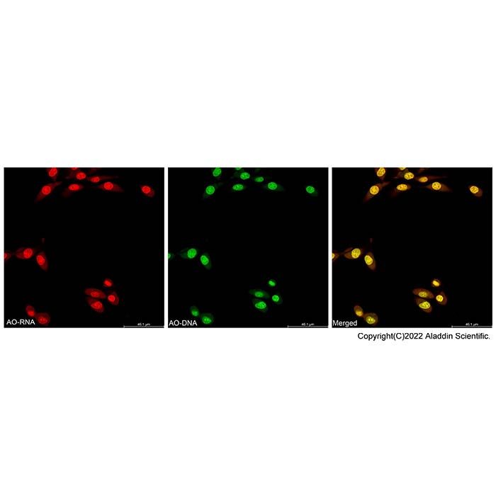
Acridine Orange (AO) is a metachromatic fluorescent dye whose emission color varies depending on the target it binds to:
When binding to double-stranded DNA: It intercalates between base pairs and emits green fluorescence upon excitation (Ex 488 nm, Em 530 nm).
When binding to single-stranded RNA or lysosomes: It attaches via electrostatic interactions and emits orange-red fluorescence (Em 640 nm).
Under a fluorescence microscope, Acridine Orange permeates the membranes of normal cells, staining the nucleus with uniform green or yellow-green fluorescence. In apoptotic cells, due to chromatin condensation and fragmentation into apoptotic bodies, AO stains them with intense, condensed yellow-green fluorescence or fragmented yellow-green particles. In necrotic cells, the yellow-green fluorescence is reduced or absent.
Acridine Orange is often used in combination with Propidium Iodide (PI) for dual staining. Since PI stains only dead cells, producing orange-red fluorescence, this method allows differentiation among normal, apoptotic, and necrotic cells.
Components
A1456513 | Component | 50 Test | 100 Test | Storage Condition | Quantity Per Test | A1456513A | Dilution Buffer | 10 mL | 50 mL | 2-8℃ | 0.1 mL per 0.5-1.0 × 10⁶ cells | A1456513B | AO Staining Solution | 100 μL | 500 μL | 2-8℃, Protect from light. Do not freeze | 1 μL per 0.5-1.0 × 10⁶ cells |
Note: The recommended number of cells to stain per test is 0.5-1.0 × 10⁶ cells. |
Procedure
1. Preparation of Acridine Orange Staining Solution
b. Mix the AO Staining Solution with the Dilution Buffer at a ratio of 1:1000 to prepare the working solution. For example, add 10 μL of AO Staining Solution to 10 mL of Dilution Buffer to obtain 10 mL of Acridine Orange staining solution.
2. Staining with Acridine Orange
a. For adherent cells:
(a) Gently aspirate the culture medium from the plate. Rinse with PBS for about 10 seconds, then remove PBS.
(b) Add Acridine Orange staining solution and incubate at room temperature for 5 minutes. Remove the staining solution and rinse with PBS for about 10 seconds. Repeat the rinse once.
Note: For adherent cells cultured in a 6-well plate with a confluence exceeding 80%, it is recommended to add the staining working solution at a volume of 1 mL per well. This volume can be optimized based on the specific experimental system.
(c) Incubate at room temperature for 5 minutes.
(c) Add an appropriate amount of cell culture medium, staining buffer, or other suitable solution to cover the well bottom. Observe under a microscope. Depending on the detection requirements, green fluorescence can be observed at Ex/Em = 488/530 nm, and red fluorescence can be observed at Ex/Em = 540/640 nm. Alternatively, measure fluorescence intensity using a fluorescence microplate reader with bottom-reading capability.
b. For suspension cells:
(a) Take 1 mL of cell suspension. Centrifuge at 500g for 5 minutes at room temperature. Gently aspirate the medium, resuspend in PBS, and centrifuge again at 500g for 5 minutes. Remove PBS.
(b) Add an appropriate amount of Acridine Orange staining solution to achieve a cell density of approximately 10⁶ cells/mL.
(c) Incubate at room temperature for 5 minutes.
(d) A drop of the sample was directly applied onto a glass slide, covered with a coverslip, and examined under a microscope. Depending on the detection requirements, green fluorescence can be observed at Ex/Em = 488/530 nm, and red fluorescence can be observed at Ex/Em = 540/640 nm. Alternatively, after staining, analyze directly by flow cytometry or measure fluorescence with a microplate reader.
Note: Centrifugation to remove staining solution can reduce background fluorescence. For suspension cells or adherent cells in suspension, consider reducing the AO staining solution concentration by 2–5 times and shortening the staining time to 2 minutes.
Precautions
1. AO Staining Solution is toxic. Handle with care.
2. For your safety and health, wear a lab coat and disposable gloves.
3. Fluorescent dyes are susceptible to quenching. It is recommended to complete detection on the same day after staining.