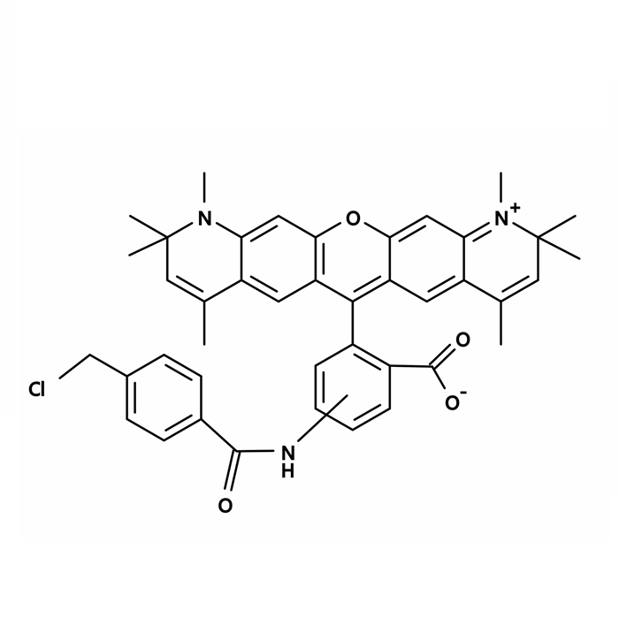
CytoTrace Red CMTPX is chemically same to CellTracker CMTPX. CytoTrace Red CMTPX freely passes through cell membranes into cells, where it is transformed into cell-impermeant reaction products. CytoTrace Red CMTPX dye is retained in living cells through several generations. The dye is transferred to daughter cells but not adjacent cells in a population. CytoTrace Red CMTPX dye is designed to display fluorescence for at least 72 hours, and the dye exhibits ideal tracking properties. Additionally, the excitation and emission spectra of Red CMTPX Red CMPTX dye are well separated from GFP (green fluorescent protein) spectra allowing for multiplexing.
Sample Assay Protocol
Note: The following is the recommended protocol. It only provides a guideline, should be modified according to the specific needs.
1. Prepare 2-10 mM DMSO stock solution
Add 36 uL DMSO into a 50 μg vial to make 2 mM stock solution (1 mg/ml is equivalent to 1.46 mM)
Note: The stock solution should be used promptly; any remaining solution should be aliquoted and frozen at ≤ -20 °C. Avoid repeated freeze-thaw cycles, and protect from light.
2. Prepare dye working solutionPrepare a 1 to 20 μM dye working solution right before use by diluting the DMSO stock solution from Step 1 with Hanks and 20 mM Hepes buffer (HHBS) or the buffer of your choice, pH 7. Mix them well by vortexing.
3. Analyze cells with a flow cytometer or a fluorescence microscope:
3.1 Treat cells with test compounds for a desired period of time.
3.2 Centrifuge the cells to get 2-10 × 10⁵ cells per tube.
3.3 Resuspend cells in 500 μL of the dye working solution (from Step 2).
3.4 Incubate cells with a dye solution at room temperature or 37 °C for 15 to 30 min, protected from light.
3.5 Remove the dye working solution from the cells, wash the cells with HHBS or buffer of your choice. Resuspend cells in 500 μL of pre-warmed HHBS or medium to get 2-10 × 10⁵ cells per tube.
3.6 Monitor the fluorescence changes at Ex/Em = 577/602 nm by flow cytometry or fluorescence microscope.
Note: For bacterial cells staining: Staining is most efficient when stock solution is diluted 1:800 in nutrient broth preconditioned by overnight growth with the test bacteria, but fresh nutrient broth or PBS may also be used. Bacterial suspensions should be diluted with PBS to 10⁵ - 10⁷ organisms per ml. Bacteria may be stained by applying one ml of solution to .45 um filter (25mm) and vacuum filtering to remove solution, then adding 1ml of dye solution and incubating 5 - 10 minutes at room temperature.

 ZJ26F0332516