
Chromatin open sequencing
Chromatin open sequencing
Chromatin accessibility is also commonly understood as the degree to which chromatin is open.The replication of DNA, as well as its transcription, requires that the tightly packed structure of chromatin be opened to allow the binding of regulatory proteins, such as transcription factors and cofactors, to DNA. This part of the chromatin that is opened is called open chromatin, and the property that allows binding of regulatory proteins is called chromatin accessibility.
ATAC-seq (Assay for Transposase Accessible Chromatin with high-throughput sequencing), a high-throughput sequencing technology that utilizes the transposase Tn5 to study chromatin accessibility, is currently the method of choice for sequencing chromatin accessibility.
Principle
ATAC-Seq technology works by taking advantage of the ability of the transposase Tn5 to recognize DNA bound to open chromatin regions, cleave it, and attach specific sequences to its ends. As shown in the figure below, the transposon complexes carrying known DNA sequence tags (i.e. Tn5 transposase with red and blue sequence tags) are added to the nucleus and incubated together, and then amplified by PCR using the known sequence tags to form a library, which is then sequenced and aligned with the genome to obtain the information of the open chromatin region.
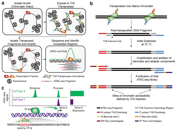
ATAC transcription and library construction process Nat Protoc. 2022 Jun;17(6):1518-1552.
Appliance
(1) mapping chromatin openness; (2) searching for key transcription factors that regulate biological processes; (3) searching for target genes regulated by transcription factors; (4) identification and discovery of enhancers; (5) searching for the localization of nucleosomes; and (6) exploring regulatory mechanisms associated with disease.
Operation method
Open chromatin assay
Principle
ATAC-Seq technology works by taking advantage of the ability of the transposase Tn5 to recognize DNA bound to open chromatin regions, cleave it, and attach specific sequences to its ends. As shown in the figure below, the transposon complex carrying known DNA sequence tags (i.e., Tn5 transposase with red and blue sequence tags) is added to the nucleus and incubated together, and then PCR amplification using the known sequence tags is performed to form a library, which can be sequenced and compared with the genome to obtain the information of the open region of chromatin.The ATAC transcription and library construction process Nat Protoc. 2022 Jun;17(6)(a). 2022 Jun;17(6):1518-1552.
Materials and Instruments
Sample Preparation: Move Sample Preparation: (1) Cell samples: a) Wash the cells twice with PBS, and the adherent cells were digested into suspension with 0.5% trypsin; b) Abort digestion with DMEM containing 10% FBS; c) Centrifuge the suspended cells at 500 g for 5 minutes at 4 °C, and carefully aspirate all the supernatant; d) Resuspend the cell precipitate with freezing solution (60% complete medium + 30% FBS + 10% DMSO) (v/v), dispense the amount of cells as specified above, and transfer to a cryopreservation tube; e) Place the cryopreservation tubes in the programmed cooling freezer box, put them into the -80 degree refrigerator for gradient cooling and temporary storage; f) Send the dry ice to the sequencing company; a) Remove fresh tissue samples and remove excess tissue and other impurities; b) Transfer the tissue sample to a new centrifuge tube and rinse it with pre-cooled PBS solution. c) Dry the tissue with dust-free paper and, if the tissue is large, cut it into small pieces or slices (about 50 mg per portion) on ice; place them in 2 ml screw-cap cryopreservation tubes ready to be labeled with the name of the sample; d) Place them quickly in liquid nitrogen. d) Quickly freeze in liquid nitrogen for 30 mins, then transfer to a -80°C refrigerator for storage; e) Freeze in dry ice and send to sequencing company; 1, Tissue (cell) nucleus extraction
① Sample to be tested (can be cells, animal tissues, plant tissues, etc.)
② 1x PBS
③ Freezing solution
④ Cell nucleus extraction kit (Dithiothreitol (DTT), Ribolock RNase Inhibitor),
⑤ Enzyme-free EP tubes
⑥ BSA
(2) Animal/plant tissue samples:
(1) According to the instructions of the cell nucleus isolation kit to complete
(2) Add BSA into LB solution, the final concentration is 1%, ready to use.
(3) Take 2 ml EP tube and add 1 ml LB.
(4) Add lysis solution to the sample, homogenizer break up and grinding
(5) Fully lysed on ice for 5-10 minutes.
(6) Remove impurities with a 40 μm cell sieve and centrifuge at 500 g for 5 minutes at 4 ℃.
(7) Discard the supernatant, add 300 μl of LB solution, resuspend, and transfer to a new EP tube.
(8) Add 300 μl of PB1 solution, blow and mix well.
(9) Slowly add 600 μl of PB2 solution to the bottom of the solution to stratify the solution.
(10) Slowly add 600 μl of PB3 solution to the bottom of the solution, allowing the solution to stratify.
(11) Centrifuge at 3000 g for 20 minutes at 4 ℃.
(12) The nuclei are located at the junction of the PB2 and PB3 solutions, and the top 600 μl of supernatant and 500 μl of liquid above the nucleus are removed sequentially.
(13) Add NB solution and blow to mix, pass through a cell sieve and centrifuge at 500 g for 5 minutes at 4 ℃.
(14) Aspirate the supernatant slowly, add 0.5 mL of NB solution and resuspend.
(15) centrifuge at 500 g for 5 minutes at 4 ℃, remove the supernatant, add 50 μl of NB solution, and resuspend the nuclei by blowing.
(16) Take 5 μl of nuclei suspension, stain with Taipan blue, and use for nuclei counting and microscopic observation.
2, Tn5 enzyme transposition reaction
(1) Configure the following Interrupting Mix in a PCR tube:
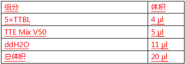
(2) Resuspend the nuclei with the Interrupting Mix and mix gently with a pipette;
(3) React at 37℃ for 30 min (the time can be extended according to the result of Agilent 2100);
3, DNA purification, library construction and on-line sequencing
(1) Purify DNA using VAHTS DNA Clean Beads (return to room temperature before use):
(2) Mix the magnetic beads with the PCR products and incubate for 5 min at room temperature;
(3) Place the PCR tubes on a magnetic rack and rinse twice with 200 μl of freshly prepared 80% ethanol, then carefully remove the supernatant;
(4) Open the lid and dry for 5 min;
(5) After the magnetic beads were dried, the DNA on the beads was eluted with 22 μl of double-distilled water;
(6) The library was then amplified by PCR with P5/P7 primers;
(7) The obtained library was purified by VAHTS DNA Clean Beads;
(8) The concentration of the library was measured by Q-bit and the fragment distribution of the library was detected by Agilent 2100;
(9) The library was sequenced by a sequencing company.
4, Bioinformatics analysis (according to individual needs)
Caveat
1. In the process of sample preparation, the pre-configured cell cryopreservation solution should be placed on ice to prevent the newly configured cryopreservation solution from overheating and damaging the cells, and then used after the cryopreservation solution returns to room temperature.
2. It is best to prepare 1 to 2 additional samples for backup when sending samples, in order to prevent errors in subsequent experiments can not be remedied.
3. All reagents should be added with protease inhibitor, RNase inhibitor and DTT in the nucleus extraction experiments.
4. In the nucleus extraction experiments, add BSA to the LB solution at a final concentration of 1%, which should be used immediately.
For more product details, please visit Aladdin Scientific website.