Negative staining, also known as negative contrast staining, was discovered by Hall. It is a staining technique in contrast to conventional staining (i.e., positive staining). Its principle relies on encasing low-electron-density specimens with heavy metal salts to raise the electron density surrounding the samples. This creates mass-thickness differences among fine structures, amplifies contrast via scattered and absorbed electrons, and renders bright structural features of the specimen against a dark background.Common negative staining solutions include phosphotungstic acid, ammonium molybdate, and India ink, among which 1–3% phosphotungstic acid is the most widely used.
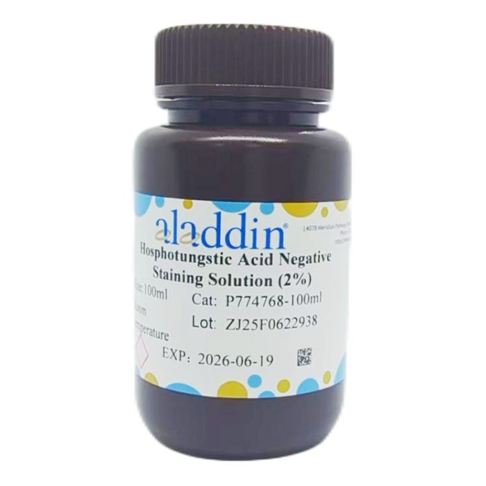
2% phosphotungstic acid negative staining solution is suitable for visualizing macromolecules, bacteria, viruses, protozoa, bacteriophages, organelles, nucleic acid macromolecules, protein crystals, and other macromolecular materials. Stained specimens exhibit transparent bright outlines, while the background appears black in micrographs.
Instructions for Use
I. Drop Staining Method
1. Concentrate the sample via low-speed centrifugation (2000 g, 10 min) or alternative methods to prepare a suspension with appropriate concentration and purity.
2. Drop the sample suspension directly onto a copper grid coated with support film, and let it stand for 3–5 min.
3. Blot excess liquid from the edge of the droplet with filter paper and allow slight drying.
4. Add negative staining solution dropwise and incubate for 2–3 min.
5. Remove excess staining solution, air-dry naturally, and observe under an electron microscope.
II. Floating Staining Method
1. Concentrate the sample via low-speed centrifugation (2000 g, 10 min) or alternative methods to prepare a suspension with appropriate concentration and purity.
2. Float the support-film-coated copper grid on the sample droplet to adsorb the sample.
3. Float the grid on the negative staining solution for 1–2 min.
4. Remove excess staining solution, air-dry naturally, and observe under an electron microscope.
Precautions
1. The target sample should be as fresh as possible.
2. The sample must form a homogeneous suspension with suitable purity and concentration; otherwise, specific and distinct binding reactions with the stain cannot be achieved.
3. Wear a lab coat and disposable gloves during operation for personal safety and health protection.
Staining Results:
Specimen
| transparent bright areas
|
background
| Black |

 ZJ26F0736956