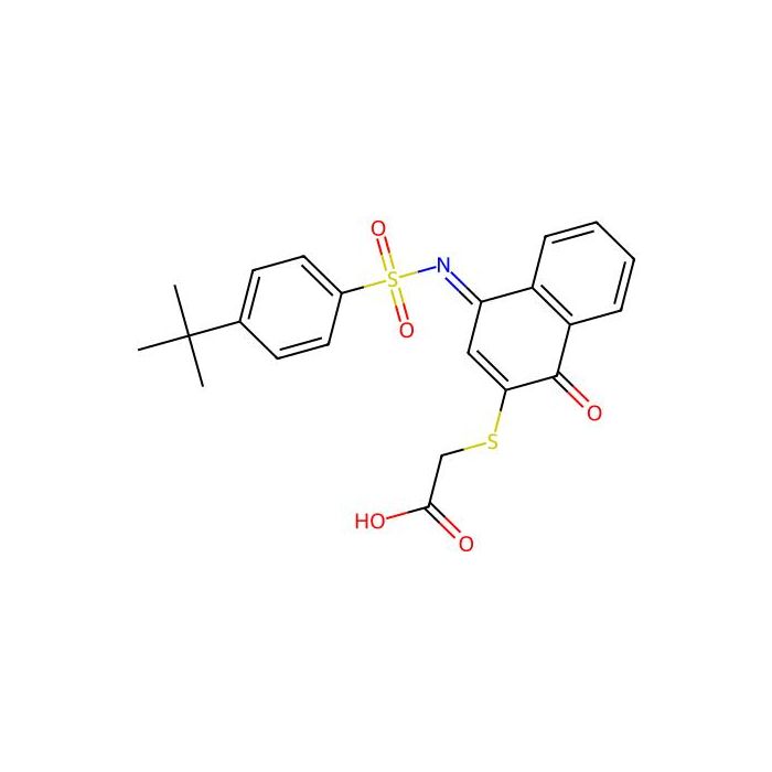
Determine the necessary mass, volume, or concentration for preparing a solution.
KPT-6566 - ≥98% , CAS No.881487-77-0
Disponible para pedir
GRADE & PURITY ≥98%
Storage
Store at 2-8°C,Protected from light,Argon charged
Shipped In
Wet ice

≥98% for sensitive chromatographic and analytical workflows requiring minimal baseline interference.
Store at 2-8°C,Protected from light,Argon charged Ships Wet ice Check lot-specific COA for exact specifications.
SDS, COA, datasheet, and spec sheet available for download. Lot-specific COA accessible via lot number lookup.
Cited in 0 peer-reviewed publications across chromatography, organic synthesis, and cross-coupling reactions.
KPT-6566 is a selective and covalent prolyl isomerase PIN1 inhibitor, covalently binds to the catalytic site of PIN1, selectively inhibits and degrades PIN1 . KPT-6566 shows an IC 50 value of 640 nM and a K i value of 625.2 nM for PIN1 PPIase domain . KPT-6566 can be used for the research of cancer
In Vitro
KPT-6566 (1-5 μM; 0-8 d) inhibits WT fibroblasts proliferation. KPT-6566 (0-10 μM; 48 h) inhibits normal breast epithelial cells and cancer cells viability via a PIN1-dependent manner. KPT-6566 (0-10 μM; 48 h) affects hyperphosphorylated pRB level, Cyclin D1 and PIN1 concentration. KPT-6566 (2.5-5 μM; 48 h) inhibits the mut-p53, NOTCH1 and NRF2 pathways. KPT-6566 (0-5 μM; 48 h) induces DNA damage via a PIN1-dependent way. MCE has not independently confirmed the accuracy of these methods. They are for reference only. Cell Proliferation AssayCell Line: Immortalized fibroblasts derived from WT and Pin1 KO mouse embryos Concentration: 1 and 5 μM Incubation Time: 0-8 days Result: Dose-dependently inhibited proliferation of WT fibroblasts, while showed no effect on Pin1 KO fibroblasts. Cell Viability AssayCell Line: MCF10A, HMEC, HeLa, LNCaP, SKOV-3, PANC-1, PC-3, MDA-MB-468 and MDA-MB-231 cells Concentration: 0-10 μM Incubation Time: 48 hours Result: Inhibited normal breast epithelial cells and cancer cells viability even at a low concentration and increased the concentration of PIN1 in MDA-MB-468, SKOV-3, PC-3, LNCaP and PANC-1. Western Blot AnalysisCell Line: Immortalized fibroblasts derived from WT and Pin1 KO mouse embryos and PIN1 KO MDA-MB-231 cells Concentration: 0-10 μM Incubation Time: 48 hours Result: Decreased hyperphosphorylated pRB and Cyclin D1 levels, dose- and time-dependently promoted degradation of PIN1 . Western Blot AnalysisCell Line: MDA-MB-231, MCF10A, MDA-MB-468 and MDA-MB-231 cells Concentration: 0, 2.5 and 5 μM Incubation Time: 48 hours Result: Dose-dependently increased H2AX phosphorylation and caused H2AX phosphorylation in MCF10A, MDA-MB-468 and MDA-MB-231 cell lines. Increased H2AX phosphorylation while other inhibitors such as ATRA, PiB and Juglone disabled to induce H2AX phosphorylation at same concentration. Achieved DNA damage through formation of DNA adducts. RT-PCRCell Line: MDA-MB-231 cells Concentration: 2.5 and 5 μM Incubation Time: 48 hours Result: Dose-dependently inhibitd the activation of mut-p53 and NOTCH1 pathways which are controlled by PIN1. Inhibitd the expression of cFOS HO1, NQO1, TXNRD1 and DNAJAB, and induced cellular responses to oxidative stress.
In Vivo
KPT-6566 (5 mg/kg; i.p. once a day for 26 days) shows no toxicity to mice . MCE has not independently confirmed the accuracy of these methods. They are for reference only. Animal Model: 6-week-old female mice with 1 million of MDA-MB-231Luc 6 cells injection Dosage: 5 mg/kg Administration: Intraperitoneal injection; 5 mg/kg once a day; for 26 days Result: Exibited no sign of local or systemic and organ toxicity by post mortem morphologic analyses.
Form:Solid
IC50& Target:IC50: 640 nM (PIN1 PPIase), Ki: 625.2 nM (PIN1 PPIase)
Comprehensive hazard, handling, storage, and regulatory compliance document.
Download SDS →Lot-specific quality data. Enter your lot number to retrieve the exact COA.
Look up COA →Full quality attributes and acceptance criteria for this grade.
View spec sheet →| Solubilidad | DMSO : 19.23 mg/mL (43.36 mM; Need ultrasonic) |
|---|