GRADE & PURITYBioReagent?BioReagent grade — tested suitable for life-science and molecular-biology use. Use for cell culture, assays, and biochemical work needing biological compatibility.
BioReagent BioReagent for sensitive chromatographic and analytical workflows requiring minimal baseline interference.
🌡
Storage & shipping
Store at 2-8°C,Do not freeze Ships Wet ice,Do not freeze Check lot-specific COA for exact specifications.
📋
Quality documents
SDS, COA, datasheet, and spec sheet available for download. Lot-specific COA accessible via lot number lookup.
📚
Literature proof
Cited in 0 peer-reviewed publications across chromatography, organic synthesis, and cross-coupling reactions.
Descripción general
Mouse CD3⁺ Cell Removal Beads are generated by covalently coupling anti-mouse CD3 antibody to magnetic beads. They can be used for rapid and efficient depletion of CD3⁺ cells from single‑cell suspensions such as mouse splenocytes.
Applications
Depletion of CD3⁺ cells from mouse spleen and lymph nodes.
Beads Volume (suspension)
Total Cell Number
250 μL
5 × 10⁸ cells
1 mL
2 × 10⁹ cells
Protocol (Using Depletion of CD3⁺ Cells from Mouse Spleen as an Example)
Prepare single‑cell suspension
Grind the spleen on a 70 μm cell strainer, rinse the strainer with pre‑chilled PBS, collect the cell suspension into a 50 mL centrifuge tube, and centrifuge at 500 g for 5 min.
Lyse red blood cells
After centrifugation, discard the supernatant. Add 5 mL of ACK red blood cell lysis buffer and lyse at room temperature for 5 min. Then add 20 mL PBS and centrifuge at 500 g for 5 min. If spleens from multiple mice are pooled for the same batch, increase the volume of red blood cell lysis buffer accordingly, following the manufacturer's instructions.
Note: The red blood cell lysis step can be adjusted in volume and time depending on the lysis buffer used. A small amount of residual red blood cells will not affect subsequent depletion or cell purity.
Resuspend splenocytes
After centrifugation, discard the supernatant and resuspend the splenocytes in PBS. Filter the cell suspension through a 70 μm cell strainer and count the cells. After counting, centrifuge at 500 g for 5 min.
Note: The cell suspension must be filtered through a cell strainer to remove tissue and cell clumps; otherwise, subsequent cell depletion purity may be affected.
Adjust cell concentration
After centrifugation, discard the supernatant and resuspend the cells in Isolation Buffer to a density of 2×10⁸ cells/mL.
Note: Isolation buffer is PBS containing 2 mM EDTA and 2% fetal bovine serum (FBS), or PBS containing 2 mM EDTA and 0.5% BSA. The buffer must be pre‑filtered through a 0.22 μm filter.
Incubate cells with beads
Add 500 μL of cell suspension (1×10⁸ cells) to the bottom of a 1.5 mL centrifuge tube, then add 50 μL of washed and resuspended beads (Mouse CD3⁺ Cell Removal Beads must be washed before use: select a pipette with an appropriate volume range, thoroughly resuspend the beads by pipetting, avoiding bubbles. Transfer the required volume of beads to a 1.5 mL centrifuge tube, add 1 mL of Isolation Buffer, centrifuge at 10,000 g for 1 min or use a magnetic separator for 3 min, then discard the supernatant. Add 1 mL of Isolation Buffer, wash the beads once more, and then resuspend the beads in the same volume of Isolation Buffer as originally taken. For example, if 50 μL of beads are taken, resuspend them in 50 μL of Isolation Buffer after washing). Mix the liquid in the tube by pipetting, and incubate at room temperature for 30 min using a rotator.
Note: For more cells, increase the volume of Mouse CD3⁺ Cell Removal Beads proportionally. For example, to process 1.5×10⁸ cells, add 750 μL of cell suspension and 75 μL of Mouse CD3⁺ Cell Removal Beads in a 1.5 mL centrifuge tube. For ≤5×10⁷ cells, adjust the cell suspension volume to 250 μL and add 25 μL of Mouse CD3⁺ Cell Removal Beads.
Magnetic separation
After incubation, add Isolation Buffer to bring the total volume to 3 mL (the liquid can be transferred from the 1.5 mL centrifuge tube to a FACS tube in several steps). Mix thoroughly by pipetting up and down 10 times (avoid vigorous shaking or inverting). Then place the FACS tube on a magnetic separator and let stand for 5 min.
Collect target cells
After magnetic separation, the target cells are in the supernatant. Carefully collect the supernatant (avoid taking up any beads), centrifuge at 500 g for 5 min, discard the supernatant, and collect the cells.
Resuspend for further use
Wash the cells as required for your experiment, then resuspend them in the desired buffer or culture medium for subsequent molecular or cell biology experiments.
Depletion Performance
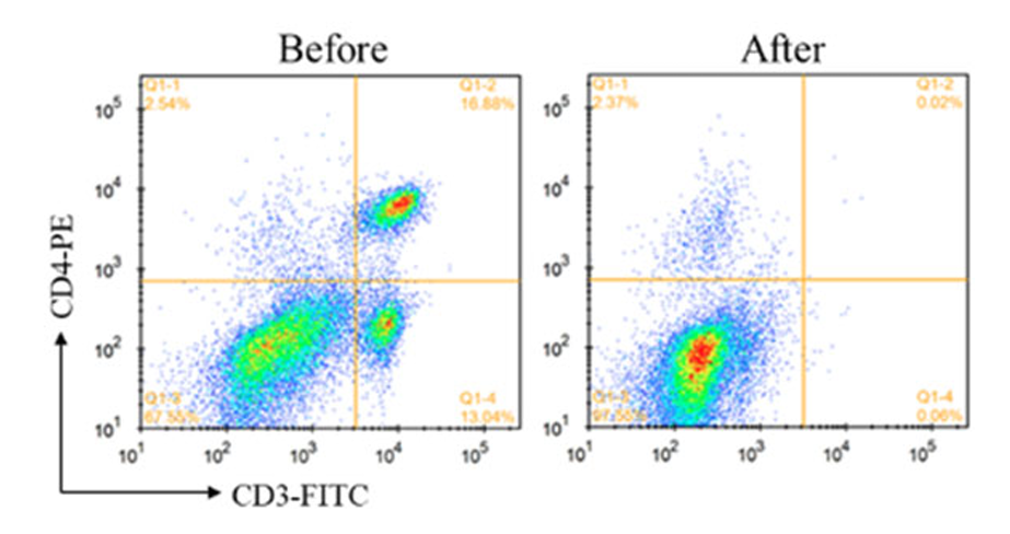
CD3⁺ cells were depleted from C57BL/6 mouse splenocytes. Cells before and after depletion were stained with FITC anti‑mouse CD3 antibody (clone 145‑2C11) and PE anti‑mouse CD4 antibody (clone RM4‑4) and analyzed by flow cytometry. The purity of CD3⁺ T cells before and after depletion was 29.92% and 0.08%, respectively.
Precautions
Avoid freezing the beads during use and storage.
Low‑retention pipette tips and centrifuge tubes are recommended to avoid loss of beads due to adsorption.
Before taking the beads, select a pipette with an appropriate volume range, thoroughly resuspend the beads by pipetting, and avoid generating bubbles during pipetting.
For research use only.
Specifications
Especificaciones y pureza
BioReagent
Estabilidad y almacenamiento
Store at 2-8℃ long term (12 months). Do not freeze.
Condiciones de almacenamiento de almacenamiento
Store at 2-8°C,Do not freeze
Enviado en
Wet ice,Do not freeze
Este producto requiere envío en cadena de frío. Los servicios terrestres y otros servicios económicos no están disponibles.
Grado
BioReagent
Product Line
Productos biológicos
Documentation
📋 Safety Data Sheet (SDS)
Comprehensive hazard, handling, storage, and regulatory compliance document.
Determine the necessary mass, volume, or concentration for preparing a solution.
Dilution Calculator
Determine the dilution needed to prepare a stock solution.
Reconstitution Calculator
Reseñas
Reseñas de cliente
Preguntas frecuentes
What does BioReagent mean?
BioReagent indicates that the product has been processed and tested for biological and biochemical applications. Specifications may include endotoxin limits, microbial controls, sterility, low DNase/RNase/protease activity, and verified biological activity where relevant.
How should this product be stored?
Store at 2–8 °C. Do not freeze. Freezing may cause irreversible damage to this material.
How is this product shipped?
This product ships chilled on wet ice. Unpack on arrival and transfer it to the storage condition stated above.
What documentation is provided?
Available product documentation, including Certificates of Analysis (COA), Safety Data Sheets (SDS), and specification sheets, is shown in the product document area. Document availability and access follow the current site policy.
Need help choosing the grade?
Our grade selection guide covers purity, stabilizer status, and application suitability for all variants in our catalog.
We use cookies to ensure the website functions properly and, where permitted, to improve your experience. You can manage your preferences at any time in Settings. Learn more in our Cookie Policy.
Shall we send you a message when we have discounts available?
Remind me later
Thank you! Please check your email inbox to confirm.
Products are supplied to verified businesses, institutions, and qualified professionals for research and development use only. Not for use in humans, animals, diagnosis, or therapy.


 ZJ26F0838370