SYBR Green I Gel Staining Solution, 10000x - 10000 x in DMSO , CAS No.163795-75-3

CAS: 163795-75-3 Cat. No.: S171397 Peso molecular: 509.7 Número EC: 838-836-0
Disponible para pedir
GRADE & PURITY 10000 x in DMSO
Synonyms
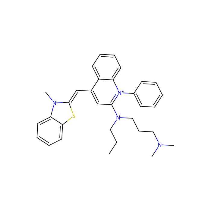
2-{[3-(dimethylamino)propyl](propyl)amino}-4-[(E)-(3-methyl-1,3-benzothiazol-2(3H)-ylidene)methyl]-1-phenylquinolinium | 2-{(E)-[2-{[3-(dimethylamino)propyl](propyl)amino}-1-phenylquinolin-4(1H)-ylidene]methyl}-3-methyl-1,3-benzothiazol-3-ium | N,N-dimeth
Storage
Store at -20°C,Argon charged
Shipped In
Ice chest + Ice pads
 ·  off list, applied to all prices below.
Size
Estado
Price
Qty
1ml
S171397-1ml
2

697,90US$

872,90US$
Guardar 175,00 US$ (20.05%)
Enter a quantity for the sizes you want to add.
🧪

Why this grade

10000 x in DMSO for sensitive chromatographic and analytical workflows requiring minimal baseline interference.

🌡

Storage & shipping

Store at -20°C,Argon charged Ships Ice chest + Ice pads Check lot-specific COA for exact specifications.

📋

Quality documents

SDS, COA, datasheet, and spec sheet available for download. Lot-specific COA accessible via lot number lookup.

📚

Literature proof

Cited in 15 peer-reviewed publications across chromatography, organic synthesis, and cross-coupling reactions.

Descripción general

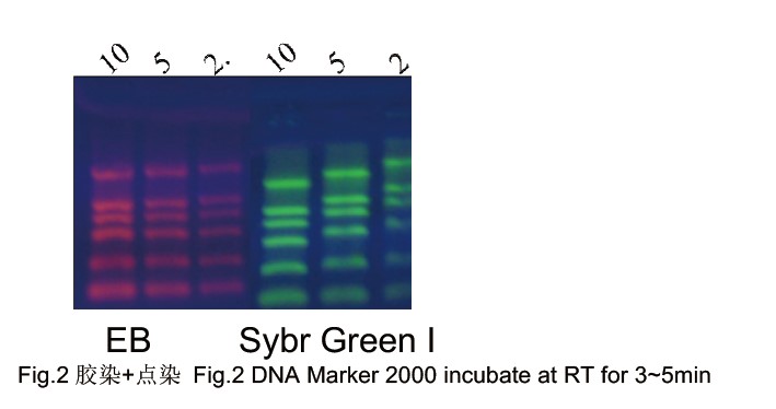
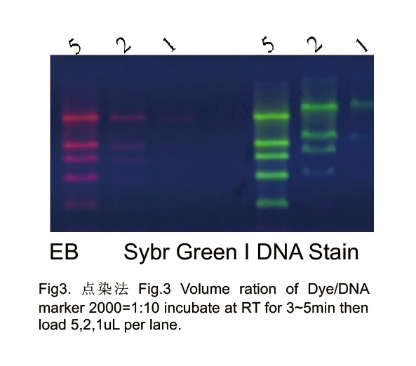
Sybr Green I and Sybr Green II Nucleic Acid Stains are produced by Dr.Chimin Du, are a kind of novel generation of fluorescent nucleic acid gel stains designed to replace the highly toxic ethidium bromide (EtBr). Sybr Green I is nontoxic and more sensitive than EtBr. Gels can be visualized under UV or Visible Light.

Features of Sybre Green Nucleic Acid Stains
1.  Safety: Sybr Green is nontoxic and noncarcinogenic.
2.  Ultra-sensitivity: It allows the visualization of as little as 20pg dsDNA, around 5-10 times more sensitive than EtBr under
UV and 8-20 times more sensitive than EtBr in Visible Light.
3.  Convenience: NO need to rinse or wash gels. Add stain before load samples. Visualize gels under UV or Visible Light to avoid UV damage on DNA/RNA.
4.  Wide range:   suitable for agrose gel or PAGE.
5.  No impact for the next experiments such as RT, PCR, enzyme digestion, and ligation.
6.  Strong signal and no background
Directions for the Agrose Gels stained by Sybr Green
Protocol 1: Pre-cast Protocol (Add dye in the gel)
1.1 Prepare molten agarose gel solution using your standard protocol.
1.2 Add 1~3µl Sybr Green Nucleic Acid Stain per 50ml gel when the gel cool down to 50℃and mix thoroughly.
1.3 Cast the gel and allow it to solidify. Any leftover gel solution may be stored and reheated later for additional gel casting.
1.4 Load samples and run the gels using your standard protocol.
1.5 DNA stained with SYBR Green I stain can be readily visualized using a UV or blue-light sources (emit at 450, 473, 488, or 532 nm). Image the stained gel with the transilluminator and photograph the gel using.
Protocol 2: Post-staining Protocol (Stain Nucleic Acid after electrophoresis by adding dye in the gel stain solution)
2.1 Make gels: Do not add any nucleic acid stain when make gels.
2.2 Run gels as usual according to your standard protocol.
2.3 Prepare Sybr Green Nucleic Acid Staining solution: Dilute Sybr Green Nucleic Acid Stain with TAE or TBE (TBE (89 mM Tris base, 89 mM
boric acid, 1 mM EDTA, pH 8) and TAE (40 mM Trisacetate,1 mM EDTA, pH 8) on ratio 1:10000. Stain gels in the dark for 10-30min. Staining time depends on gel concentration and thickness. PAGE can be stained directly on the glass. Let staining solution cover PAGE gels for
30min. please use glassware to store staining solution or silicified glassware because stain will absorb on the glass.
2.4 Visualize gels a UV or blue-light sources.
2.5 Exact molecular weight can be measured by this method but the dyes are used much more in this way.
Protocol 3: Stain nucleic acid before electrophoresis (add dye in the loading buffer)
3.1.   Prepare working solution: Dilute 10 µl Sybr Green Stain with 1ml running buffer TBE or TAE. This solution is stable up to one month at 4℃
3.2.   Make gels: based on the routine method. Do not add any DNA/RNA stain in the gel.
3.3.   Stain Nucleic Acid: Add 1µl Sybr Green I Nucleic Acid Stain working solution to 10µ mixture of sample and loading buffer, let it stay at RT for
3-5min for stain binding to nucleic acid completely. Normally, 1µL working solution is enough for one sample loading, and 1ml Sybr Green I Nucleic Acid Stain is enough to load 10,000 samples.
3.4.   Stain markers: Mix 5µL Marker and 1µL Sybr Green I Nucleic Acid Stain working solution thoroughly, let it stay at RT for 5min to let Sybr
Green I Stain and DNA/RNA binding completely.
3.5.   Load samples and run gels.
3.6.   Visualize gels in UV or Visible Light to avoid UV damage on DNA/RNA.
*The big DNA fragments (>2Kb) will move slowly when bind to the stain. So please stain in DNA after electrophoresis or add stain in gels to measure molecular weight exactly.
Notes :
1.  Do not run gels over 2 hrs. Or smeared bands appeared because Sybr Green I Stain will dissociate from DNA/RNA.
2.  Sybr Green I Stain can dissociate from nucleic acids in ethanol.
3.  Please stain nucleic acid in the gel or after electrophoresis to check the exact molecular weight of fragments when compared with molecular weight markers.
4.  Please use EP tubes and other plastic wares in Sybr Green I Stain storage, dilution, and staining. Sybr Green I Stain can bind to glassware.


Specifications

Sinónimos
2-{[3-(dimethylamino)propyl](propyl)amino}-4-[(E)-(3-methyl-1, 3-benzothiazol-2(3H)-ylidene)methyl]-1-phenylquinolinium | 2-{(E)-[2-{[3-(dimethylamino)propyl](propyl)amino}-1-phenylquinolin-4(1H)-ylidene]methyl}-3-methyl-1, 3-benzothiazol-3-ium | N, N-dimeth
Especificaciones y pureza
10000 x in DMSO
Condiciones de almacenamiento de almacenamiento
Store at -20°C, Argon charged
Enviado en
Ice chest + Ice pads
Este producto requiere envío en cadena de frío. Los servicios terrestres y otros servicios económicos no están disponibles.
Nombres e identificadores
Pubchem Sid504765463
Pubchem Sid Urlhttps://pubchem.ncbi.nlm.nih.gov/substance/504765463
Sonrisas canónicasCCCN(CCCN(C)C)C1=[N+](C2=CC=CC=C2C(=C1)C=C3N(C4=CC=CC=C4S3)C)C5=CC=CC=C5
IUPAC NameN,N-dimethyl-N'-[4-[(E)-(3-methyl-1,3-benzothiazol-2-ylidene)methyl]-1-phenylquinolin-1-ium-2-yl]-N'-propylpropane-1,3-diamine
InChIKeyCGNLCCVKSWNSDG-UHFFFAOYSA-N
INCHI1S/C32H37N4S/c1-5-20-35(22-13-21-33(2)3)31-23-25(24-32-34(4)29-18-11-12-19-30(29)37-32)27-16-9-10-17-28(27)36(31)26-14-7-6-8-15-26/h6-12,14-19,23-24H,5,13,20-22H2,1-4H3/q+1
Isómeros SMILES CCCN(CCCN(C)C)C1=[N+](C2=CC=CC=C2C(=C1)/C=C/3\N(C4=CC=CC=C4S3)C)C5=CC=CC=C5
Peso molecular 509.7

Documentation

📋 Safety Data Sheet (SDS)

Comprehensive hazard, handling, storage, and regulatory compliance document.

Download SDS →

✅ Certificate of Analysis (COA)

Lot-specific quality data. Enter your lot number to retrieve the exact COA.

Look up COA →

📊 Datasheet

Quick-reference summary of product specifications and applications.

View datasheet →

🔬 Specification Sheet

Full quality attributes and acceptance criteria for this grade.

View spec sheet →

Advanced Data

Taxonomic Classification

Taxonomy Tree

KingdomOrganic compounds
SuperclassOrganoheterocyclic compounds
ClaseQuinolines and derivatives
SubclassPhenylquinolines
Intermediate Tree Nodes Not available
Direct ParentPhenylquinolines
Alternative Parents Aminoquinolines and derivatives  Benzothiazoles  Dialkylarylamines  Aryl thioethers  Aminopyridines and derivatives  Pyridinium derivatives  Benzene and substituted derivatives  Thiazoles  Heteroaromatic compounds  Trialkylamines  Ketene acetals  Azacyclic compounds  Organopnictogen compounds  Hydrocarbon derivatives  Organic cations  
Molecular FrameworkAromatic heteropolycyclic compounds
Substituents Phenylquinoline - Aminoquinoline - Benzo-thiazole - 1,2-benzothiazole - 1,3-benzothiazole - Aryl thioether - Dialkylarylamine - Aminopyridine - Benzenoid - Monocyclic benzene moiety - Pyridinium - Pyridine - Thiazole - Heteroaromatic compound - Tertiary aliphatic amine - Ketene acetal or derivatives - Tertiary amine - Azacycle - Hydrocarbon derivative - Organopnictogen compound - Amine - Organonitrogen compound - Organic nitrogen compound - Organic cation - Aromatic heteropolycyclic compound
DescripciónThis compound belongs to the class of organic compounds known as phenylquinolines. These are heterocyclic compounds containing a quinoline moiety substituted with a phenyl group.
External Descriptors cyanine dye - tertiary amine - quinolines - benzothiazolium ion
Estructura 3D
Modelo de Estructura Química Interactiva


Certificados (CoA, COO, BSE/TSE y tabla de análisis)
C of A & Other Certificates(BSE/TSE, COO):
Analytical Chart:

Find and download the COA for your product by matching the lot number on the packaging.

4 results found

Lot NumberCertificate TypeFechaArticulo
K2222779Certificate of AnalysisSep 04, 2025 S171397
I2526408Certificate of AnalysisJun 11, 2025 S171397
C2503269Certificate of AnalysisFeb 24, 2025 S171397
G2207301Certificate of AnalysisJun 09, 2022 S171397
Propiedades químicas y físicas
SensibilidadLight sensitive;Moisture sensitive
Peso molecular509.700 g/mol
XLogP38.200
Hydrogen Bond Donor Count0
Hydrogen Bond Acceptor Count4
Rotatable Bond Count9
Exact Mass509.274 Da
Monoisotopic Mass509.274 Da
Topological Polar Surface Area38.900 Ų
Heavy Atom Count37
Formal Charge1
Complexity734.000
Isotope Atom Count0
Defined Atom Stereocenter Count0
Undefined Atom Stereocenter Count0
Defined Bond Stereocenter Count1
Undefined Bond Stereocenter Count0
The total count of all stereochemical bonds1
Covalently-Bonded Unit Count1
Preguntas frecuentes y artículos
Types, Mechanisms, and Selection Principles of Nucleic Acid Dyes for Gel Electrophoresis
Understanding Cyanine Dyes: Structure & Mechanism, Channel Selection, Troubleshooting Essentials, and a Product Selection Roadmap (with 6 Category Tables)
Fluorescent Probes: A Complete Beginner’s Guide — Definitions & Emission Mechanisms, Signal Readouts, Classification Framework, Selection Roadmap, and Product Tables (Tables 1–4)
Role of ROX Passive Reference Dye in Real-Time Quantitative Fluorescence PCR: Functions and Practical Considerations
Deoxyribonuclease (DNase): DNA Hydrolytic Cleavage Properties, Methodological Value, and Multi-Context Applications
Agarose Precast Gels: Composition, Advantages, and Application Workflows for Standardized Nucleic Acid Electrophoresis Matrices
Micrococcal Nuclease (MNase): A Technical Review of Enzymatic Properties, Reaction Control, and Research and Biomanufacturing Applications
Comparative Guide and Selection Framework for Common Fluorophores
Citations of This Product
Referencias
1. Chengjie Duan, Yan Chen, Zhiqiang Hou, Dayong Li, Jin Jiao, Weihao Sun, Yang Xiang.  (2023)  Heteromultivalent scaffolds fabricated by biomimetic co-assembly of DNA–RNA building blocks for the multi-analysis of miRNAs.  Journal of Materials Chemistry B,  11  (7): (1478-1485).  [PMID:36723144] [10.1039/D2TB02663E]
2. Zhang Kai, Cao Jinxuan, Wu Yongxiang, Hu Futao, Li Tianhua, Wang Ying, Gan Ning.  (2019)  A fluorometric aptamer method for kanamycin by applying a dual amplification strategy and using double Y-shaped DNA probes on a gold bar and on magnetite nanoparticles.  MICROCHIMICA ACTA,  186  (2): (1-9).  [PMID:30666478] [10.1007/s00604-018-3207-6]
3. Hailiang Huang, Shuo Shi, Xuyue Zheng, Tianming Yao.  (2015)  Sensitive detection for coralyne and mercury ions based on homo-A/T DNA by exonuclease signal amplification.  BIOSENSORS & BIOELECTRONICS,      [PMID:25950941] [10.1016/j.bios.2015.04.076]
4. Kexin Xu, Jinhui Zhu, Tong Zhang, Guodong Sui.  (2024)  A phosphorylated guanidine chitosan and UiO-66-NH2 modified magnetic nanoparticle platform for enrichment and detection of multiple bacteria.  TALANTA,      [PMID:38924986] [10.1016/j.talanta.2024.126435]
5. Shurui Tao, Yi Long, Guozhen Liu.  (2024)  Entropy-Driven Molecular Beacon Assisted Special RCA Assay with Enhanced Sensitivity for Room Temperature DNA Biosensing.  Biosensors-Basel,  14  (12): (618).  [PMID:39727883] [10.3390/bios14120618]
6. Weiyi Lu, Bin Yang, Zihua Guo, Zhen Tian, Wenhui He, Tiancheng Yang.  (2024)  Machine learning-based fluorescence sensor array: Accurate discrimination, quantitative assay of phenothiazine drugs via versatile DNA probes.  SENSORS AND ACTUATORS B-CHEMICAL,      [PMID:] [10.1016/j.snb.2024.135330]
7. Shan He, Yiyu Chen, Huiting Lian, Xuegong Cao, Bin Liu, Xiaofeng Wei.  (2024)  Photocatalytic colorimetry and fluorescence dual-signal biosensor for instant determination of terminal deoxynucleotidyl transferase.  SENSORS AND ACTUATORS B-CHEMICAL,      [PMID:] [10.1016/j.snb.2024.135957]
8. Yi Long, Shurui Tao, Dongni Shi, Xingyu Jiang, Tian Yu, Yingxi Long, Libing Song, Guozhen Liu.  (2024)  Special RCA based sensitive point-of-care detection of HPV mRNA for cervical cancer screening.  Aggregate,      [PMID:] [10.1002/agt2.569]
9. Rongrong Huang, Yue Xu, Peipei Wan, Tong Zhu, Weili Heng, Wenjun Miao.  (2024)  Thylakoid-based green preparation of porous microneedles for antibiotic residues detection in food samples.  ANALYTICA CHIMICA ACTA,      [PMID:39266197] [10.1016/j.aca.2024.343181]
10. Guobin Huang, Weiting Xu, Jinxin Chi, Chenchen Lin, Jiabin Wang, Xucong Lin.  (2025)  tFNA-aptasensor integrating DNA-framework confined affinity recognition and HCR amplification towards accurate profiling of microRNA for COPD analysis.  MICROCHEMICAL JOURNAL,      [PMID:] [10.1016/j.microc.2025.114603]
11. Li Peng, Jian Zhu, Bin Yang, Huimin Hao, Shuyan Lou.  (2022)  A green photocatalytic-biosensor for colorimetric detection of pesticide (carbaryl) based on inhibition of acetylcholinesterase.  TALANTA,      [PMID:35533565] [10.1016/j.talanta.2022.123525]
12. Tian Yu, Siyi Zou, Yingxi Long, Yourong Ou, Shixian Liu, Tongjia Kang, Libing Song, Congcong Sun, Guozhen Liu.  (2025)  Glass fiber-interfaced CRISPR/Cas biosensing adaptable for diverse biomarker detection.  TRENDS IN BIOTECHNOLOGY,      [PMID:40447514] [10.1016/j.tibtech.2025.05.001]
13. Tingyuan Pang, Shuting Ouyang, Jia Li, Xiaorui Liu, Wenying Shu, Wenhui Gao, Junren Li, Qi Song, Jianing Huang, Jun Yang, Xiaoshun Jian, Xiaole Wang, Meimei Zhang.  (2025)  Photocatalytic DNA chemiluminescent biosensor via cooperative hybridization for miR-21 assay with both good sensitivity and selectivity.  MICROCHEMICAL JOURNAL,      [PMID:] [10.1016/j.microc.2025.114218]
14. Zhenzhen Xie, Wei Jia, Xingzhen Li, Lei Pan, Shihao Xu, Changlong Jiang.  (2025)  Fluorescent biosensor based on aptamer-cDNA hybridization for Cd2+ detection and molecular mechanism investigation.  MICROCHEMICAL JOURNAL,      [PMID:] [10.1016/j.microc.2025.115614]
15. Chen Lingling, Chen Chen.  (2026)  Cyclic reverse transcription cooperating self-priming cyclic amplification for sensitive and label-free non-small cell lung cancer related microRNA analysis.  Journal of Analytical Science and Technology,  17  (1): (12).  [PMID:] [10.1186/s40543-026-00533-4]
Calculadoras de soluciones
Reseñas

Reseñas de cliente

Shall we send you a message when we have discounts available?

Remind me later

Thank you! Please check your email inbox to confirm.

Oops! Notifications are disabled.