Embryonic Reproductive System Detection Laboratory
Embryonic Reproductive System Detection Laboratory
Alkaline phosphatase staining of primordial germ cells (germinal cristae) specifically demonstrates the localization and number of embryonic primordial germ cells and is the classical method for routine analysis of embryonic germ cells.
Operation method
Alkaline phosphatase staining of the genital cristae
Materials and Instruments
Equipment: Move The basic procedure for alkaline phosphatase staining of the genital cristae can be divided into the following steps: Caveat For germinal ridge analysis, mouse embryos from embryonic day 11.5 to embryonic day 14.5 are selected. Care should be taken to prevent damage to the tissues.AP staining solutions should be prepared and used on site. For more product details, please visit Aladdin Scientific website.
SPF grade laboratory animal housing environment, detector pins, stereomicroscope
Reagents:
① PBS buffer
② 4% paraformaldehyde
③ Naphthol AS-MX stock (- 20 ℃, Sigma, No. 855-20ml)
④ Fast red TR (4 ℃, Sigma, No. F8764-1g)
⑤ 70% ethanol
⑥ 70% glycerol
A Male and female mice were used in a combined cage for the experiment, after which they were brought to the animal room before 10 o'clock in the morning every day, and the presence of vaginal plugs in the vaginas was tested for both experimental and control female mice to verify the presence of mating behavior. The presence of pessary was recorded. After the pessary was detected, the female mice were numbered and the embryos were removed at embryonic day 11.5 and embryonic day 14.5 for analysis.
B The embryos were removed in pre-cooled PBS, and the germinal cristae were removed under a stereomicroscope. The germinal ridge is generally located on the medial side of the lower limb, on both sides of the spine, and connected to the middle kidney.
C The removed germinal ridge was fixed with 4% paraformaldehyde at 4 ℃ for 1~2 hours. Wash with deionized water 3 times for 3 minutes each time.
D Add freshly prepared AP staining solution (Naphthol AS-MXstock lg, Fast red TR 40 μl, millQ 1 ml completely dissolved, the amount of preparation depends on the needs of the experiment) and stain for about 10 minutes, during which time, pay close attention to the staining situation, and then stop if there is obvious staining.
E Wash with deionized water 3 times for 3 minutes each time. E Wash with deionized water 3 times for 3 minutes each time.
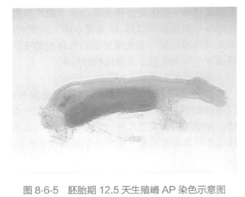
F 70% glycerol transparent and then observe and take pictures, the staining results of normal genital cristae are as follows (E13.5d, bar = 0.1 mm, the red color represents genital cristae, and the outer tissue is the middle kidney). An example of the results is shown in Figure 8-6-5.