
p38 MAPK Signaling Pathway: Activation Mechanisms, MK2 Functional Output, and Experimental Interpretation
p38 MAPK Signaling Pathway: Activation Mechanisms, MK2 Functional Output, and Experimental Interpretation
p38 MAPK is an important MAPK branch involved in cellular responses to inflammation, pathogen recognition, and environmental stress. Its canonical signaling is driven by the MAP3K–MKK3/6–p38 cascade and influences inflammatory output, stress adaptation, and cell fate through MK2–HSP27, MK2–TTP, and nuclear transcriptional regulation.
Keywords: p38 MAPK; MAPK14; MKK3/MKK6; MK2; HSP27; TTP; ERK/JNK; p38 inhibitors
1 Components and Isoforms of the p38 MAPK Signaling Axis
1.1 p38α, p38β, p38γ, and p38δ
(1) p38α
p38α is encoded by MAPK14 and is the predominant p38 isoform studied in inflammation and stress responses. Following dual phosphorylation at Thr180/Tyr182 by MKK3/6, p38α can further activate MK2, MSK, and multiple nuclear substrates, forming the canonical p38α–MK2 functional axis.
(2) p38β
p38β is encoded by MAPK11 and shares high structural homology with p38α, with partial overlap in upstream kinases and substrates. Classical inhibitors such as SB203580 and SB202190 generally act on both p38α and p38β, making it difficult to distinguish the independent contribution of each isoform using these tools alone.
(3) p38γ and p38δ
p38γ and p38δ are encoded by MAPK12 and MAPK13, respectively. p38γ is relatively abundant in skeletal muscle, whereas p38δ is more involved in signaling in certain immune, epithelial, and endocrine tissues. Both show relatively low sensitivity to classical p38α/β inhibitors such as SB203580.
Table 1 Major Differences Among p38 MAPK Isoforms
Isoform | Encoding Gene | Expression and Functional Characteristics | SB203580 Sensitivity | Major Research Directions |
p38α | MAPK14 | Broadly expressed; forms the classical functional axis with MK2 | High | Inflammation, immunity, stress, cell cycle |
p38β | MAPK11 | Partially overlaps with p38α in substrates and pharmacological characteristics | High | Neural, inflammatory, and stress signaling |
p38γ | MAPK12 | Relatively abundant in skeletal muscle | Low | Muscle, metabolism, and tissue-specific signaling |
p38δ | MAPK13 | Expressed in some immune, epithelial, and endocrine tissues | Low | Inflammation, differentiation, and tissue-specific regulation |
1.2 MKK3/MKK6 and Major Upstream MAP3Ks
(1) MKK3/MKK6
MKK3 and MKK6 are the principal direct upstream MAP2Ks of p38 and sequentially phosphorylate Thr180 and Tyr182 within the p38 activation loop. Dual-site phosphorylation shifts the activation loop into a catalytically competent conformation, enabling p38 to efficiently phosphorylate downstream substrates.
(2) TAK1
Following TLR or IL-1R activation, IRAK–TRAF6-associated complexes form K63-linked ubiquitin signaling platforms, through which TAB2/TAB3 recruit and activate TAK1. TAK1 subsequently branches into MKK3/6–p38, MKK4/7–JNK, and IKK–NF-κB pathways, serving as an important convergence node through which inflammatory stimuli simultaneously initiate multiple signaling pathways.
(3) ASK1
Under resting conditions, reduced Thioredoxin binds ASK1 and restricts its activation. When ROS levels increase, Thioredoxin becomes oxidized and dissociates from ASK1. Activated ASK1 then transmits signals to MKK3/6–p38 and MKK4/7–JNK, causing oxidative stress to frequently generate parallel p38/JNK responses.
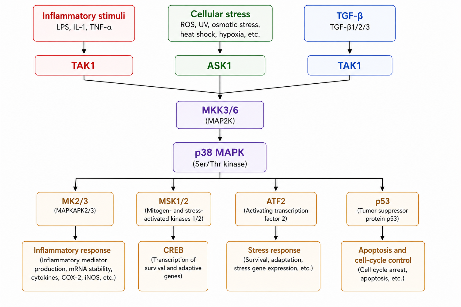
Figure 1. Upstream activation, core kinase cascade, and major functional outputs of the p38 MAPK signaling pathway
2 Activation Mechanisms of p38 MAPK
2.1 Inflammatory Cytokine and Pattern-Recognition Receptor Signaling
(1) TLR4–TAK1–p38
After LPS binds the TLR4–MD-2 receptor complex, MyD88-associated signaling recruits IRAK and TRAF6 and activates TAK1 through a ubiquitin-based signaling platform. TAK1 subsequently enters the MKK3/6–p38 cascade while simultaneously initiating inflammatory branches including JNK and NF-κB.
(2) IL-1R–TAK1–p38
IL-1R and some TLRs share the MyD88–IRAK–TRAF6 signaling architecture. Following IL-1 binding to its receptor, signaling converges with TLR pathways at the TAK1–MKK3/6 level, allowing different inflammatory stimuli to generate similar p38 activation patterns.
(3) TNFR–p38
Following TNF-α binding to TNFR1, a membrane-associated complex containing components such as RIPK1, TRAF, and cIAP can form and recruit upstream kinases including TAK1. This complex simultaneously controls p38/JNK, NF-κB, and cell-death-related signaling, and the functional output of TNFR depends on complex composition and signal duration.
2.2 Oxidative Stress, Osmotic Stress, and DNA Damage
(1) ROS–ASK1–p38
H₂O₂, mitochondrial ROS, and other oxidative stimuli can release ASK1 from Thioredoxin-mediated inhibition, allowing ASK1 to continuously transmit signals to p38 and JNK. Transient ROS exposure mainly induces adaptive responses, whereas sustained oxidative conditions can additionally cause mitochondrial and DNA damage.
(2) Hyperosmotic Stress
Hyperosmotic conditions cause cellular dehydration and volume reduction while altering ion concentrations and Actin Cytoskeleton tension. Stress-responsive MAP3Ks such as TAO and ASK can transmit these changes to MKK3/6–p38, after which MK2–HSP27 participates in cytoskeletal remodeling.
(3) DNA Damage
Ultraviolet radiation, topoisomerase inhibitors, and ROS can simultaneously initiate DNA-damage responses and stress MAPK signaling. p38 further influences cell-cycle regulatory nodes such as p53 and Cdc25, linking damage signals to cell-cycle arrest, repair, or cell death.
2.3 p38 Phosphorylation and Signal Termination
(1) Dual Phosphorylation of the TGY Motif
The p38 activation loop contains a Thr–Gly–Tyr (TGY) motif, and canonical activation depends on dual phosphorylation at Thr180 and Tyr182. Experimentally, detection of p-p38 Thr180/Tyr182 primarily reflects whether the activation loop has been acted on by upstream MAP2Ks.
(2) TAB1-Mediated Noncanonical Activation
In addition to participating in the TAK1 complex, TAB1 can directly bind p38α. Under certain stress conditions, this interaction can induce conformational changes and autophosphorylation of p38α, allowing it to acquire activity in the absence of classical MKK3/6 input.
(3) DUSP-Mediated Signal Termination
Dual-specificity phosphatases such as DUSP1/MKP-1 can remove phosphate groups from both Thr and Tyr residues within the p38 activation loop. Inflammatory and stress stimuli can also induce DUSP expression, creating a “rapid activation–delayed shutdown” temporal pattern of p38 signaling.
3 Major Downstream Functional Modules of p38 MAPK
3.1 p38–MK2 Signaling Axis
(1) MK2 Activation
p38α/β can form stable complexes with MK2. Activated p38 further phosphorylates MK2 and changes its catalytic state and subcellular localization. MK2 thereby transmits p38 signaling to cytoskeletal regulation and post-transcriptional control of inflammation.
(2) MK2–HSP27
MK2 can phosphorylate HSP27 at sites including Ser15, Ser78, and Ser82, with Ser82 being one commonly detected site. Following phosphorylation, the oligomeric state of HSP27 and its interaction with Actin change, contributing to microfilament reorganization, maintenance of cell morphology, and migration.
(3) MK2–TTP
TTP recognizes AU-rich elements in the 3′UTRs of certain inflammatory mRNAs, including TNF and IL6, and promotes their degradation. Following phosphorylation by MK2, TTP shows enhanced binding to 14-3-3, reducing TTP-mediated mRNA degradation and extending the half-lives of these inflammatory transcripts.
3.2 ATF2 and Nuclear Transcriptional Regulation
(1) ATF2
The N-terminal transcriptional activation region of ATF2 can be phosphorylated by p38 and JNK, with representative sites including Thr69 and Thr71. Phosphorylated ATF2 forms transcriptional complexes with proteins such as Jun and regulates expression of inflammation- and stress-related genes.
(2) MSK–CREB
Both p38 and ERK can activate MSK1/2, after which MSK acts on CREB Ser133 and certain chromatin-associated proteins. This module creates a clear shared nuclear transcriptional output between p38 and ERK.
(3) p53 and MEF2
Under DNA-damage conditions, p38 can strengthen p53-associated cell-cycle checkpoints. During myogenic differentiation, p38 can enhance MEF2- and MyoD-associated transcriptional programs. Differences in the transcription factors available in different cell types allow the same p38 kinase to generate different functional outcomes.
3.3 Inflammatory Mediator Expression and mRNA Stability
(1) Transcriptional Initiation
Following TLR or IL-1R stimulation, NF-κB and other transcription factors first promote expression of inflammatory genes such as TNF and IL6, with p38 participating through ATF2 and MSK. This stage primarily determines how much inflammatory transcript is produced.
(2) Transcript Stability
After transcription, p38–MK2–TTP continues to determine the degradation rate of certain short-lived inflammatory mRNAs. When MK2 reduces the mRNA-destabilizing function of TTP, existing transcripts such as TNF persist longer within the cell and continue to enter translation.
(3) Protein Output
Final levels of inflammatory mediators are jointly determined by transcriptional production, mRNA stability, and protein translation. A prominent role of p38–MK2 is to prolong the lifetime of certain inflammatory messages rather than simply “switching on” inflammatory genes.
4 Major Cellular Functions Mediated by p38 MAPK
4.1 Inflammation and Immune Responses
The p38α–MK2 axis can convert short-lived receptor-derived kinase signals into sustained inflammatory output. After TLR and IL-1R activate p38 through TAK1–MKK3/6, MK2 suppresses TTP-mediated degradation of inflammatory mRNAs, while ATF2 and MSK participate in nuclear transcription, thereby regulating both the intensity and duration of inflammation.
4.2 Stress Adaptation and Cell Survival
Under transient oxidative or hyperosmotic stress, MK2–HSP27 promotes Actin Cytoskeleton remodeling and helps cells restore structural homeostasis. When ROS or DNA damage persists, ASK1–p38/JNK signaling progressively integrates with p53, cell-cycle checkpoints, and mitochondrial damage, and the cellular response may shift from adaptation toward growth inhibition, senescence, or death.
4.3 Cell Cycle, Apoptosis, and Differentiation
(1) Cell-Cycle Checkpoints
Following DNA damage, p38–MK2 can influence cell-cycle regulatory proteins such as the Cdc25 family, reduce CDK activation, and delay cell-cycle transitions. Transient checkpoint arrest provides time for DNA repair, whereas prolonged arrest may progress into senescence or cell-death programs.
(2) Apoptosis
Strong oxidative stress can sustain ASK1-mediated activation of p38 and JNK and intersect with p53 and mitochondrial apoptotic networks. Once cellular injury exceeds adaptive capacity, mitochondrial integrity and the Caspase cascade are altered, and cells progressively enter apoptosis.
(3) Differentiation
In myogenic cells, p38 can enhance MEF2- and MyoD-associated transcriptional complexes, promote muscle-specific gene expression, and facilitate exit from the proliferative state. The relatively high expression of p38γ in muscle tissue further distinguishes this process from the canonical p38α–MK2 pattern in inflammatory cells.
5 Crosstalk Between p38 MAPK, JNK, and ERK
5.1 Shared Stress Network of p38 and JNK
(1) Shared MAP3Ks
ASK1 can transmit ROS signals simultaneously into the MKK3/6–p38 and MKK4/7–JNK pathways, while TAK1 can connect both branches in inflammatory contexts. Simultaneous increases in p38 and JNK therefore commonly arise from signal branching from shared upstream components.
(2) Shared ATF2
Both p38 and JNK can phosphorylate the transcriptional activation region of ATF2. After inhibition of one pathway, ATF2 may still receive input from the other MAPK branch, making p-ATF2 unsuitable as a p38-specific indicator.
5.2 Nuclear Convergence of p38 and ERK
(1) MSK1/2
Although p38 and ERK have different canonical upstream pathways, both can activate MSK1/2. MSK subsequently regulates CREB and chromatin proteins, allowing growth-factor and inflammation/stress signaling to converge in the nucleus.
(2) Multi-MAPK Combinatorial Responses
TLR, TNFR, and some cytokine receptors can simultaneously generate ERK, JNK, and p38 responses. The final transcriptional program depends on the magnitude and duration of activation across all three branches rather than a simple correspondence of “ERK for proliferation and p38/JNK for stress.”
5.3 DUSP and Feedback Remodeling
(1) DUSP Selectivity
Different DUSPs have different substrate preferences for p38, JNK, and ERK. DUSP1 can markedly limit sustained p38/JNK activation, whereas other DUSP members show varying degrees of selectivity toward ERK.
(2) Pathway Compensation
Long-term inhibition of one MAPK branch can alter DUSP expression, receptor feedback, and allocation of shared upstream signals, causing compensatory changes in other MAPKs. Increased ERK or JNK after drug treatment does not necessarily indicate that the drug directly activates these pathways.
6 Experimental Investigation and Interpretation of p38 MAPK Signaling
6.1 Detection of p-p38 and Total p38
(1) Detection Time Window
p-p38 usually rises rapidly during the early phase after stimulation and then gradually declines as DUSP-mediated negative feedback develops. LPS, Anisomycin, H₂O₂, and hyperosmotic stimulation have different peak times, and time-course experiments can distinguish rapid peaks, sustained activation, and recovery phases.
(2) p-p38/Total p38
p-p38 Thr180/Tyr182 reflects activation-loop phosphorylation, whereas total p38 reflects protein abundance. Acute stimulation mainly alters phosphorylation status, whereas chronic inflammation, differentiation, or stress may also alter expression of MAPK14 and other isoforms.
(3) Interpretation Boundary of p-p38
Persistence of Thr180/Tyr182 phosphorylation on p38 does not necessarily mean that its ATP-binding pocket can still normally support substrate phosphorylation. Direct catalytic inhibitors may leave p-p38 unchanged while markedly suppressing downstream outputs such as MK2.
6.2 p-MK2, p-HSP27, and Functional Validation
(1) p-MK2
p-MK2 is closer than p-p38 to the actual catalytic output of the canonical p38α/β pathway. Concurrent increases in p-p38 and p-MK2 indicate that activation has been transmitted from p38 to the downstream kinase level.
(2) p-HSP27
p-HSP27 further reflects whether MK2 has transmitted the signal to a functional substrate. Sequential changes in p-p38, p-MK2, and p-HSP27 can form a continuous evidence chain of “activation loop–downstream kinase–functional substrate.”
(3) Functional Endpoints
Inflammatory models can measure TNF-α and IL-6; stress models can assess ROS, cytoskeletal changes, and survival status; and DNA-damage models can combine cell-cycle distribution, p53, and Caspase measurements. Functional endpoints determine whether signaling changes are actually translated into cellular phenotypes.
6.3 Inhibitor Experiments and Pathway Specificity
(1) p-p38 Does Not Decrease After SB203580
SB203580 enters the ATP-binding region of p38α/β and inhibits substrate phosphorylation but does not prevent MKK3/6 from continuing to act on Thr180/Tyr182. Experimentally, p-p38 may remain unchanged or even increase while p-MK2 and p-HSP27 have already markedly decreased.
(2) Differences Between Doramapimod and SB203580
Doramapimod binds p38 differently, can stabilize specific inactive conformations, and covers a broader range of p38 isoforms. These conformational changes may also influence the action of upstream kinases on p38, causing its effects on p-p38 itself to differ from those of SB203580.
(3) Dissection of MAPK Pathways
When LPS, TNF-α, or ROS simultaneously activates p38 and JNK, tools targeting p38, JNK, and the MEK/ERK pathway can be used separately to compare corresponding downstream readouts. Key mechanisms can also be examined through knockdown or knockout of MAPK14 or other target isoforms, allowing pharmacological and genetic evidence to validate each other.
7 Products Related to p38 MAPK Signaling Research
7.1 Compounds Related to p38 MAPK Inhibition
Name | CAS No. | Major Targeting Position | Research Direction |
SB203580 | p38α/p38β catalytic inhibition | Functional validation of p38–MK2/HSP27 | |
SB202190 | p38α/p38β inhibition | Inflammatory and stress-related p38 research | |
Doramapimod (BIRB796) | Multiple p38 isoforms | p38 conformational inhibition and isoform comparison | |
Neflamapimod (VX-745) | p38α | p38α and neuroinflammation research | |
Talmapimod (SCIO-469) | p38α-related | p38α and inflammatory mediator regulation | |
PH-797804 | p38α | Selective functional research of p38α | |
Losmapimod | p38α/β-related | Inflammation and tissue stress | |
Skepinone-L | p38α | Selective catalytic inhibition of p38α | |
TAK-715 | p38α-related | p38 inflammatory signaling research | |
VX-702 | p38α-related | Pharmacological mechanisms of p38α | |
Ralimetinib (LY2228820) | p38α/β-related | Tumor and stress signaling research |
7.2 Compounds Related to p38 Activation and Stress Models
Name | CAS No. | Major Stimulation Direction | Research Direction |
Anisomycin | Stress MAPK activation | Positive stimulation of p38/JNK | |
Hydrogen Peroxide | ROS | ASK1–p38 oxidative stress | |
Sodium Arsenite | Oxidative and proteotoxic stress | p38/JNK stress responses | |
D-Sorbitol | Hyperosmotic stress | Osmotic-pressure-induced p38 activation | |
D-Mannitol | Hyperosmotic conditions | Osmotic pressure and MAPK responses | |
Doxorubicin Hydrochloride | DNA damage, ROS | p38 and DNA-damage responses | |
Etoposide | DNA damage | p38 and cell-cycle checkpoints | |
Camptothecin | DNA damage | p38/JNK and apoptosis research | |
Menadione | ROS generation | Oxidative stress and MAPK | |
Staurosporine | Apoptosis and kinase perturbation | p38 and cell-death networks | |
4-Nitroquinoline 1-oxide | DNA damage, oxidative stress | DNA-damage responses | |
Paraquat Dichloride | ROS-related oxidative stress | Oxidative injury and stress MAPKs | |
Thapsigargin | Disruption of ER Ca²⁺ homeostasis | Endoplasmic-reticulum stress and p38 | |
Tunicamycin | Endoplasmic-reticulum stress | Crosstalk between UPR and p38/JNK |
7.3 Compounds for Studying p38 Upstream Signaling, MK2, and Parallel MAPKs
Name | CAS No. | Major Target Node | Research Direction |
Selonsertib (GS-4997) | ASK1 | ROS–ASK1–p38/JNK research | |
PF-3644022 | MK2 | p38–MK2 and post-transcriptional inflammatory regulation | |
SP600125 | JNK | Functional dissection of p38/JNK | |
JNK-IN-8 | JNK | Validation of JNK dependence | |
FR180204 | ERK1/2 | Comparison of p38 and ERK pathways | |
U0126 | MEK1/2 | Blockade of the MEK–ERK axis | |
PD98059 | MEK1-related | Mechanistic control for the ERK pathway | |
SCH772984 | ERK1/2 | Validation of ERK dependence | |
CGP57380 | MNK-related | Research on MAPK–MNK–eIF4E output |
p38 MAPK forms isoform- and time-dependent signaling patterns through different upstream inputs and converts these signals into specific cellular functions through MK2 and nuclear transcriptional networks. Distinguishing p38 phosphorylation, MK2 functional output, and the final cellular phenotype is critical for resolving the mechanism of p38 signaling.
For more related articles, please see below:
[2] Wnt/β-Catenin Signaling Pathway
[4] Metabolic signaling pathway
[5] Wnt Signaling
[7] JAK-STAT Cell Signaling Pathway
[8] PD-1/PD-L1 Signaling Pathway
[9] Granzyme A and Granzyme B Signaling Pathways: Cytotoxic Effects and Immune Regulatory Mechanisms
[10] CTLA4 Signaling Pathway: Molecular Basis, Immunoregulatory Mechanisms, and Disease Associations
[11] VEGF Signaling Pathway: Receptor Lineage, Activation Mechanisms, and Biological Effects
[14] RANK Signaling Pathway: Molecular Components, Activation Mechanisms, and Biological Effects
[15] Ras-Raf-MEK-ERK Signaling
References
[1] Fukunaga R, et al. MNK1, a new MAP kinase-activated protein kinase, isolated by a novel expression screening method for identifying protein kinase substrates.
[2] Fabian MA, et al. A small molecule-kinase interaction map for clinical kinase inhibitors. Nat Biotechnol. 2005 Mar;23(3):329-36.
[3] Kuma Y, et al. BIRB796 inhibits all p38 MAPK isoforms in vitro and in vivo. J Biol Chem. 2005 May 20;280(20):19472-9.
[4] Wagner G, et al. Small molecular anti-cytokine agents. Med Res Rev. 2006 Jan;26(1):1-62.
[5] Natarajan SR, et al. P38 MAP kinase inhibitors: evolution of imidazole-based and pyrido-pyrimidin-2-one lead classes.
[6] Raingeaud J, et al. Pro-inflammatory cytokines and environmental stress cause p38 mitogen-activated protein kinase activation by dual phosphorylation on tyrosine and threonine.
[7] Kuliopulos A, et al. Effect of selective inhibition of the p38 MAP kinase pathway on platelet aggregation. Thromb Haemost. 2004 Dec;92(6):1387-93. (VX-702)
[8] Hope HR, et al. Anti-inflammatory properties of a novel N-phenylpyridinone inhibitor of p38 mitogen-activated protein kinase: preclinical-to-clinical translation. J Pharmacol Exp Ther. 2009 Dec;331(3):882-95.
[9] Xing L, et al. Structural bioinformatics-based prediction of exceptional selectivity of p38 MAP kinase inhibitor PH-797804. Biochemistry. 2009 Jul 14;48(27):6402-11.
[10] Chaudhary O, et al. Inhibition of p38 MAPK in combination with ART reduces SIV-induced immune activation and provides additional protection from immune system deterioration. PLoS Pathog. 2018 Aug 30;14(8):e1007268.
[11] Cicenas J, et al. JNK, p38, ERK, and SGK1 Inhibitors in Cancer. Cancers (Basel). 2017 Dec 21;10(1). pii: E1.