
Caspase Immunofluorescence Staining Protocol
Caspase Immunofluorescence Staining Protocol
Licia Miller Product Manager
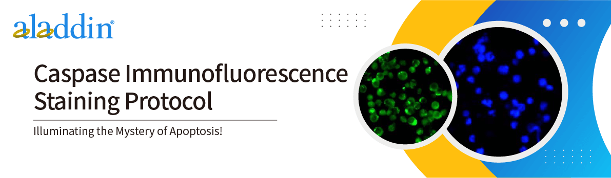
Apoptosis is a tightly regulated cell death mechanism, the core of which is the activation of caspases. Immunofluorescence staining is an effective technique that can simultaneously detect caspases and other apoptosis-related proteins in cell samples.
This protocol is intended as a general guide and may require optimization for specific experiments depending on the samples and antibodies used.
General process
Materials required
- Primary antibody against caspase, e.g. recombinant caspase - 3 antibody , rabbit anti- (Ab093013)
- Prepare samples and mount on slides
- Triton X-100
- PBS
- Blocking buffer (PBS/0.1% Tween 20 + 5% serum)
- Conjugated secondary antibody , e.g. Goat anti-rabbit IgG H&L ( Alexa Fluor® 488 ) ( Ab138205 )
- Mounting medium
- Humidification chamber
Experimental Steps
1. Incubate the fixed samples in PBS/0.1% Triton X-100 at room temperature for 5 minutes to permeabilize the membrane.
2. Wash three times with PBS, each time at room temperature for 5 minutes.
3. Aspirate the slides and add 200 μL of blocking buffer (PBS/0.1% Tween 20 + 5% serum).
3.1 Place the slides flat in a humidified chamber and incubate at room temperature for 1-2 hours.
3.2 Then rinse once with PBS.
NOTE: We recommend using serum from the secondary antibody host species (or a closely related species), e.g., if using a goat anti-rabbit conjugate, use goat serum in the blocking buffer.
4. Add 100 μL of primary antibody diluted 1:200 in blocking buffer. Then incubate the slides overnight at 4°C in a humidified chamber.
Tip: You can prepare slides without primary antibody as negative controls.
5. The next day, wash the slides three times with PBS/0.1% Tween 20 at room temperature for 10 minutes each time.
6. Aspirate slides and add 100 μL of appropriate secondary antibody (diluted 1:500 in PBS).
6.1 Place the slides flat in a humidified chamber, away from light, and incubate at room temperature for 1-2 hours.
6.2 Wash three times with PBS/0.1% Tween 20, 5 minutes each time, and protect from light.
7. Drain the liquid, mount the slides in permanent or aqueous mounting medium, and observe under a fluorescence microscope.
Application
1) Cell apoptosis research: Study the activation status of caspases during apoptosis and understand the molecular mechanism of cell death.
2) Drug screening and toxicity testing: Evaluate the effects of drugs or compounds on cell apoptosis and screen candidate drugs that induce or inhibit cell apoptosis activity.
3) Cell signal transduction research: Study the process by which cell signaling pathways affect the activation of caspases and cell apoptosis.
4) Developmental Biology: Studying the role of caspases in programmed cell death during embryonic development and tissue morphogenesis.
5) Immunology research: Study the regulatory role of caspases on immune responses in immune cells such as T cells and B cells.
For more product details, please visit Aladdin Scientific website.