EdU Cell Proliferation Detection Kit (AF488)

Cat. No.: E1373491
Zu bestellen verfügbar
QUALITÄT & REINHEIT Bioactive ? Bioactive grade — verified to retain biological activity in functional assays. Use when the molecule must be functionally active, not just pure. Biological Stain ? Biological stain grade — dyes characterized for staining cells and tissues. Use in histology and microscopy where staining consistency matters. for Fluorescence analysis ? Fluorescence-analysis grade — very low fluorescent impurities for clean spectra. Use in fluorescence assays where background interferes. for Microscopy ? Microscopy grade — reagents/stains suited to sample prep and imaging. Use in microscopy where clarity and low background are needed.
★
Größe
Deutschland (EU)
USA*
Preis
Menge
50T
E1373491-50T
Auf Bestellung · 8–12 Wochen
95,36€
100T
E1373491-100T
—
2 Auf Lager
164,78€
200T
E1373491-200T
Auf Bestellung · 8–12 Wochen
260,23€
Enter a quantity for the sizes you want to add.
🧪
Warum diese Qualität

Bioactive, Biological Stain, for fluorescence analysis, for microscopy Bioactive,Biological Stain,für die Fluoreszenzanalyse,für die Mikroskopie für empfindliche chromatographische und analytische Workflows, die minimale Basislinieninterferenzen erfordern.

🌡

Lagerung & Versand

Protected from light,Store at -20°C Versand Ice chest + Ice pads. Genaue Spezifikationen finden Sie im chargenspezifischen COA.

📋

Qualitätsdokumente

SDS, COA, Datenblatt und Spezifikationsblatt stehen zum Download bereit. Chargenspezifische COA über Chargennummernsuche abrufbar.

📚

Literaturbeleg

In 0 peer-reviewten Publikationen zu Chromatographie, organischer Synthese und Kreuzkupplungsreaktionen zitiert.

Übersicht

Product Introduction

Cell proliferation assays are widely used in the evaluation of cell viability, genotoxicity, and the efficacy of antitumor drugs. Direct detection of DNA synthesis in cells is considered the most accurate method for assessing cell proliferation. EdU (5-ethynyl-2′-deoxyuridine) is a novel thymidine (thymine deoxyribonucleoside) analogue. During DNA synthesis, EdU can be incorporated into newly synthesized DNA in place of thymidine. The ethynyl group on EdU can undergo a covalent reaction with fluorescently labeled small-molecule azide probes (such as Azide AF 488, Azide AF 555, Azide AF 594, Azide AF 647, etc.) via Cu(I)-catalyzed click chemistry, forming a stable triazole ring. This reaction is highly efficient and is referred to as the Click reaction. Through this process, newly synthesized DNA is labeled with the corresponding fluorescent probes, enabling the detection of proliferating cells using appropriate fluorescence detection equipment. 

Usage Protocol

1. Preparation

1) Preparation of Click Additive Solution: 

For a 50-test kit: Add 12.5 mL of pre-chilled deionized water to the tube. Mix thoroughly until completely dissolved to obtain the Click Additive Solution. For a 100-test kit: Add 25 mL of pre-chilled deionized water to the tube. Mix thoroughly until completely dissolved to obtain the Click Additive Solution. For a 200-test kit: Add 50 mL of pre-chilled deionized water to the tube. Mix thoroughly until completely dissolved to obtain the Click Additive Solution. After preparation, aliquot the solution as needed and store at -20°C. If a white precipitate forms after dissolution, invert the tube repeatedly until it is fully dissolved before use. If the solution turns brown, it indicates degradation of the active component; discard it.

2) Upon initial dissolution of the Click Reaction Buffer, aliquot it according to the number of samples per experiment and store at -20°C.

2. EdU Labeling of Cells

It is recommended to use a final EdU concentration of 10 μM (1×). A 1:500 dilution of EdU (10 mM) in cell culture medium yields a 2× EdU working solution (20 μM). Mix an equal volume of pre-warmed (37°C) 2× EdU working solution (20 μM) with the cell suspension to achieve a final 1× EdU concentration. Incubate in a 37°C, 5% CO₂ incubator. Factors such as cell culture medium, cell density, cell type, and other experimental conditions may affect labeling efficiency. Therefore, the optimal EdU concentration and labeling duration must be empirically determined based on the cell type under investigation.

3. Fixation and Permeabilization

1) Harvest cells and centrifuge at 300 ×g for 5 min. Wash cells twice with  PBS containing 2% FBS.

2) Fix cells with 4% paraformaldehyde solution. Mix thoroughly and incubate for 15 min at room temperature protected from light.

3) Collect cells and centrifuge at 300 × g for 5 min. Wash cells twice.

4) Resuspend cells in PBS containing 0.1% Triton X-100. Mix well and incubate for 15 min at room temperature.

5) Centrifuge at 300 × g for 5 min and wash cells twice.

4. Fluorescent Labeling

1) This protocol is based on a 500 μL reaction system per 2 × 10⁶ cells. The volume of the Click reaction mixture can be adjusted according to the experimental sample size.

2) Centrifuge the cells at 300 ×g for 5 minutes. Add 500 μL of Click reaction mixture per sample, mix gently, and incubate for 30 minutes at room temperature protected from light.

3) After the reaction, wash the cells twice with PBS containing 2% FBS.

4) Dilute the DAPI Staining Solution (1000×) to 1× using PBS containing 2% FBS. Add 250 μL of the diluted DAPI solution to each sample and incubate for 5 minutes at room temperature.

5) Add an additional 250 μL of PBS containing 2% FBS, mix gently, and proceed to detection using an appropriate flow cytometry instrument

Precautions

1. Strictly adhere to the component order and volumes specified in the table above when preparing the Click reaction mixture, as deviations may affect subsequent experimental results.

2. The Click reaction mixture must be used within 15 minutes of preparation.

3. To avoid fluorescence quenching, perform detection as soon as possible after sample preparation.

Spezifikationen

Synonyme
VF 488 Click-iT EdU Universal Cell Proliferation Detection Kit
Spezifikationen & Reinheit
Bioactive, Biological Stain, for fluorescence analysis, for microscopy
Note
Bioactive, Biological Stain, für die Fluoreszenzanalyse, für die Mikroskopie
Hinweis
This product can replace A598377.
Anwendung
Cell proliferation, Flow Cytometry
Empfindlichkeit
light-sensitive
Lagerung und Versand
Storage
Protected from light,Store at -20°C
Verschickt in
Ice chest + Ice pads
Stabilität und Lagerung
Store at -20℃ long term (12 months)
Contents & Storage

E1373491

Component

50 T

100 T

200 T

Storage conditions

Quantity Per Test

E1373491A

EdU(10 mM)

100 μL

200 μL

400 μL

-20℃.Store in the dark.

2 μL  per 1.0-2.0 × 10⁶ cells

E1373491B

AF488 azide

100 μL

200 μL

400 μL

-20℃.Store in the dark.

2 μL  per 1.0-2.0 × 10⁶ cells

E1373491C

Click Reaction Buffer

13 mL

26 mL

52 mL

-20℃.Store in the dark.

243 μL  per 1.0-2.0 × 10⁶ cells

E1373491D

CuSO₄

250 μL

500 μL

1000 μL

-20℃.

5 μL  per 1.0-2.0 × 10⁶ cells

E1373491E

Click Additive

248 mg

496 mg

992 mg

-20℃.Store in the dark.

250 μL  per 1.0-2.0 × 10⁶ cells

E1373491F

DAPI Staining Solution(1000×)

25 μL

50 μL

100 μL

-20℃.Store in the dark.

0.5 μL  per 1.0-2.0 × 10⁶ cells

Bilder
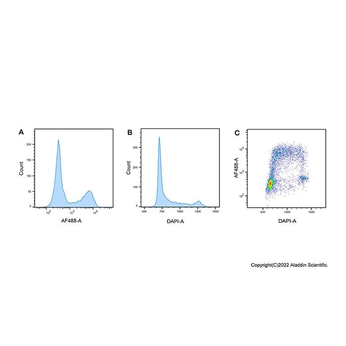
Flow Cytometry: EdU Cell Proliferation Detection Kit (AF488)(E1373491)
Jurkat (human T-cell leukemia) cells were treated with 10 µM EdU (E131265) for 2 hours and detected according to the recommended staining protocol. The incorporated EdU was then conjugated to AF 488 azide via a click reaction and analyzed by flow cytometry using 488 nm (for AF 488) and 405 nm (for DAPI) excitation. (A) Histogram of EdU-AF 488 signal, distinguishing EdU-positive S-phase cells from cells in other cell cycle phases. (B) DNA content histogram (DAPI) resolving the G0/G1, S, and G2/M populations. (C) Dot plot of EdU incorporation versus DNA content, allowing for direct quantification of S-phase cells.

Dokumentation

📋 Sicherheitsdatenblatt (SDS)

Umfassendes Dokument zu Gefahren, Handhabung, Lagerung und Einhaltung regulatorischer Vorschriften.

<<<<<<< HEAD SDS herunterladen → ======= SDS herunterladen → >>>>>>> release248-p3

✅ Analysezertifikat (COA)

Chargenspezifische Qualitätsdaten. Geben Sie Ihre Chargennummer ein, um das genaue COA abzurufen.

COA nachschlagen →

📊 Datenblatt

Kurzübersicht über Produktspezifikationen und Anwendungen.

Datenblatt ansehen →

🔬 Spezifikationsblatt

Vollständige Qualitätsmerkmale und Akzeptanzkriterien für diese Qualität.

Spezifikationsblatt ansehen →

Erweiterte Daten

Zertifikate (CoA, COO, BSE/TSE und Analyse-Diagramm)
C of A & Other Certificates(BSE/TSE, COO):
Analytical Chart:

Find and download the COA for your product by matching the lot number on the packaging.

6 results found

Lot NumberCertificate TypeDatumArtikel
ZJ26F0838357ZJ26F0838357Certificate of AnalysisAug 31, 2026 E1373491
ZJ26F0838356ZJ26F0838356Certificate of AnalysisAug 28, 2026 E1373491
ZJ26F0838355ZJ26F0838355Certificate of AnalysisAug 28, 2026 E1373491
ZJ26F0231967ZJ26F0231967Certificate of AnalysisFeb 06, 2026 E1373491
ZJ26F0231966ZJ26F0231966Certificate of AnalysisFeb 06, 2026 E1373491
ZJ26F0231965ZJ26F0231965Certificate of AnalysisFeb 06, 2026 E1373491
FAQs und Artikel
Biological Stain
Choosing Reagents ‘for Fluorescence Analysis’: A Practical Guide
EdU Cell Proliferation Assay Kit
Staining Principles and Methods for Cell Death, Proliferation, and Metabolic Viability
Application Comparison of the Neutral Red Assay and the MTT/CCK-8 Assays in Cytotoxicity Testing
Culture Systems, Phenotypic Characterization, and Translational Research Applications of Tumor Organoids
Applications of the MCM Protein Family in DNA Replication Licensing, Replication Stress, and Tumor Proliferation Research
Lösungsrechner
Bewertungen

Kundenbewertungen

Dürfen wir Sie über Rabattaktionen informieren?

Später erinnern

Vielen Dank! Bitte überprüfen Sie Ihren E-Mail-Posteingang, um zu bestätigen.

Hoppla! Benachrichtigungen sind deaktiviert.