
Analysis of the Canonical and Non-Canonical NF-κB Pathways: Activation Mechanisms, Detection Methods, and Research Tool Selection
Analysis of the Canonical and Non-Canonical NF-κB Pathways: Activation Mechanisms, Detection Methods, and Research Tool Selection
1. Why Is NF-κB a Core Pathway in Inflammation and Immunity Research?
Nuclear factor κB (nuclear factor kappa-light-chain-enhancer of activated B cells, NF-κB) is one of the most important transcriptional regulatory systems by which cells respond to infection, inflammation, and stress stimuli. Its core role is to convert extracellular or intracellular danger signals into gene-expression responses in the nucleus.
When cells are exposed to stimuli such as tumor necrosis factor-α (TNF-α), interleukin-1β (IL-1β), lipopolysaccharide (LPS), viral or bacterial components, oxidative stress, and DNA damage, they need to rapidly initiate a series of defensive responses, including inflammatory cytokine secretion, chemokine production, immune-cell recruitment, anti-apoptotic responses, and tissue repair. NF-κB is an important transcriptional switch for these responses. The importance of NF-κB can be summarized in three aspects:
Core Question | Role of NF-κB |
How do cells recognize danger stimuli? | Upstream signals from TNF receptors, IL-1 receptors, Toll-like receptors, antigen receptors, and other systems are transmitted to the NF-κB pathway. |
How do cells rapidly generate an inflammatory response? | After nuclear translocation, NF-κB induces the expression of inflammation- and chemotaxis-related genes such as TNF, IL6, IL1B, CXCL8, and CCL2. |
How do cells change their fate? | NF-κB can regulate genes related to anti-apoptosis, cell survival, immune regulation, and tissue injury. |
NF-κB is not only a physiological pathway required for host defense against infection and for tissue repair, but also a pathway that is frequently aberrantly activated in chronic inflammation, autoimmune disease, tumor progression, and therapeutic resistance. When evaluating NF-κB, it should not simply be judged as “beneficial” or “harmful”; instead, its activation intensity, duration, cell type, and disease context should be considered.
2. NF-κB Is a Family of Transcription Factors
NF-κB is not a single protein, but a family of transcription factors. In mammals, the NF-κB/Rel family mainly consists of five members:
Member | Common Name | Characteristics |
RelA | p65 | A transcriptionally activating subunit commonly detected in the canonical NF-κB pathway |
RelB | RelB | Mainly involved in the non-canonical NF-κB pathway |
c-Rel | c-Rel | Closely associated with immune-cell activation and lymphocyte function |
NF-κB1 | p105/p50 | p105 is a precursor protein that is processed to generate p50 |
NF-κB2 | p100/p52 | p100 is a precursor protein that is processed to generate p52 |
These members usually function as homodimers or heterodimers. In the canonical NF-κB pathway, the p65/p50 dimer is commonly observed. Under resting conditions, p65/p50 is bound by inhibitor of κB (IκB) proteins and retained in the cytoplasm. After stimulation, IκB is degraded, and p65/p50 is released and enters the nucleus to initiate target-gene transcription.
RelA/p65, RelB, and c-Rel contain transcriptional activation domains and can directly promote gene transcription. p50 and p52 lack typical transcriptional activation domains; their homodimers often act as transcriptional repressors or require co-regulatory factors such as BCL3 to participate in transcriptional regulation. When p50 or p52 forms heterodimers with members containing transcriptional activation domains, such as p65, RelB, or c-Rel, these dimers are generally more capable of mediating transcriptional activation. When studying the canonical pathway, p65/p50 is usually the focus; when studying the non-canonical pathway, p100 processing, p52 generation, and RelB nuclear translocation should be emphasized.
3. The Canonical NF-κB Pathway: Five Steps from Stimulation to Nuclear Translocation
Before discussing the detailed mechanisms, an overall understanding can first be established through the diagram below. The core of the canonical NF-κB pathway is the IKK–IκBα–p65/p50 axis; the core of the non-canonical pathway is the NIK–IKKα–p100/p52–RelB axis. Although both pathways belong to the NF-κB signaling system, they differ in upstream stimuli, key molecules, and major biological functions.
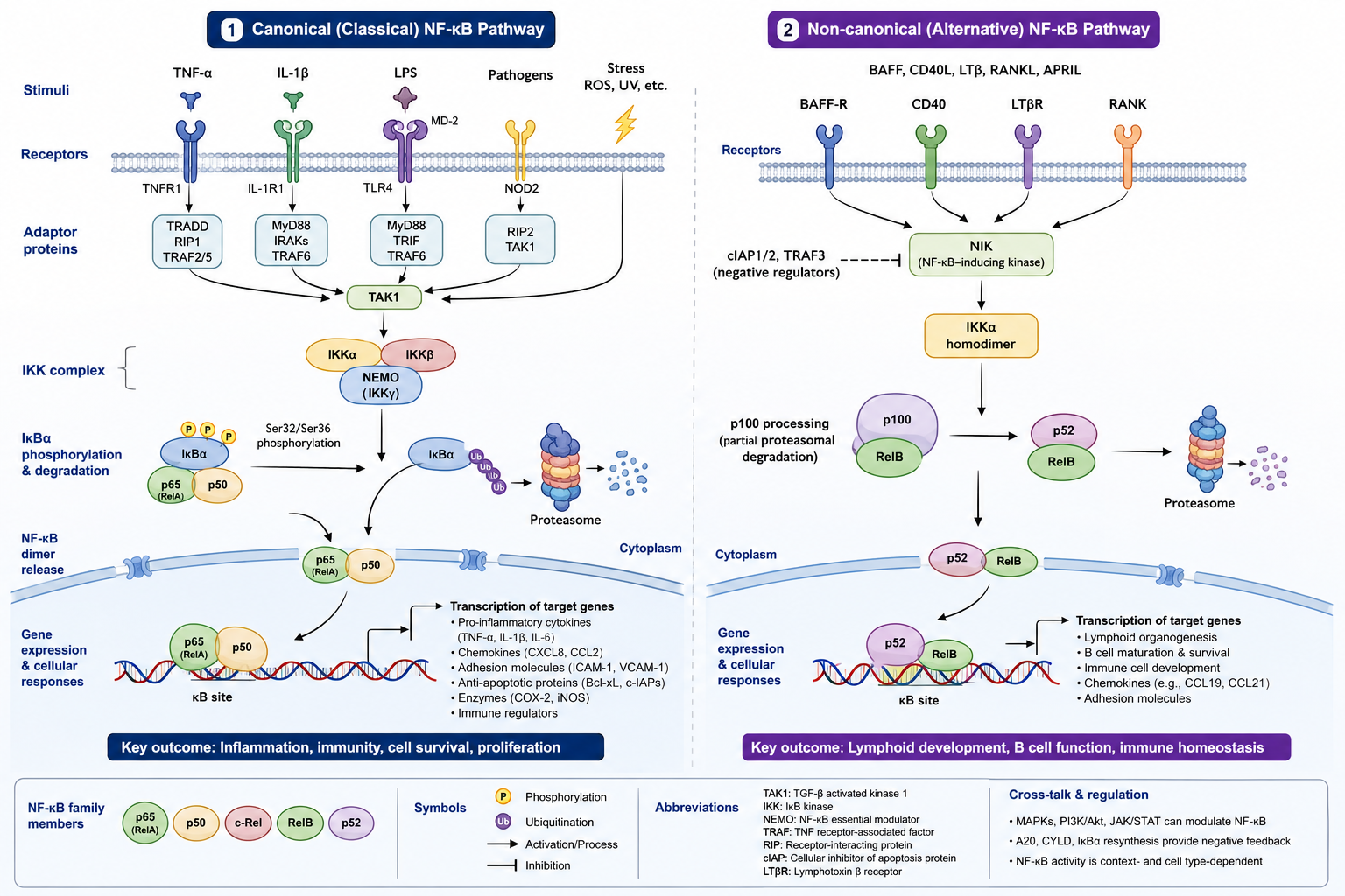
NF-κB signaling pathway core mechanism diagram. In the canonical NF-κB pathway, stimuli such as TNF-α, IL-1β, and LPS activate the IKK complex through receptors and adaptor proteins, inducing phosphorylation, ubiquitination, and proteasomal degradation of IκBα. This releases the p65/p50 dimer and allows it to enter the nucleus, initiating the expression of target genes such as inflammatory cytokines, chemokines, adhesion molecules, anti-apoptotic proteins, and negative feedback factors. In contrast, the non-canonical NF-κB pathway centers on stable accumulation of NIK, activation of IKKα, processing of p100 into p52, and nuclear translocation of p52/RelB. It is mainly involved in lymphoid-organ development, B-cell function, and regulation of immune homeostasis.
The canonical NF-κB pathway is the most common mode of NF-κB activation in inflammation and immunity research. Its core process is:
Stimulatory signal → IKK complex activation → IκBα phosphorylation and degradation → p65/p50 nuclear translocation → target-gene transcription.
3.1 Step 1: Inflammatory or Infectious Signals Are Recognized by Receptors
The canonical NF-κB pathway can be initiated by multiple types of stimuli. Common stimuli include:
Stimulus | Common Receptor or Recognition System | Research Context |
TNF-α | TNF receptor | Inflammation, tumor microenvironment, cell death and survival |
IL-1β | IL-1 receptor | Inflammatory amplification, immune-cell activation |
LPS | Toll-like receptor 4 (TLR4) | Macrophage inflammation models, endotoxin responses |
T-cell/B-cell antigen stimulation | T-cell receptor/B-cell receptor | Adaptive immune activation |
Oxidative stress, DNA damage | Multiple stress-sensing systems | Cell injury, aging, tumor therapy response |
Although these stimuli have different upstream receptors, in the canonical pathway they often ultimately converge on the IκB kinase (IKK) complex.
It should be noted that oxidative stress and DNA damage can affect NF-κB activity, but their upstream mechanisms depend more strongly on cell type, stimulus intensity, and model conditions. They should not be assumed to be completely equivalent to the standard canonical NF-κB activation process induced by TNF-α, IL-1β, or LPS.
3.2 Step 2: The IKK Complex Is Activated
The IKK complex is a key relay node in the canonical NF-κB pathway. It is mainly composed of IKKα, IKKβ, and NF-κB essential modulator (NEMO, also known as IKKγ). Among these components, IKKβ plays a central role in canonical inflammatory signaling, whereas NEMO mainly serves regulatory and scaffold functions.
Upstream stimuli do not directly push p65 into the nucleus. Instead, they first activate IKK through adaptor proteins, ubiquitination complexes, and kinase cascades. After activation, IKK acts directly on IκBα.
3.3 Step 3: IκBα Is Phosphorylated, Ubiquitinated, and Degraded
IκBα is the major inhibitory protein in the canonical NF-κB pathway. Under resting conditions, IκBα binds p65/p50 and masks its nuclear localization signal, keeping NF-κB in the cytoplasm.
After stimulation, IKK mediates phosphorylation of IκBα at Ser32 and Ser36. Phosphorylated IκBα is then recognized by E3 ubiquitin ligases and undergoes K48-linked polyubiquitination, followed by degradation by the 26S proteasome. After IκBα degradation, the nuclear localization signal of p65/p50 becomes exposed, and the dimer enters the nucleus.
Mechanistic outline:
TNF-α / IL-1β / LPS
↓
Receptor and adaptor-protein complex
↓
Activation of the IKKα–IKKβ–NEMO complex
↓
Phosphorylation of IκBα at Ser32/Ser36
↓
K48-linked polyubiquitination of IκBα
↓
26S proteasomal degradation of IκBα
↓
Release and nuclear translocation of p65/p50
This process is a key molecular event in the canonical NF-κB pathway. In experiments, detection of IκBα degradation, increased p-IκBα, and p65 nuclear translocation is commonly used to determine whether the canonical pathway has been activated.
3.4 Step 4: p65/p50 Translocates into the Nucleus
After IκBα degradation, the p65/p50 dimer translocates from the cytoplasm into the nucleus. p65 nuclear translocation is an important marker of canonical NF-κB pathway activation, but it is still only one part of the activation process. After p65 enters the nucleus, it must also bind κB-binding sequences in the promoter or enhancer regions of target genes and cooperate with transcriptional co-regulators in order to effectively initiate target-gene transcription.
3.5 Step 5: Target-Gene Expression Changes
The ultimate function of NF-κB is to regulate gene expression. Typical target genes include:
Functional Category | Representative Genes or Molecules | Biological Outcome |
Inflammatory cytokines | TNF, IL6, IL1B | Amplification of inflammatory responses |
Chemokines | CXCL8, CCL2, CCL5 | Recruitment of immune cells |
Adhesion molecules | ICAM1, VCAM1, SELE | Promotion of leukocyte adhesion and migration |
Anti-apoptotic molecules | Certain anti-apoptotic BCL2 family members, such as BCL2, BCL2L1/Bcl-xL, and BCL2A1; BIRC/IAP family; CFLAR/c-FLIP | Enhancement of cell survival |
Negative feedback factors | NFKBIA/IκBα, TNFAIP3/A20 | Limitation of sustained NF-κB signaling |
Inflammatory enzymes | PTGS2/COX-2, NOS2/iNOS | Participation in inflammatory mediator generation |
To determine whether NF-κB is truly exerting its function, it is not sufficient to examine only p65 phosphorylation or nuclear translocation; downstream target genes and functional phenotypes should also be assessed.
4. The Non-Canonical NF-κB Pathway: NIK, IKKα, p100/p52, and RelB
The non-canonical NF-κB pathway differs from the canonical pathway. Its main features are not IκBα degradation and rapid p65/p50 nuclear translocation, but rather stable accumulation of NF-κB-inducing kinase (NIK), activation of IKKα, processing of p100 into p52, and nuclear translocation of p52/RelB.
The non-canonical pathway is often triggered by specific members of the TNF receptor superfamily, such as B-cell activating factor receptor (BAFF receptor, BAFFR), CD40, lymphotoxin β receptor (LTβR), and receptor activator of NF-κB (RANK).
The main sequence of the non-canonical pathway is as follows:
BAFFR / CD40 / LTβR / RANK
↓
Stable accumulation of NIK
↓
Activation of IKKα
↓
Phosphorylation and partial proteasomal processing of p100
↓
Formation of p52
↓
Nuclear translocation of p52/RelB
↓
Regulation of genes related to B-cell maturation, lymphoid-organ development, and immune homeostasis
p100 has a dual role in the non-canonical pathway. On the one hand, it is the precursor protein of p52; on the other hand, it also functions as an inhibitor of RelB. After pathway activation, p100 is processed into p52, and p52 forms a dimer with RelB and enters the nucleus.
The differences between the canonical and non-canonical pathways can be summarized as follows:
Comparison Dimension | Canonical NF-κB Pathway | Non-Canonical NF-κB Pathway |
Main stimuli | TNF-α, IL-1β, LPS, antigen-receptor stimulation | BAFFR, CD40, LTβR, RANK, etc. |
Key kinases | IKKβ and the NEMO-dependent IKK complex | NIK and IKKα |
Key inhibitory/precursor protein | IκBα | p100 |
Main dimer | p65/p50 | p52/RelB |
Response characteristics | Rapid, typical inflammatory response | Slower, associated with immune development and homeostasis |
Major functions | Inflammatory cytokine expression, immune activation, cell survival | B-cell maturation, lymphoid-organ formation, immune homeostasis |
5. What Downstream Responses Does NF-κB Regulate?
The downstream responses of NF-κB can be divided into five categories, each corresponding to a defined biological outcome.
5.1 Inflammatory Cytokine Expression
NF-κB can induce the expression of pro-inflammatory genes such as TNF, IL6, and IL1B. These factors further stimulate immune cells, endothelial cells, and tissue cells, forming an inflammatory amplification response. During acute infection, this response helps eliminate pathogens; during chronic inflammation, sustained cytokine expression can aggravate tissue injury.
5.2 Chemokines and Immune-Cell Recruitment
NF-κB can promote the expression of chemokines such as CXCL8, CCL2, and CCL5. These molecules participate in the recruitment of neutrophils, monocytes, T cells, and other immune cells, and are an important reason for immune-cell infiltration at inflammatory sites.
5.3 Adhesion-Molecule Expression
NF-κB can regulate the expression of adhesion molecules such as ICAM1, VCAM1, and SELE, making it easier for leukocytes to bind to vascular endothelial cells and cross the vessel wall into inflamed tissues. This process is beneficial for host defense against infection, but in chronic inflammation and vascular inflammation it may promote persistent tissue injury.
5.4 Cell Survival and Anti-Apoptosis
NF-κB can induce anti-apoptosis-related genes such as BCL2 family members, BIRC/IAP family members, and CFLAR/c-FLIP, thereby increasing cell survival capacity in inflammatory or stress environments. In normal immune responses, this helps maintain cellular function; in tumors, sustained NF-κB activation may enhance the anti-apoptotic capacity of tumor cells and reduce therapeutic sensitivity.
5.5 Negative Feedback Regulation
NF-κB can also induce negative feedback factors such as NFKBIA/IκBα and TNFAIP3/A20. Newly synthesized IκBα can re-bind NF-κB and return it from the nucleus to the cytoplasm; A20 limits sustained NF-κB activation by regulating upstream ubiquitination signals. These negative feedback mechanisms help control the intensity and duration of inflammatory responses.
6. Aberrant NF-κB Activation and Disease
The disease relevance of NF-κB stems from one central issue: under normal conditions, it helps cells respond to infection and injury; under abnormal conditions, it can keep inflammation, survival signaling, and immune responses in a chronically activated state.
6.1 Chronic Inflammation
In chronic inflammatory diseases such as rheumatoid arthritis, inflammatory bowel disease, and asthma, sustained activation of NF-κB can lead to long-term expression of inflammatory cytokines, chemokines, and adhesion molecules, driving persistent immune-cell infiltration and tissue damage.
6.2 Cancer
NF-κB can participate in multiple stages of tumor initiation and progression, including promoting tumor-cell survival, enhancing anti-apoptotic capacity, maintaining an inflammatory tumor microenvironment, promoting angiogenesis, and affecting therapeutic resistance. The role of NF-κB is not exactly the same across different tumor types, but sustained NF-κB activation is often associated with tumor-related inflammation and therapeutic tolerance.
6.3 Autoimmune Diseases
NF-κB participates in immune-cell activation, cytokine production, and the maintenance of immune homeostasis. If regulation of the NF-κB pathway becomes dysregulated, it may promote abnormal immune activation and damage to self-tissues.
6.4 Infection and Immune Evasion
Pathogen infection usually activates NF-κB and induces anti-infective inflammatory responses. Some viruses and bacteria can also affect host immune responses by enhancing, suppressing, or altering NF-κB signaling, thereby promoting immune evasion or chronic infection.
6.5 Metabolic Inflammation and Neuroinflammation
In conditions such as obesity, insulin resistance, lipotoxicity, and neurodegenerative diseases, NF-κB can connect metabolic stress, oxidative stress, immune-cell activation, and tissue functional damage. Therefore, it is also an important pathway in studies of metabolic inflammation and neuroinflammation.
7. How Should the NF-κB Pathway Be Studied?
7.1 Pathway Activation Level: Examine IκBα, p65, and Nuclear Translocation
Common detection methods include:
Method | Detection Content | Suitable Question |
Western blot | p-IKKα/β, p-IκBα, IκBα, p-p65, total p65 | Whether the IKK–IκBα–p65 axis is activated |
Nuclear/cytoplasmic protein fractionation | Nuclear p65 and cytoplasmic p65 | Whether p65 translocates from the cytoplasm into the nucleus |
Immunofluorescence | Co-localization of p65 with the nucleus | Whether p65 nuclear translocation occurs and whether cellular heterogeneity is present |
Electrophoretic mobility shift assay (EMSA) | DNA-binding capacity of NF-κB | Whether NF-κB has DNA-binding activity |
Western blot can show phosphorylation and degradation of key proteins; immunofluorescence can directly visualize p65 nuclear translocation; nuclear/cytoplasmic protein fractionation can quantitatively compare nuclear p65 levels. Combining these three approaches is more reliable than detecting p-p65 alone.
7.2 Transcriptional Output Level: Examine Reporter Assays, qPCR, and ChIP-qPCR
Method | Detection Content | Suitable Question |
NF-κB luciferase reporter assay | NF-κB-dependent reporter-gene activity | Whether NF-κB produces transcriptional activity |
Reverse transcription quantitative PCR (RT-qPCR) | mRNA levels of TNF, IL6, IL1B, CXCL8, NFKBIA, and other genes | Whether downstream target genes are upregulated |
Chromatin immunoprecipitation qPCR (ChIP-qPCR) | Whether p65 binds to promoters or enhancers of target genes | Whether a specific gene may be directly regulated by p65 |
The NF-κB luciferase reporter assay reflects transcriptional activity, RT-qPCR reflects target-gene mRNA output, and ChIP-qPCR is more suitable for verifying the binding of p65 to regulatory regions of specific genes.
7.3 Functional Level: Examine Inflammatory Factors and Cellular Phenotypes
Method | Detection Content | Suitable Question |
Enzyme-linked immunosorbent assay (ELISA) | Secreted proteins such as TNF-α, IL-6, and CXCL8; IL-1β secretion should be interpreted in the context of inflammasome activation/protein processing | Whether transcriptional changes are converted into inflammatory cytokine secretion |
Flow cytometry | Immune-cell activation markers, apoptosis, cell cycle | Whether NF-κB affects cellular states |
Cell-based functional assays | Survival, migration, invasion, phagocytosis, cytotoxic killing, etc. | Whether NF-κB changes have functional significance |
Among these, NF-κB usually mainly promotes IL1B transcription and pro-IL-1β production; cleavage and secretion of mature IL-1β often also depend on inflammasome/caspase-1-mediated processing and release. Therefore, detection of IL-1β secretion should be interpreted in combination with whether a second stimulus is present and with the specific cell model used.
A complete evidence chain is recommended as follows:
Treatment factor or stimulus
↓
p-IKK / p-IκBα / IκBα degradation
↓
p65 nuclear translocation
↓
NF-κB reporter activity or enhanced p65 DNA binding
↓
Upregulation of NF-κB target-gene mRNA
↓
Inflammatory cytokine secretion or changes in cellular function
↓
Validation of causality using inhibitors or genetic interventions
To demonstrate that a drug, gene, or treatment factor acts through NF-κB, the evidence should include at least four types of data: pathway indicators, transcriptional output, functional phenotypes, and intervention-based validation.
8. How Should NF-κB-Related Inhibitors and Experimental Tools Be Selected?
When selecting NF-κB tools, the research objective should first be clarified. Different tools act at different positions and therefore answer different questions.
8.1 Establishing NF-κB Activation Models
Research Objective | Common Stimulus | Application Notes |
Rapid inflammatory activation | TNF-α | Applicable to multiple cell types; suitable for observing IκBα degradation and p65 nuclear translocation |
IL-1-related inflammation | IL-1β | Suitable for studying IL-1R-mediated inflammatory signaling |
Macrophage inflammation model | LPS | Commonly used in RAW 264.7 cells, THP-1-derived macrophages, and primary macrophages |
Antigen-receptor-related activation | TCR/BCR stimulation | Suitable for studies of T-cell and B-cell activation |
Oxidative-stress-related activation | H₂O₂ or ROS-inducing models | Suitable for studying crosstalk between stress and inflammatory pathways |
The choice of stimulus should match the cell type. For example, an LPS model requires cells to have the corresponding TLR4 recognition and downstream signaling capacity, whereas a TNF-α model is more suitable for observing rapid activation of the canonical NF-κB pathway.
8.2 Inhibiting IκBα Phosphorylation
BAY 11-7082 is commonly used to inhibit cytokine-induced IκBα phosphorylation, thereby blocking activation of the canonical NF-κB pathway. It is suitable for verifying whether a treatment factor depends on IκBα phosphorylation and canonical NF-κB activation. However, BAY 11-7082 should not be simply regarded as a fully specific NF-κB inhibitor. In experiments, IκBα phosphorylation, p65 nuclear translocation, and target-gene expression should be examined simultaneously to avoid drawing conclusions based solely on inhibitor results. Because BAY 11-7082 may also affect deubiquitinases, inflammasomes, or cell-death-related processes, causal validation is recommended in combination with genetic intervention or another NF-κB tool with a different mechanism of action.
8.3 Inhibiting p65 Nuclear Translocation
JSH-23 is a commonly used inhibitor of NF-κB p65 nuclear translocation. It is characterized by inhibition of p65 nuclear translocation and NF-κB transcriptional activity, without markedly affecting IκBα degradation. Therefore, JSH-23 is more suitable for answering the following question: even if upstream IκBα degradation has already occurred, can blocking p65 nuclear entry reduce downstream inflammatory gene expression?
8.4 Inhibiting Proteasome-Mediated IκB Degradation
MG132 is a 26S proteasome inhibitor that blocks degradation of ubiquitinated proteins, including proteasomal degradation of IκB, and can therefore indirectly inhibit NF-κB activation. However, MG132 has a broad range of effects and can affect the degradation of many intracellular proteins, the cell cycle, endoplasmic reticulum stress, and apoptosis. Therefore, MG132 is more suitable as an auxiliary mechanistic tool and should not be used alone as specific evidence for NF-κB inhibition.
8.5 Oxidative-Stress-Related Interventions
Tools such as N-acetylcysteine (NAC) and pyrrolidine dithiocarbamate (PDTC) are often used to explore the relationship between reactive oxygen species (ROS) and NF-κB activation.
These tools may affect NF-κB through antioxidant effects, metal-ion chelation, or other forms of cellular stress regulation. If NAC or PDTC reduces NF-κB activity, this only indicates that the redox state may be involved in the process; it does not directly prove that all effects are specifically mediated by NF-κB.
8.6 Genetic Interventions
To strengthen causal evidence, small interfering RNA (siRNA), short hairpin RNA (shRNA), CRISPR-Cas9, overexpression, or dominant-negative tools can be used to intervene in key nodes such as RELA/p65, NFKB1/p50, IKBKB/IKKβ, IKBKG/NEMO, and NFKBIA/IκBα.
A more reliable study design is one in which the results of small-molecule inhibitors and genetic interventions are consistent, while downstream target genes and functional phenotypes change in the same direction.
9. Common Pitfalls in NF-κB Research
9.1 Increased p65 Phosphorylation Does Not Equal Complete NF-κB Activation
p-p65 is an important indicator, but it alone does not represent the complete function of NF-κB. Whether NF-κB exerts transcriptional effects also depends on whether p65 translocates into the nucleus, binds DNA, initiates target-gene transcription, and whether target-gene products produce functional effects.
9.2 IκBα Degradation Does Not Mean That All Downstream Inflammatory Factors Will Increase
IκBα degradation indicates release of inhibition in the canonical pathway, but downstream gene expression is also influenced by cell type, chromatin accessibility, co-transcription factors, stimulation duration, and negative feedback regulation. Therefore, IκBα degradation should be interpreted together with p65 nuclear translocation and target-gene detection.
9.3 NF-κB Inhibition Is Not Always Beneficial
NF-κB participates in host defense against infection, tissue repair, and cellular protection. Excessive inhibition of NF-κB may weaken immune defense or affect tissue recovery. Whether NF-κB should be inhibited depends on disease type, timing of intervention, cell type, and degree of activation.
9.4 Different NF-κB Inhibitors Are Not Interchangeable
BAY 11-7082, JSH-23, MG132, PDTC, and other tools act at different positions. They affect IκBα phosphorylation, p65 nuclear translocation, proteasomal degradation, or redox status, respectively. In experimental design, tools should be selected according to the research question, rather than grouping all tools broadly as “NF-κB inhibitors.”
9.5 NF-κB Outputs Differ Among Cell Types
Macrophages, endothelial cells, epithelial cells, fibroblasts, T cells, B cells, and tumor cells do not have identical NF-κB target-gene profiles. The same stimulus may induce different downstream responses in different cells; therefore, conclusions from other cell models should not be directly applied without validation.
10. Classification Table of Representative Chemicals for NF-κB Signaling Pathway Research
Table 1. Pathway Activation Modeling and Upstream Receptor-Signaling Intervention
Category | CAS No. | Aladdin Catalog No. | Name | Specification or Purity | Product Features and Applications |
TLR4 inflammatory stimulation model | 93572-42-0 | Lipopolysaccharide (LPS) | Derived from Escherichia coli O55:B5, purified by trichloroacetic acid extraction | Used to establish TLR4-mediated NF-κB activation models; suitable for studies of macrophage inflammatory responses, IκBα degradation, p65 nuclear translocation, and expression of inflammatory factors such as TNF-α, IL-6, and IL-1β | |
PKC-related pathway activation model | 16561-29-8 | P167764 | Phorbol 12-myristate 13-acetate (PMA) | Moligand™, ≥98% | Used to induce protein kinase C-related signaling activation; suitable for detection of NF-κB pathway activation, immune-cell differentiation, inflammatory cytokine expression, and transcriptional activity |
TLR4 signaling inhibition | 243984-11-4 | TAK-242, TLR4 signaling inhibitor | Moligand™, ≥98% | Used to inhibit downstream TLR4 signaling; suitable for verifying whether LPS-induced activation of the IKK–IκBα–p65 axis depends on the TLR4 pathway |
Table 2. Inhibition of the IKK/IκBα Axis and the Non-Canonical NIK Pathway
Category | CAS No. | Aladdin Catalog No. | Name | Specification or Purity | Product Features and Applications |
IκBα phosphorylation inhibition | 19542-67-7 | BAY 11-7082, IκBα phosphorylation inhibitor / NF-κB inhibitor | Moligand™, ≥98% | Used to interfere with IκBα phosphorylation; suitable for studying IκBα degradation, p65 release, p65 nuclear translocation, and inflammatory gene transcription in the canonical NF-κB pathway | |
IκBα phosphorylation inhibition | 196309-76-9 | BAY 11-7085, irreversible inhibitor of TNF-α-induced IκBα phosphorylation | ≥98% (HPLC) | Used to inhibit TNF-α-induced IκBα phosphorylation; suitable for studies of canonical NF-κB pathway activation, p65 nuclear translocation, and downstream inflammatory cytokine expression | |
IKK-1/IKK-2 inhibition | 445430-58-0 | BMS 345541, IKK-2 and IKK-1 inhibitor | ≥99% | Used to inhibit IKK-1- and IKK-2-related activity; suitable for analyzing IKK complex-mediated IκBα phosphorylation, NF-κB transcriptional output, and inflammatory responses | |
IKK complex inhibition | 873225-46-8 | IKK-16 (IKK inhibitor VII) | ≥99% | Used to interfere with IKK complex activity; suitable for studies of IκBα phosphorylation, p65 nuclear translocation, and NF-κB target-gene expression under TNF-α, IL-1β, or LPS stimulation | |
IKK complex inhibition | 1186195-62-9 | IKK-16, IκB kinase inhibitor | ≥98% | Used to inhibit IκB kinase-related signaling; suitable for validation of the IKK–IκBα–p65 axis in the canonical NF-κB pathway and intervention in inflammatory models | |
IKKβ inhibition | 978-62-1 | IMD 0354, IKKβ inhibitor | ≥99% | Used to inhibit IKKβ-mediated IκBα phosphorylation; suitable for studies of TNF-α-, IL-1β-, or LPS-induced NF-κB activation and inflammatory cytokine expression | |
IKKβ inhibition | 783348-36-7 | MLN120B, IκB kinase β (IKKβ) inhibitor | Moligand™, ≥98% | Used to study the role of IKKβ in the canonical NF-κB pathway; suitable for studies of inflammatory cytokine transcription, cell survival, and tumor-associated inflammatory signaling | |
IKKβ inhibition | 507475-17-4 | TPCA-1, IκB kinase inhibitor | Moligand™, ≥98% | Used to interfere with IKKβ-related signaling; suitable for detecting IκBα phosphorylation, p65 nuclear translocation, and expression of inflammatory factors such as TNF-α or IL-6 | |
IKKβ inhibition | 431898-65-6 | PS-1145 | ≥98% | Used to inhibit the IKKβ-mediated canonical NF-κB pathway; suitable for studies of IκBα degradation, p65 nuclear translocation, and target-gene transcription under TNF-α or LPS stimulation | |
IKKβ inhibition | 354812-17-2 | SC-514, selective, reversible, ATP-competitive IKKβ/IKK-2 inhibitor | ≥98% | Used to interfere with IKKβ-related signaling; suitable for studies of NF-κB-dependent inflammatory gene expression, such as IL-6, IL-8, and COX-2 | |
Natural-product-derived IKK/NF-κB modulation | 524-12-9 | Wedelolactone | Moligand™, ≥98% (HPLC) | Used to study regulation of the IKK/NF-κB axis by natural products; suitable for inflammatory models, p65 activation, inflammatory cytokine expression, and evaluation of anti-inflammatory activity | |
Non-canonical NIK pathway inhibition | 1660114-31-7 | NIK SMI1 | Moligand™, ≥98% | Used to inhibit NIK-related signaling; suitable for studies of p100 processing, p52/RelB nuclear translocation, B-cell function, and immune homeostasis in the non-canonical NF-κB pathway |
Table 3. p65 Nuclear Translocation, Transcriptional Activity, and Natural-Product-Derived NF-κB Modulation
Category | CAS No. | Aladdin Catalog No. | Name | Specification or Purity | Product Features and Applications |
NF-κB transcriptional activity inhibition | 545380-34-5 | QNZ (EVP4593), NF-κB inhibitor | Moligand™, ≥98% | Used to inhibit NF-κB transcriptional activity; suitable for NF-κB reporter assays, TNF-α production, inflammatory cytokine expression, and pathway-output detection | |
p65 nuclear translocation inhibition | 749886-87-1 | 4-Methyl-N1-(3-phenylpropyl)-1,2-benzenediamine | ≥98% (HPLC) | Used to inhibit p65 nuclear translocation and NF-κB transcriptional activity; suitable for distinguishing IκBα degradation from transcriptional regulation after p65 nuclear entry | |
Sesquiterpene lactone NF-κB modulation | 20554-84-1 | Parthenolide | Analytical standard, Moligand™, ≥98% | Used to study the effects of sesquiterpene lactone compounds on NF-κB activation, inflammatory cytokine expression, tumor-associated inflammatory signaling, and cell-survival-related processes | |
Diterpene lactone NF-κB modulation | 5508-58-7 | Andrographolide | Moligand™, ≥98% | Used to study the effects of natural diterpene lactone compounds on NF-κB-related inflammatory responses, immune regulation, p65 activation, and cytokine expression | |
Steroidal lactone NF-κB modulation | 5119-48-2 | Withaferin A | ≥98% (HPLC) | Used to study regulation of IKK/NF-κB signaling, inflammatory responses, tumor-cell survival, and anti-apoptosis-related pathways by steroidal lactone compounds | |
Phenolic acid ester NF-κB modulation | 104594-70-9 | Caffeic acid phenethyl ester | ≥97% | Used to study regulation of NF-κB activation, oxidative-stress-related inflammation, p65 transcriptional activity, and inflammatory cytokine expression by phenolic acid ester compounds |
Table 4. Proteasome/IκBα Degradation and Oxidative-Stress Interventions
Category | CAS No. | Aladdin Catalog No. | Name | Specification or Purity | Product Features and Applications |
Irreversible proteasome inhibition | 868540-17-4 | Carfilzomib | Moligand™, ≥99% | Used to inhibit proteasome activity; suitable for studies of the ubiquitin–proteasome system, IκBα degradation, NF-κB signaling regulation, and tumor-cell survival | |
Reversible proteasome inhibition | 133407-82-6 | MG-132, reversible proteasome inhibitor | Moligand™, ≥98% | Used to block 26S proteasome-mediated protein degradation; suitable for mechanistic validation of IκBα degradation, p65 release, accumulation of ubiquitinated proteins, and the canonical NF-κB pathway | |
Reversible proteasome inhibition | 179324-69-7 | Bortezomib (PS-341), reversible proteasome inhibitor | Moligand™, ≥98% | Used to interfere with 20S/26S proteasome-related protein degradation; suitable for blocking IκBα degradation, regulating the NF-κB pathway, accumulating ubiquitinated proteins, and studying tumor-cell survival | |
Reactive oxygen species intervention | 616-91-1 | N-Acetyl-L-cysteine (NAC) | PharmPure™, USP, Moligand™, European Pharmacopoeia (Ph. Eur.), ≥98.5% | Used to regulate reactive oxygen species-related cellular stress; suitable for studying the relationship between oxidative stress and NF-κB activation, IκBα degradation, p65 activation, and inflammatory cytokine expression | |
Redox-related NF-κB inhibition | 5108-96-3 | Ammonium pyrrolidinedithiocarbamate (APDC) | Moligand™, ≥99% | Used to study the relationship among redox status, metal-ion chelation, and NF-κB activation; suitable for inflammatory models, p65 activation detection, and cytokine expression analysis |
Note: The products listed above are representative Aladdin products for scientific research and formulation-related studies. For additional product specifications, grades, and certificate of analysis (COA) information, please search by product name, CAS number, or catalog number on the Aladdin website.
References
[1] Guo Q, Jin Y, Chen X, et al. NF-κB in biology and targeted therapy: new insights and translational implications. Signal Transduction and Targeted Therapy. 2024;9:53. doi:10.1038/s41392-024-01757-9.
[2] Kanehisa Laboratories. KEGG PATHWAY: NF-kappa B signaling pathway. KEGG PATHWAY Database. Accessed July 9, 2026.
[3] Christian F, Smith EL, Carmody RJ. The Regulation of NF-κB Subunits by Phosphorylation. Cells. 2016;5(1):12. doi:10.3390/cells5010012.
[4] Sun S-C. Non-canonical NF-κB signaling pathway. Cell Research. 2011;21:71–85. doi:10.1038/cr.2010.177.
[5] Sun S-C. The non-canonical NF-κB pathway in immunity and inflammation. Nature Reviews Immunology. 2017;17:545–558. doi:10.1038/nri.2017.52.
[6] Dolcet X, Llobet D, Pallares J, Matias-Guiu X. NF-kB in development and progression of human cancer. Virchows Archiv. 2005;446:475–482. doi:10.1007/s00428-005-1264-9.
[7] Oeckinghaus A, Hayden MS, Ghosh S. Crosstalk in NF-κB signaling pathways. Nature Immunology. 2011;12:695–708. doi:10.1038/ni.2065.
[8] Mendez JM, Keestra-Gounder AM. NF-κB-dependent Luciferase Activation and Quantification of Gene Expression in Salmonella Infected Tissue Culture Cells. Journal of Visualized Experiments. 2020;(155):e60567. doi:10.3791/60567.
[9] Cell Signaling Technology. BAY 11-7082 Product Information. Product No. 78679. Accessed July 9, 2026.
[10] Shin HM, Kim MH, Kim BH, Jung SH, Kim YS, Park HJ, Hong JT, Min KR, Kim Y. Inhibitory action of novel aromatic diamine compound on lipopolysaccharide-induced nuclear translocation of NF-κB without affecting IκB degradation. FEBS Letters. 2004;571(1–3):50–54. doi:10.1016/j.febslet.2004.06.056.
[11] InvivoGen. MG-132 peptide aldehyde: 26S proteasome inhibitor. Product Information. Accessed July 9, 2026.
[12] Schroder K, Tschopp J. The inflammasomes. Cell. 2010;140(6):821–832. doi:10.1016/j.cell.2010.01.040.
More related articles are listed below:
JAK-STAT Cell Signaling Pathway
Noncanonical Functions of Epigenetic Enzymes and Regulation of Mitochondrial Metabolism
Transposase-Based Epigenomic Sequencing Strategies: Principles and Recent Progress of CUT&Tag
Regulatory Framework and Biological Effects of the GSK3 Signaling Pathway
RANK Signaling Pathway: Molecular Components, Activation Mechanisms, and Biological Effects
CTLA4 Signaling Pathway: Molecular Basis, Immunoregulatory Mechanisms, and Disease Associations
Molecular Mechanisms and Biological Functions of the Fas Pathway
Type Stratification, Signal Transduction, and Effector Networks of the Interferon (IFN) Pathway
Wnt/β-Catenin Signaling Pathway