
Apoptosis Signaling Pathways and Regulated Cell Death Research
Apoptosis Signaling Pathways and Regulated Cell Death Research
Apoptosis is a form of programmed cell death regulated by genes and protein networks. It mainly functions to eliminate senescent, damaged, abnormally proliferating, or functionally imbalanced cells. Typical apoptosis is characterized by cell shrinkage, chromatin condensation, DNA fragmentation, caspase cascade activation, and apoptotic body formation. Abnormal apoptosis is closely associated with tumors, autoimmune diseases, ischemic injury, AIDS, neurodegenerative diseases, and other pathological conditions.
Keywords: apoptosis; extrinsic apoptotic pathway; mitochondrial apoptotic pathway; caspase; Bcl-2 family; IAP; p53; ferroptosis; necroptosis; pyroptosis
1 Basic Logic of Apoptosis Research
1.1 Biological Significance of Apoptosis
Apoptosis is not merely a cell death phenomenon, but an important regulatory mechanism for maintaining tissue homeostasis, developmental morphogenesis, and immune balance. Under physiological conditions, apoptosis eliminates aged cells, cells with severe DNA damage, abnormally activated immune cells, and potential tumor cells. Under pathological conditions, either excessive or insufficient apoptosis may lead to disease.
(1) Insufficient apoptosis
Insufficient apoptosis allows damaged cells, abnormally proliferating cells, or autoreactive immune cells to survive persistently, and is commonly associated with tumorigenesis, therapeutic tolerance, and autoimmune lymphoproliferation.
(2) Excessive apoptosis
Excessive apoptosis can cause massive loss of tissue cells and is commonly observed in ischemia-reperfusion injury, neurodegenerative diseases, virus infection-related cell depletion, and drug-induced toxic injury.
(3) Apoptosis and tissue homeostasis
Apoptotic cells are usually engulfed and cleared by macrophages or neighboring cells. If clearance is delayed, late-stage apoptotic cells may undergo secondary necrosis, release cellular contents, and induce inflammatory responses.
1.2 Distinguishing Apoptosis from Other Forms of Regulated Cell Death
Cell death research should not classify all death phenomena as apoptosis. Ferroptosis, necroptosis, pyroptosis, cuproptosis, paraptosis, and other forms of regulated cell death differ in triggering mechanisms, molecular markers, and detection indicators.
Table 1 Comparison of Apoptosis and Other Forms of Regulated Cell Death
Cell Death Form | Core Mechanism | Typical Molecules | Common Interpretation Indicators |
Apoptosis | caspase cascade, mitochondrial outer membrane permeabilization, DNA fragmentation | caspase-8, caspase-9, caspase-3, Bax, Bcl-2, PARP | Annexin V, cleaved caspase-3, cleaved PARP, TUNEL |
Necroptosis | RIPK1/RIPK3/MLKL-mediated membrane rupture-associated death | RIPK1, RIPK3, p-MLKL | p-MLKL, membrane integrity disruption, Nec-1 sensitivity |
Pyroptosis | Inflammatory lytic death mediated by inflammasomes and Gasdermin | NLRP3, caspase-1, GSDMD, IL-1β | GSDMD cleavage, IL-1β release, LDH release |
Ferroptosis | Iron-dependent accumulation of lipid peroxidation | GPX4, SLC7A11, ACSL4, TFRC | Lipid ROS, MDA, 4-HNE, iron ions, GPX4 reduction |
Cuproptosis | Copper-induced aggregation of mitochondrial lipoylated proteins and proteotoxic stress | FDX1, DLAT, LIAS | Copper-dependent cell death and abnormal mitochondrial metabolism |
Paraptosis | Non-caspase-dependent cell death often accompanied by ER/mitochondrial swelling | MAPK, IGF1R-related signaling | Cytoplasmic vacuolization, caspase insensitivity, ER/mitochondrial dilation |
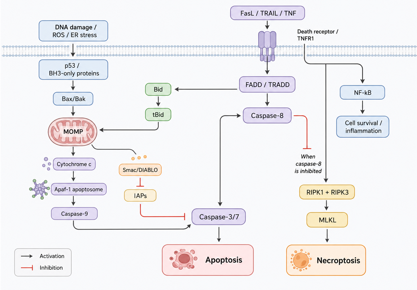
Figure 1 Simplified schematic diagram of apoptosis and necroptosis signaling pathways
2 Extrinsic Apoptotic Pathway
2.1 Death Receptor-Mediated Signal Initiation
(1) Mechanistic core
The extrinsic apoptotic pathway is mediated by cell membrane death receptors (DRs). After death ligands such as FasL, TNF-α, and TRAIL bind to their corresponding receptors, intracellular death domains of the receptors are clustered, and the adaptor protein FADD and initiator caspase-8 are recruited to form the death-inducing signaling complex (DISC).
(2) Key molecules
Typical molecules include Fas/CD95, TNF receptor, TRAIL receptor, FADD, caspase-8, and c-FLIP. After DISC formation, caspase-8 is activated and further initiates the effector caspase cascade.
(3) Result interpretation
If FasL, TNF-α, or TRAIL treatment induces caspase-8 cleavage, caspase-3 activation, and PARP cleavage, activation of the extrinsic apoptotic pathway is usually supported. If death receptors are upregulated but caspase-8 is not activated, insufficient DISC assembly, c-FLIP inhibition, or switching to alternative pathways such as necroptosis should be considered.
2.2 Caspase-8 and the Effector Caspase Cascade
(1) Mechanistic core
Caspase-8 is an important initiator caspase in the extrinsic apoptotic pathway. After activation, it can directly cleave and activate caspase-3, caspase-6, and caspase-7. It can also cleave Bid into tBid, amplifying death receptor signals into the mitochondrial pathway.
(2) Experimental indicators
Common indicators include cleaved caspase-8, cleaved caspase-3, cleaved caspase-7, cleaved PARP, Annexin V positivity, and DNA fragmentation.
(3) Result interpretation
Caspase-3 cleavage is a commonly used apoptosis marker, but it cannot distinguish the extrinsic pathway from the intrinsic pathway by itself. If caspase-8 is activated before caspase-9 and blockade of death receptors reduces apoptosis, dominance of the extrinsic pathway is better supported.
2.3 Dual Effects of TNF Signaling
(1) Mechanistic core
Binding of TNF-α to TNF receptors does not necessarily induce apoptosis. TNFR signaling can form different complexes. A membrane-proximal complex can activate NF-κB and pro-survival signaling. When intracellular conditions change, a death complex containing FADD and caspase-8 can form and induce apoptosis.
(2) Experimental indicators
TNF receptor, FADD, caspase-8, NF-κB activation, IκBα degradation, cell survival, and inflammatory factor expression should be detected simultaneously.
(3) Result interpretation
Increased TNF-α does not directly indicate that cells are undergoing apoptosis. If TNF signaling mainly activates NF-κB and induces survival factor expression, cells may show inflammatory activation rather than apoptosis. Only when caspase-8 and downstream effector caspases are activated is TNF-related apoptosis supported.
Table 2 Key Nodes in the Extrinsic Apoptotic Pathway
Signaling Node | Representative Molecules | Main Role | Interpretation Focus |
Death ligands | FasL, TNF-α, TRAIL | Initiate death receptor signaling | Increased ligand level does not equal apoptosis; receptors and caspases should be evaluated together |
Death receptors | Fas, TNFR, TRAILR | Transmit membrane receptor death signals | DISC formation and downstream caspase activation should be observed |
Adaptor protein | FADD | Connects receptors with caspase-8 | FADD loss or abnormality can weaken extrinsic apoptosis |
Initiator caspase | caspase-8 | Activates effector caspases or cleaves Bid | Can connect extrinsic and mitochondrial pathways |
Effector caspases | caspase-3, caspase-6, caspase-7 | Execute apoptotic morphology and substrate cleavage | Cleaved caspase-3 is commonly used as a core readout |
3 Intrinsic Mitochondrial Apoptotic Pathway
3.1 Bcl-2 Family Regulation of Mitochondrial Outer Membrane Permeabilization
(1) Mechanistic core
The intrinsic apoptotic pathway is mainly determined by mitochondrial outer membrane permeabilization (MOMP). Bcl-2 family proteins are the core regulators of this process. Pro-apoptotic members include Bid, Bim, Bax, and Bak, whereas anti-apoptotic members include Bcl-2, Bcl-xL, and Mcl-1.
(2) Key mechanism
After activation, Bax and Bak can form pores in the mitochondrial outer membrane and promote cytochrome c release. Anti-apoptotic Bcl-2 family proteins can inhibit Bax/Bak activation, thereby preventing initiation of the mitochondrial pathway.
(3) Result interpretation
An increased Bax/Bcl-2 ratio often suggests that cells are more prone to apoptosis, but it cannot replace functional validation. More reliable evidence includes loss of mitochondrial membrane potential, cytochrome c release, caspase-9 activation, and caspase-3/PARP cleavage.
3.2 Cytochrome c and Apoptosome Formation
(1) Mechanistic core
After cytochrome c is released from mitochondria, it can form the apoptosome with Apaf-1 and procaspase-9, further activating caspase-9. Caspase-9 then activates caspase-3 and caspase-7, completing the execution phase of apoptosis.
(2) Experimental indicators
Mitochondrial membrane potential, cytosolic cytochrome c, Apaf-1, cleaved caspase-9, cleaved caspase-3, and cleaved PARP are commonly detected.
(3) Result interpretation
A decrease in mitochondrial membrane potential does not always represent apoptosis; it may also occur in necrosis, ferroptosis, or metabolic failure. Therefore, mitochondrial indicators should be interpreted together with caspase activation, Annexin V, TUNEL, and related results.
3.3 Bid Links the Extrinsic and Intrinsic Pathways
(1) Mechanistic core
After activation of caspase-8 in the extrinsic pathway, Bid can be cleaved into tBid. tBid translocates to mitochondria and promotes Bax/Bak activation, thereby amplifying death receptor signaling into mitochondrial apoptotic signaling.
(2) Experimental indicators
tBid, mitochondrial translocation of Bax, cytochrome c release, and simultaneous activation of caspase-8 and caspase-9 are commonly detected.
(3) Result interpretation
If death receptor stimulation simultaneously triggers caspase-8 activation and mitochondrial injury, the extrinsic pathway may amplify apoptosis through Bid. If caspase-8 inhibition reduces mitochondrial injury, this connection is further supported.
4 Apoptosis Execution Phase and Detection Interpretation
4.1 Caspase-3, Caspase-6, and Caspase-7
(1) Mechanistic core
Caspase-3, caspase-6, and caspase-7 are the major proteases in the execution phase of apoptosis. They cleave structural proteins, repair proteins, and regulatory proteins, causing cell shrinkage, DNA fragmentation, cytoskeletal remodeling, and apoptotic body formation.
(2) Experimental indicators
Cleaved caspase-3, cleaved caspase-7, cleaved PARP, DNA ladder, TUNEL, Annexin V, and nuclear morphology are commonly detected.
(3) Result interpretation
Annexin V positivity can indicate phosphatidylserine externalization, but early apoptosis, late apoptosis, and some non-apoptotic membrane changes can all affect results. More reliable apoptosis interpretation should combine caspase cleavage, PARP cleavage, and cellular morphology.
4.2 PARP Cleavage and Interruption of DNA Repair
(1) Mechanistic core
PARP participates in DNA damage repair. During apoptosis, caspase-3 can cleave PARP, stopping energy-consuming DNA repair processes and promoting irreversible progression of apoptosis.
(2) Experimental indicators
Cleaved PARP, total PARP, γH2AX, TUNEL, and cell viability are commonly detected.
(3) Result interpretation
Cleaved PARP is a common marker of apoptosis, but PARP also participates in DNA damage repair and energy depletion associated with necrotic cell death. If only PARP changes are observed, caspase-3 and Annexin V/TUNEL should be combined for interpretation.
4.3 Regulation by IAPs and Survivin
(1) Mechanistic core
IAPs, or inhibitors of apoptosis proteins, can inhibit caspase activity and restrict apoptosis execution. Survivin is a member of the IAP family and is often highly expressed in tumor cells. It is associated with cell division, anti-apoptosis, and therapeutic tolerance.
(2) Experimental indicators
XIAP, cIAP1/2, Survivin, cleaved caspase-3, cell cycle distribution, drug sensitivity, and apoptotic proportion are commonly detected.
(3) Result interpretation
Increased IAP or Survivin usually indicates stronger anti-apoptotic capacity, but whether it truly causes therapeutic tolerance requires validation through IAP inhibition, restored caspase activation, and changes in drug sensitivity.
Table 3 Apoptosis Detection Indicators and Interpretation Points
Detection Indicator | Main Significance | Notes |
Annexin V | Phosphatidylserine externalization; commonly used for early apoptosis detection | PI or 7-AAD should be combined to distinguish early apoptosis from late-stage death |
cleaved caspase-3 | Effector caspase activation | A key apoptosis indicator, but upstream pathway source should be interpreted together |
cleaved caspase-8 | Initiation of extrinsic pathway | Can connect to the mitochondrial pathway through Bid |
cleaved caspase-9 | Initiation of intrinsic mitochondrial pathway | Should be combined with mitochondrial membrane potential and cytochrome c release |
cleaved PARP | caspase-mediated PARP cleavage | Does not independently represent the complete apoptotic mechanism |
TUNEL | DNA break labeling | Severe necrosis or other DNA breaks may also yield positive signals |
Decreased mitochondrial membrane potential | Mitochondrial functional impairment | Non-apoptotic mitochondrial injury should be excluded |
5 p53, c-Myc, and Apoptosis Regulation
5.1 p53-Mediated Damage Response
(1) Mechanistic core
p53 is a key fate-regulating factor after DNA damage, replication stress, and cellular stress. Under mild to moderate damage, p53 can induce p21 expression and cause cell cycle arrest. When damage is severe or repair fails, p53 can induce expression of pro-apoptotic genes such as Bax, PUMA, and NOXA, promoting mitochondrial apoptosis.
(2) Regulatory node
MDM2 promotes p53 ubiquitination and degradation and is an important negative regulator of the p53 pathway. Abnormalities in the MDM2/p53 axis can allow cells to evade damage-induced apoptosis.
(3) Result interpretation
Increased p53 does not necessarily equal apoptosis. p53-mediated apoptosis is supported only when p53 activation is accompanied by upregulation of pro-apoptotic target genes, mitochondrial injury, and caspase activation. If p21 is mainly induced, the outcome may be dominated by cell cycle arrest or senescence.
5.2 c-Myc and Apoptosis Sensitivity
(1) Mechanistic core
c-Myc promotes cell growth and proliferation, but under nutrient deprivation, DNA damage, or insufficient survival signaling, it can also increase cellular sensitivity to apoptosis. Its function is strongly context-dependent.
(2) Experimental indicators
c-Myc, cell proliferation, p53, Bim, Bax, caspase activation, and cell death proportion are commonly detected.
(3) Result interpretation
Increased c-Myc may represent proliferative drive, but it may also make cells more prone to apoptosis under stress. Interpretation of c-Myc results must consider survival signaling, nutritional status, and DNA damage background.
6 Apoptosis and Disease Mechanisms
6.1 Tumors
(1) Mechanistic characteristics
Tumor cells often evade apoptosis by upregulating Bcl-2, Bcl-xL, Mcl-1, IAPs, and Survivin, or by p53 inactivation, weakened death receptor signaling, or abnormal caspase expression. Apoptosis escape can promote tumor progression and therapeutic tolerance.
(2) Research indicators
Bcl-2 family proteins, p53, MDM2, IAPs, caspase cleavage, PARP cleavage, colony formation, drug IC50, and resistant cell models are commonly detected.
(3) Result interpretation
A decrease in tumor cell viability after drug treatment does not equal apoptosis induction. To prove that apoptosis participates in drug efficacy, caspase-dependent evidence should be observed, and cell death should be partially reversed by caspase inhibitors.
6.2 Autoimmunity and Lymphoproliferation
(1) Mechanistic characteristics
Immune cells need to eliminate abnormally activated or autoreactive clones through apoptosis. Abnormal Fas/FasL signaling can impair lymphocyte clearance and promote autoimmunity and lymphoproliferation.
(2) Research indicators
Fas, FasL, caspase-8, T/B cell apoptosis proportion, inflammatory factors, and abnormal lymphocyte expansion are commonly detected.
(3) Result interpretation
Increased lymphocyte number does not necessarily result from enhanced proliferation; it may also result from insufficient apoptotic clearance. Proliferation and apoptosis indicators should be detected simultaneously.
6.3 AIDS and Infection-Related Cell Death
(1) Mechanistic characteristics
Viral infection can induce immune cell loss through multiple mechanisms, including immune activation, death receptor signaling, mitochondrial injury, and inflammatory cell death. In AIDS, CD4⁺ T cell reduction is associated with multiple cell death mechanisms.
(2) Research indicators
T cell apoptosis, Fas/FasL, caspase activation, mitochondrial membrane potential, inflammatory factors, and viral replication levels are commonly detected.
(3) Result interpretation
Cell death in infection models is not necessarily all apoptosis. Apoptosis, pyroptosis, necroptosis, and direct viral lytic effects should be distinguished.
6.4 Ischemic Injury and Neurodegenerative Diseases
(1) Mechanistic characteristics
Ischemia, oxidative stress, Ca²⁺ overload, mitochondrial dysfunction, and protein aggregation can all induce apoptosis in neurons or tissue cells. In neurodegenerative diseases, apoptosis is often intertwined with oxidative stress, ferroptosis, inflammation, and protein homeostasis abnormalities.
(2) Research indicators
Mitochondrial membrane potential, Bax/Bcl-2, cleaved caspase-3, TUNEL, ROS, ferroptosis indicators, and neuronal survival are commonly detected.
(3) Result interpretation
In nervous system injury, TUNEL positivity or increased caspase does not explain all cell death. If lipid peroxidation, GPX4 reduction, and iron accumulation are also present, ferroptosis should be considered as a co-participating mechanism.
7 Apoptosis-Targeted Intervention Strategies
7.1 Therapeutic Strategies That Promote Apoptosis
(1) Bcl-2 inhibition
Inhibition of Bcl-2, Bcl-xL, or Mcl-1 can release anti-apoptotic protection and make tumor cells more prone to mitochondrial apoptosis. This strategy is commonly used in tumors highly dependent on anti-apoptotic Bcl-2 family proteins.
(2) IAP inhibition
IAP inhibitors can release caspases from inhibition and increase cellular sensitivity to death receptor signaling or chemotherapeutic drugs.
(3) MDM2/p53 regulation
In a p53 wild-type background, MDM2 inhibition can stabilize p53 and enhance damage response and apoptotic tendency. If p53 is mutated or lost, this strategy usually has limited efficacy.
(4) Death receptor activation
Strategies targeting Fas, TRAIL receptors, or TNF-related pathways can directly initiate extrinsic apoptosis, but inflammatory responses, tissue toxicity, and receptor expression differences should be considered.
7.2 Protective Strategies That Inhibit Apoptosis
(1) Caspase inhibition
In ischemia-reperfusion, neural injury, and acute toxic injury, caspase inhibition may reduce cell loss. However, long-term apoptosis inhibition may increase the risk of abnormal cell survival.
(2) Akt/PKB pathway activation
Akt signaling can promote cell survival and inhibit some pro-apoptotic factors. This strategy may be beneficial for tissue protection, but in tumor contexts it may promote therapeutic tolerance.
(3) PARP regulation
PARP participates in DNA repair and cell death. Moderate PARP inhibition may reduce energy depletion or produce synthetic lethality, but its role differs across disease contexts.
7.3 Combined Regulation with Other Cell Death Modes
(1) Apoptosis and ferroptosis
When some tumor cells are insensitive to apoptosis, inducing ferroptosis may serve as an alternative strategy. Ferroptosis research should focus on GPX4, lipid ROS, GSH, TFRC, and iron metabolism.
(2) Apoptosis and necroptosis
When caspase-8 is inhibited, cells may switch to RIPK1/RIPK3/MLKL-mediated necroptosis. In research on death receptor pathways, both caspase-dependent and RIPK-dependent routes should be considered.
(3) Apoptosis and pyroptosis
Inflammasome activation can induce pyroptosis and release inflammatory mediators such as IL-1β and IL-18. If cell death is accompanied by GSDMD cleavage and inflammatory factor release, it should not be simply classified as apoptosis.
Table 4 Apoptosis Intervention Strategies and Applicable Scenarios
Intervention Direction | Representative Targets | Expected Result | Applicable Scenario |
Promoting extrinsic apoptosis | Fas, TNFR, TRAILR, caspase-8 | Initiation of death receptor signaling | Tumor cell death and immune clearance research |
Promoting mitochondrial apoptosis | Bcl-2, Bcl-xL, Mcl-1, Bax/Bak | Induction of MOMP and caspase-9 activation | Tumor therapy and chemosensitivity research |
Releasing caspase inhibition | IAP, Survivin | Enhanced effector caspase activity | Apoptosis tolerance and tumor resistance research |
Stabilizing p53 | MDM2/p53 | Enhanced damage-induced apoptosis | p53 wild-type tumor research |
Inhibiting excessive apoptosis | caspase, PARP, mitochondrial injury | Protection of cell survival | Ischemia, neurodegenerative diseases, and toxic injury research |
Inducing non-apoptotic death | GPX4, RIPK, GSDMD, cuproptosis-related molecules | Bypassing apoptosis tolerance | Apoptosis-resistant tumors or inflammatory cell death research |
Table 5 Representative Products Related to Apoptosis and Regulated Cell Death Research
Research Module | Representative Product | CAS No. | Mechanism or Detection Positioning | Applicable Research |
Positive control for apoptosis induction | Staurosporine | Broad-spectrum kinase inhibitor commonly used to induce caspase-dependent apoptosis | Apoptosis model establishment and detection of cleaved caspase-3 and PARP cleavage | |
DNA damage-induced apoptosis | Etoposide | Topoisomerase II inhibitor that induces DNA double-strand breaks | DNA damage response, p53 pathway, and apoptosis induction research | |
DNA damage-induced apoptosis | Camptothecin | Topoisomerase I inhibitor that induces replication-associated DNA damage | γH2AX, caspase activation, and tumor cell efficacy research | |
Chemotherapy-induced apoptosis | Doxorubicin hydrochloride | DNA intercalation and topoisomerase II-related damage | Chemotherapy-induced apoptosis, p53 response, and resistance mechanism research | |
Caspase cascade inhibition | Z-VAD-FMK | Pan-caspase inhibitor | Verifying whether cell death depends on the caspase cascade | |
Caspase-3 pathway inhibition | Ac-DEVD-CHO | caspase-3-related inhibitor | Effector caspase functional validation, PARP cleavage, and apoptosis execution phase research | |
Bcl-2 family inhibition | ABT-737 | Bcl-2/Bcl-xL/Bcl-w inhibitor | Mitochondrial apoptosis, Bax/Bak dependence, and tumor cell apoptosis sensitivity research | |
Bcl-2 targeting research | Venetoclax | Selective Bcl-2 inhibitor | Bcl-2-dependent tumors, MOMP, and mitochondrial apoptosis research | |
Bcl-2/Bcl-xL inhibition | Navitoclax | Bcl-2/Bcl-xL inhibitor | Anti-apoptotic escape, BH3 mimetics, and combination efficacy research | |
Mcl-1 targeting research | S63845 | Mcl-1 inhibitor | Mcl-1-dependent survival, mitochondrial apoptosis, and resistance mechanism research | |
IAP inhibition research | Birinapant | Smac mimetic/IAP antagonist | IAP-mediated apoptosis escape, restored caspase activation, and tumor resistance research | |
IAP inhibition research | BV6 | IAP inhibitor | TNF-related cell death and switching between apoptosis and necroptosis | |
p53 stabilization research | Nutlin-3a | MDM2-p53 interaction inhibitor | p53 wild-type tumors, p21/PUMA/NOXA expression, and apoptosis induction research | |
PARP-related cell death research | Olaparib | PARP inhibitor | DNA repair inhibition, synthetic lethality, and PARP-related cell death research | |
Necroptosis inhibition | Necrostatin-1 | RIPK1 inhibitor | TNF-related necroptosis and validation of RIPK1-dependent death | |
MLKL-related necroptosis | Necrosulfonamide | MLKL inhibitor | p-MLKL-related membrane rupture-associated death and necroptosis validation | |
Ferroptosis induction | Erastin | Inhibits system xc⁻ and induces GSH depletion and lipid peroxidation | Ferroptosis models, SLC7A11/GPX4 axis, and apoptosis-resistant cell death research | |
Ferroptosis induction | RSL3 | GPX4 inhibitor | GPX4-dependent antioxidant defense, lipid ROS, and ferroptosis mechanism research | |
Ferroptosis inhibition | Ferrostatin-1 | Lipid peroxidation inhibitor | Verifying whether cell death has ferroptotic features | |
Ferroptosis/iron homeostasis research | Deferoxamine mesylate | Iron chelator | Iron-dependent death, iron overload, and oxidative injury research | |
Pyroptosis/NLRP3 research | MCC950 | NLRP3 inflammasome inhibitor | Pyroptosis, IL-1β release, and inflammasome-related cell death research | |
Caspase-1-related pyroptosis | Ac-YVAD-CMK | caspase-1 inhibitor | GSDMD cleavage, IL-1β maturation, and inflammatory cell death research | |
Cuproptosis research | Elesclomol | Copper ionophore that promotes copper-dependent cell death | Cuproptosis, mitochondrial metabolism, and tumor cell death research | |
Photodynamic cell death induction | 5-Aminolevulinic acid hydrochloride | Photosensitizer precursor that promotes PDT-related ROS generation | Photodynamic therapy, ROS-mediated apoptosis, and tumor cell killing research |
Table 6 Product Lines Related to Apoptosis and Regulated Cell Death and Application Directions
Research Direction | Product Line | Covered Object/Target | Applicable Research Direction |
Overview of apoptosis research |
| Apoptosis-related proteins, detection reagents, and regulatory molecules | Extrinsic/intrinsic apoptosis, caspase cascade, drug-induced cell death, and disease mechanism research |
Death receptor signaling research |
| TNFR and related death receptor signaling | TNF-mediated apoptosis, inflammation, survival signaling, and switching to necroptosis |
Mitochondrial apoptosis research |
| Bcl-2, Bcl-xL, Mcl-1, Bax, Bak, and others | Mitochondrial outer membrane permeabilization, anti-apoptotic escape, tumor resistance, and BH3 mimetic research |
Caspase cascade research |
| Caspase family and related cysteine proteases | Initiator caspases, effector caspases, PARP cleavage, and apoptosis execution phase research |
p53 regulation research |
| MDM2, p53, and their regulatory network | DNA damage response, p53 stability, p21/PUMA/NOXA expression, and tumor apoptosis research |
Pro-apoptotic kinase research |
| Apoptosis signal-regulating kinase 1 | Oxidative stress, JNK/p38 pathway, stress-induced apoptosis, and inflammatory injury research |
Pro-apoptotic kinase research |
| Death-associated protein kinase | Cell death, calcium/calmodulin-related signaling, tumor suppression, and neural injury research |
Anti-apoptotic mechanism research |
| XIAP, cIAP, and Survivin-related inhibitory proteins | caspase inhibition, apoptosis escape, tumor resistance, and IAP inhibitor research |
Anti-apoptotic mechanism research |
| Survivin/BIRC5 | Mitosis, anti-apoptosis, tumor proliferation, therapeutic tolerance, and prognostic marker research |
Cell proliferation and apoptosis balance research |
| c-Myc and its downstream regulatory network | Proliferative drive, stress-induced apoptosis sensitivity, tumor progression, and metabolic reprogramming research |
Protein folding and stress research |
| FK506-binding protein family | Protein folding, mTOR-related signaling, stress tolerance, and cell death regulation |
Photodynamic death induction research |
| Photosensitizers and ROS generation-related tools | Photodynamic therapy, ROS-mediated apoptosis, tumor cell killing, and mitochondrial injury research |
Thymidylate synthesis research |
| TS/TYMS and nucleotide synthesis-related targets | DNA synthesis inhibition, S phase stress, antimetabolic drugs, and apoptosis induction research |
Necroptosis research |
| RIPK1/RIPK3/MLKL-related molecules | caspase-independent death, inflammatory membrane rupture, and cell death mode switching |
RIPK signaling research |
| RIPK1, RIPK3, and related kinases | TNF signaling, necroptosis, inflammatory injury, and crosstalk among cell death pathways |
Pyroptosis research |
| Inflammasomes, caspase-1, GSDMD, and related molecules | Inflammatory cell death, IL-1β release, infectious inflammation, and tumor immunity research |
Ferroptosis research |
| GPX4, SLC7A11, ACSL4, and iron metabolism-related targets | Lipid peroxidation, iron-dependent death, neurotoxicity, and apoptosis-resistant tumors |
Antioxidant defense research |
| GPX family, especially GPX4 | GSH-dependent antioxidant defense, lipid peroxidation suppression, and ferroptosis regulation |
Iron uptake research |
| TFRC and iron transport-related targets | Iron uptake, ferroptosis sensitivity, tumor metabolism, and oxidative injury research |
Cuproptosis research |
| Copper ion homeostasis and mitochondrial lipoylated protein-related targets | Copper-dependent cell death, mitochondrial metabolism, tumor cell death, and proteotoxic stress research |
Paraptosis research |
| Non-caspase-dependent cell death-related molecules | Cytoplasmic vacuolization, ER/mitochondrial swelling, apoptosis-resistant models, and alternative death mechanisms |
PKD signaling research |
| Protein kinase D-related targets | Oxidative stress, mitochondrial function, cell survival/death balance, and signal transduction research |
The key to apoptosis research is to distinguish cell death modes, identify pathway sources, and establish a mechanistic closed loop. Only by integrating death receptor, mitochondrial, caspase, Bcl-2 family, p53/IAP regulation, and related indicators of ferroptosis, necroptosis, and pyroptosis can the true mechanism of cell death and its disease significance be accurately determined.
References
[1] Cao L, et al. J Cell Death. 2016 Dec 29;9:19-29.
[2] Dasgupta A, et al. Int J Mol Sci. 2017 Jan;18(1):23.
For more related articles, please see below:
[2] Wnt/β-Catenin Signaling Pathway
[4] Metabolic signaling pathway
[5] Wnt Signaling
[7] JAK-STAT Cell Signaling Pathway
[8] PD-1/PD-L1 Signaling Pathway
[9] Granzyme A and Granzyme B Signaling Pathways: Cytotoxic Effects and Immune Regulatory Mechanisms
[10] CTLA4 Signaling Pathway: Molecular Basis, Immunoregulatory Mechanisms, and Disease Associations
[11] VEGF Signaling Pathway: Receptor Lineage, Activation Mechanisms, and Biological Effects
[14] RANK Signaling Pathway: Molecular Components, Activation Mechanisms, and Biological Effects