

Determine the necessary mass, volume, or concentration for preparing a solution.
224,000+ productos de investigación · Triple ISO certified · COA & SDS Disponible para cada producto · Same-day shipping on in-stock items
Why this grade
for sensitive chromatographic and analytical workflows requiring minimal baseline interference.
Storage & shipping
Protected from light,Store at -20°C,Desiccated Ships Ice chest + Ice pads Check lot-specific COA for exact specifications.
Quality documents
SDS, COA, datasheet, and spec sheet available for download. Lot-specific COA accessible via lot number lookup.
Literature proof
Cited in 3 peer-reviewed publications across chromatography, organic synthesis, and cross-coupling reactions.
Descripción general
Product composition
Product name | Package |
JC-1(200×) | 100μL/tube, 5 tubes in total |
Ultra-pure water | 90mL |
JC-1 staining buffer (5×) | 80mL |
CCCP(10mM) | 20μL |
Product Introduction
Mitochondrial Membrane Potential Detection Kit (JC-1) is a kit that uses JC-1 as a fluorescent probe to quickly and sensitively detect changes in cell, tissue or purified mitochondrial membrane potential. It can be used for early apoptosis .
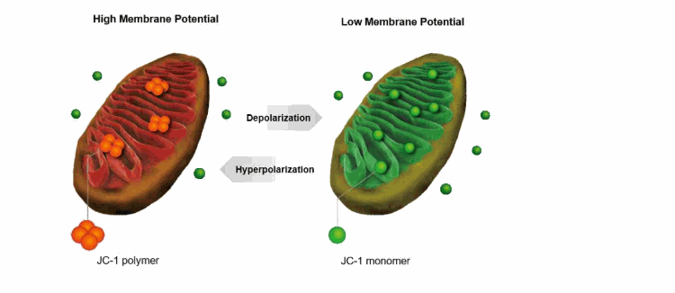
JC-1 is an ideal fluorescent probe widely used to detect mitochondrial membrane potential △Ψm. Can detect cell, tissue or purified mitochondrial membrane potential. When the mitochondrial membrane potential is high, JC-1 aggregates in the matrix of the mitochondria to form a polymer, which can produce red fluorescence; when the mitochondrial membrane potential is low, JC-1 cannot accumulate in the matrix of the mitochondria, at this time JC- 1 is a monomer, which can produce green fluorescence. In this way, it is very convenient to detect the change of mitochondrial membrane potential through the change of fluorescence color. The relative ratio of red and green fluorescence is commonly used to measure the ratio of mitochondrial depolarization.
The decrease of mitochondrial membrane potential is a landmark event in the early stage of apoptosis. The decrease of cell membrane potential can be easily detected by the transition of JC-1 from red fluorescence to green fluorescence, and the transition from red fluorescence to green fluorescence of JC-1 can also be used as an early detection indicator of cell apoptosis.
The maximum excitation wavelength of JC-1 monomer is 515nm and the maximum emission wavelength is 529nm; the maximum excitation wavelength of JC-1 polymer is 585nm and the maximum emission wavelength is 590nm. For actual observation, use the conventional settings for observing red fluorescence and green fluorescence.
This kit provides CCCP as a positive control for inducing the decrease of mitochondrial membrane potential.
For the samples in the six-well plate, the kit can detect a total of 100 samples; for the samples in the 12-well plate, the kit can detect a total of 200 samples.
Instructions for use
1. Preparation of JC-1 dyeing working solution
The amount of JC-1 staining working solution required for each well of a six-well plate is 1mL, and the amount of JC-1 staining working solution for other culture vessels can be deduced by analogy: 0.5mL JC-1 for every 500,000 to 1 million cells of cell suspension Dyeing working solution. Take an appropriate amount of JC-1 (200×) and dilute JC-1 at the ratio of adding 8mL ultrapure water to every 50μL of JC-1 (200×). Shake vigorously to dissolve and mix JC-1 thoroughly. Then add 2mL JC-1 staining buffer (5×), and mix well to become the JC-1 staining working solution.
2. Setting of positive control
Add the CCCP (10mM) provided in the kit to the cell culture medium at a ratio of 1:1000, dilute to 10μM, and treat the cells for 20 minutes. Then load JC-1 according to the following method to detect the mitochondrial membrane potential. For most cells, the membrane potential of the mitochondria will be completely lost after 20 minutes of treatment with 10μM CCCP. After staining with JC-1, it should show green fluorescence; while normal cells should show red fluorescence after staining with JC-1. For specific cells, the concentration and duration of action of CCCP may be different, so you need to refer to relevant literature to determine.
3. For suspension cells
Take 100,000 to 600,000 cells and resuspend them in 0.5 mL of cell culture medium, which can contain serum and phenol red.
Add 0.5mL JC-1 staining working solution, invert several times to mix. Incubate in a cell incubator at 37°C for 20 minutes.
During the incubation period, prepare an appropriate amount of JC-1 staining buffer (1×) at the ratio of adding 4 mL of distilled water per 1 mL of JC-1 staining buffer (5×), and place in an ice bath.
After incubation at 37°C, centrifuge at 600g 4°C for 3 to 4 minutes to pellet the cells. Discard the supernatant and try not to aspirate the cells.
Wash twice with JC-1 staining buffer (1×): add 1mL JC-1 staining buffer (1×) to resuspend the cells, centrifuge at 600g at 4°C for 3-4 minutes, pellet the cells, and discard the supernatant. Then add 1mL JC-1 staining buffer (1×) to resuspend the cells, centrifuge at 600g at 4°C for 3-4 minutes to pellet the cells and discard the supernatant.
Then resuspend it with an appropriate amount of JC-1 staining buffer (1×) and observe it with a fluorescence microscope or a laser confocal microscope. It can also be detected with a fluorescence spectrophotometer or analyzed by a flow cytometer.
4. For adherent cells
Note: For adherent cells, if you want to use a fluorescence spectrophotometer or flow cytometer to detect, you can collect the cells first, and then refer to the detection method of suspended cells after resuspension.
For one well of the six-well plate, aspirate the culture medium, wash the cells once with PBS or other appropriate solution if necessary according to the specific experiment, and add 1 mL of cell culture medium. The cell culture fluid may contain serum and phenol red.
Add 1mL JC-1 dyeing working solution and mix well. Incubate in a cell incubator at 37°C for 20 minutes.
During the incubation period, prepare an appropriate amount of JC-1 staining buffer (1×) at the ratio of adding 4 mL of distilled water per 1 mL of JC-1 staining buffer (5×), and place it in an ice bath.
After incubation at 37°C, aspirate the supernatant and wash twice with JC-1 staining buffer (1×).
Add 2 mL of cell culture medium, which can contain serum and phenol red.
Observe under fluorescence microscope or laser confocal microscope.
5. For purified mitochondria
Dilute the prepared JC-1 staining working solution with JC-1 staining buffer (1×) 5 times.
Add 0.1 mL of purified mitochondria with a total protein content of 10-100 μg to 0.9 mL of the 5-fold diluted JC-1 staining working solution.
Detect with a fluorescence spectrophotometer or a fluorescence microplate reader: directly use a fluorescence spectrophotometer to perform a time scan after mixing. The excitation wavelength is 485nm and the emission wavelength is 590nm. If you use a fluorescence microplate reader and the excitation wavelength cannot be set to 485nm, you can set the excitation wavelength within the range of 475~520nm. In addition, you can also refer to the wavelength setting in step 6 below for fluorescence detection.
Observe with a fluorescence microscope or a laser confocal microscope: the method is the same as step 6 below.
6. Fluorescence observation and result analysis
When detecting JC-1 monomer, you can set the excitation light to 490nm and the emission light to 530nm; when detecting JC-1 polymer, you can set the excitation light to 525nm and the emission light to 590nm. Note: It is not necessary to set the excitation light and emission light to the maximum excitation wavelength and maximum emission wavelength when measuring fluorescence here. If you use a fluorescence microscope to observe, you can refer to other green fluorescence settings when detecting JC-1 monomer, such as the setting when observing GFP or FITC; when detecting JC-1 polymer, you can refer to other red fluorescence, such as propidium iodide. Setting when pyridine or Cy3. The appearance of green fluorescence indicates that the mitochondrial membrane potential has decreased, and the cell is likely to be in the early stage of apoptosis. The appearance of red fluorescence indicates that the mitochondrial membrane potential is relatively normal and the state of the cells is relatively normal.
Storage Conditions
Store at -20°C and avoid light, try to avoid repeated freezing and thawing. The validity period is one year.
Ultrapure water and JC-1 staining buffer (5×) can also be stored at 4°C.
Precautions
1. JC-1 (200×) will solidify and stick to the bottom, wall or cap of the centrifuge tube at a lower temperature such as 4℃ and ice bath. It can be incubated in a water bath at 20~25℃ for a while until it is completely melted. After use.
2. JC-1 (200×) must be fully dissolved and mixed with the ultrapure water provided in the kit before adding JC-1 staining buffer (5×). Do not prepare JC-1 staining buffer (1×) before adding JC-1 (200×). In this way, JC-1 will be difficult to fully dissolve, which will seriously affect subsequent detection.
3. When washing with JC-1 staining buffer (1×) after loading JC-1, keep JC-1 staining buffer (1×) at about 4°C. The washing effect is better at this time.
4. After the JC-1 probe is loaded and washed, try to complete the follow-up test within 30 minutes. It needs to be stored in an ice bath before testing.
5. Do not mix all JC-1 staining buffer (5×) into JC-1 staining buffer (1×). You need to use JC-1 staining buffer (5×) directly during the use of this kit.
6. If it is found that there is precipitation in JC-1 staining buffer (5×), it must be completely dissolved before use. In order to promote the dissolution, it can be heated at 37°C.
7. CCCP is an inhibitor of the mitochondrial electron transport chain. It is toxic. Please take care to protect it.
8. For your safety and health, please wear lab coats and disposable gloves.
Almacenamiento y envío
Condiciones de almacenamiento de almacenamiento
Protected from light,Store at -20°C,Desiccated
Enviado en
Ice chest + Ice pads
Documentation
📋 Safety Data Sheet (SDS)
Comprehensive hazard, handling, storage, and regulatory compliance document.
Download SDS →✅ Certificate of Analysis (COA)
Lot-specific quality data. Enter your lot number to retrieve the exact COA.
Look up COA →🔬 Specification Sheet
Full quality attributes and acceptance criteria for this grade.
View spec sheet →Advanced Data
Certificados (CoA, COO, BSE/TSE y tabla de análisis)
C of A & Other Certificates(BSE/TSE, COO):
Analytical Chart:
Find and download the COA for your product by matching the lot number on the packaging.
| Lot Number | Certificate Type | Fecha | Articulo |
|---|---|---|---|
 F2611708 F2611708 | Certificate of Analysis | May 13, 2026 | M273063 |
| L2511007 | Certificate of Analysis | Nov 26, 2025 | M273063 |
| C2406535 | Certificate of Analysis | Dec 06, 2024 | M273063 |
| F2305008 | Certificate of Analysis | Mar 19, 2024 | M273063 |
| E2514347 | Certificate of Analysis | Jul 09, 2022 | M273063 |
| I2223566 | Certificate of Analysis | Jul 09, 2022 | M273063 |
Preguntas frecuentes y artículos
A Comprehensive Technical Article on Endoplasmic Reticulum and Organelle Staining Reagents
Categories: Technical articles
mtDNA Stress and cGAS–STING/AIM2 Innate Immune Activation Mediated by CRAT Deficiency
Categories: Technical articles
Oxidative Stress and Experimental Interpretation Framework for ROS, JC-1, MPTP, and Calcium Indicators in Mitochondrial Function Assessment
Categories: Technical articles
Mitochondrial Isolation: Major Methods, Applicability Boundaries, and Quality Control
Categories: Technical articles
Citations of This Product
Referencias
| 1. Ming Qian, Liang Gong, Tao Jia, Hao Liu, Qichuan Jiang, Yijie Wang, Xuefeng Wang. (2024) Cascade Nanozyme Comprising Pt-Coated Porphyrin Metal–Organic Frameworks Boosting Reactive Oxygen Species Generation for Sonodynamic Theranostics of a Tumor. ACS Applied Nano Materials, [PMID:] [10.1021/acsanm.4c04184] |
| 2. Mingbo Wu, Zhanlin Zhang, Dong Li, Xiaomiao Ruan, Jingwen Yang, Siyi Chen, Xin Li, Wenwu Ling. (2025) Integrating oxygen-boosted sonodynamic therapy and ferroptosis via engineered exosomes for effective cancer treatment. Theranostics, 15 (1): (68). [PMID:39744220] [10.7150/thno.102977] |
| 3. Yue Liu, Kai Zhou, Xinlong He, Kun Shi, Danrong Hu, Chenli Yang, Jinrong Peng, Yuqi He, Guoyan Zhao, Yi Kang, Yujun Zhang, Yue’e Dai, Min Zeng, Feier Xian, Wensheng Zhang, Zhiyong Qian. (2025) A thermo-sensitive hydrogel targeting macrophage reprogramming for sustained osteoarthritis pain relief. Acta Pharmaceutica Sinica B, [PMID:41311383] [10.1016/j.apsb.2025.07.024] |
Calculadoras de soluciones
Reseñas
Reseñas de cliente
Remind me later
Products are supplied to verified businesses, institutions, and qualified professionals for research and development use only. Not for use in humans, animals, diagnosis, or therapy.
Copyright © 2023–present Aladdin Scientific Corp. All rights reserved.