
Cytoskeleton in Cell Structure Maintenance, Migration, and Disease Mechanisms
Cytoskeleton in Cell Structure Maintenance, Migration, and Disease Mechanisms
The cytoskeleton is a dynamic fibrous network composed of microfilaments, microtubules, and intermediate filaments, mainly consisting of actin, tubulin, and different types of intermediate filament proteins. The cytoskeleton not only maintains cell morphology, cell polarity, and intracellular spatial organization, but also participates in cell migration, cell division, intracellular transport, mechanosensing, and signal transduction. It is a core system that connects structural changes in cells with functional responses.
Keywords: cytoskeleton; actin; microtubule; intermediate filament; cell migration; FAK; Paxillin; Rho GTPase; mechanotransduction
1 Basic Components of the Cytoskeleton
1.1 Microfilaments
Microfilaments are mainly formed by actin polymerization and often participate in the formation of the cell cortex, stress fibers, lamellipodia, filopodia, contractile rings, and adhesion structures in the form of F-actin. Microfilaments are highly dynamic and can rapidly polymerize and depolymerize, making them key structures for cell shape changes, migration, cytokinesis, and mechanical force transmission.
(1) Structural characteristics
G-actin can polymerize to form F-actin. F-actin is polarized, and its plus end and minus end have different polymerization dynamics. Cells regulate F-actin network morphology and contractile force through molecules such as the Arp2/3 complex, formins, cofilin, profilin, and myosin.
(2) Functional positioning
Microfilaments mainly participate in cell edge extension, focal adhesion linkage, cell contraction, membrane protrusion formation, and cytokinesis. Lamellipodia and filopodia at the leading edge of migrating cells depend on F-actin remodeling, while rear retraction depends on myosin-mediated actin contraction.
1.2 Microtubules
Microtubules are formed by polymerization of α/β-tubulin heterodimers and exhibit clear polarity and dynamic instability. Microtubules extend from the centrosome or microtubule-organizing center toward the cell periphery and participate in intracellular transport, cell polarity establishment, spindle formation, chromosome segregation, and organelle positioning.
(1) Structural characteristics
Microtubules are composed of protofilaments formed by α-tubulin and β-tubulin. Their plus ends usually show higher dynamics, while minus ends are mostly anchored at the microtubule-organizing center. Microtubule polymerization, catastrophe, and rescue are regulated by GTP hydrolysis, microtubule-associated proteins, and microtubule stability regulators.
(2) Functional positioning
Microtubules provide tracks for intracellular transport. Kinesin and dynein transport vesicles, mitochondria, protein complexes, and RNA granules along microtubules. During cell division, microtubules form the mitotic spindle to ensure correct chromosome alignment and segregation.
1.3 Intermediate Filaments
Intermediate filaments (IFs) are cytoskeletal components with strong mechanical support capacity and have a diameter between that of microfilaments and microtubules. Different cell types express different intermediate filament proteins, so intermediate filaments are often used to determine tissue origin and cell differentiation status.
(1) Structural characteristics
Intermediate filaments do not have clear polarity like microfilaments and microtubules. They mainly provide tensile resistance and structural stability. Their assembly state is regulated by phosphorylation, glycosylation, proteolysis, and other modifications.
(2) Representative proteins
Keratin is commonly found in epithelial cells, vimentin in mesenchymal cells, desmin in muscle cells, GFAP in astrocytes, neurofilament proteins in neurons, and lamins in the nuclear skeleton.
Table 1 Comparison of the Three Major Cytoskeletal Components
Component | Main Structural Proteins | Typical Structures | Main Functions | Common Research Indicators |
Microfilaments | Actin | F-actin, stress fibers, pseudopodia, contractile ring | Cell morphology, migration, contraction, cytokinesis | F-actin, Phalloidin staining, Cofilin, Myosin II |
Microtubules | α/β-Tubulin | Microtubule network, spindle, axonal microtubules | Intracellular transport, cell polarity, chromosome segregation | α-tubulin, β-tubulin, Acetylated tubulin, EB1 |
Intermediate filaments | Keratin, Vimentin, Desmin, GFAP, Neurofilament, Lamin | Cytoplasmic intermediate filament network, nuclear lamina | Mechanical support, tissue specificity, cell differentiation status | Vimentin, Keratin, Desmin, GFAP, Lamin |
2 Dynamic Regulation of the Cytoskeleton
2.1 Polymerization and Depolymerization
The cytoskeleton is not a fixed scaffold, but a dynamic system that continuously undergoes assembly, disassembly, and remodeling. Cells rapidly respond to external stimuli and internal state changes by regulating microfilament polymerization, microtubule dynamic instability, and intermediate filament reorganization.
(1) Microfilament dynamics
Actin polymerization drives extension of the cell leading edge. Cofilin-mediated severing promotes renewal of existing F-actin. The Arp2/3 complex promotes formation of branched actin networks, while formins promote elongation of linear actin bundles.
(2) Microtubule dynamics
The plus ends of microtubules continuously undergo growth, catastrophe, and rescue. Microtubule plus-end binding proteins such as EB1 can mark dynamic microtubule ends, while acetylated tubulin is commonly used to observe more stable microtubule structures.
(3) Intermediate filament reorganization
Intermediate filaments are relatively less dynamic, but they can still undergo reorganization during cell migration, division, stress, and epithelial-mesenchymal transition. Vimentin reorganization is often associated with a mesenchymal phenotype, migratory capacity, and changes in cellular mechanics.
2.2 Post-Translational Modifications
Cytoskeletal proteins can undergo phosphorylation, acetylation, detyrosination, ubiquitination, SUMOylation, O-GlcNAcylation, and other modifications. These modifications affect cytoskeletal assembly, stability, protein interactions, and cellular function.
Table 2 Common Regulatory Modes of the Cytoskeleton
Regulatory Level | Representative Molecules or Modifications | Main Role | Research Significance |
Actin polymerization | Arp2/3, formin, profilin | Promotes F-actin network formation | Cell migration, membrane protrusion, phagocytosis, and cytokinesis |
Actin depolymerization | Cofilin, gelsolin | Severs or depolymerizes F-actin | Cytoskeletal renewal and leading-edge dynamics |
Actomyosin contraction | Myosin II, MLC, ROCK | Generates cellular contractile force | Stress fibers, rear retraction, and cellular mechanics |
Microtubule stabilization | Acetylated tubulin, MAPs | Maintains stable microtubule structures | Neural axons, cilia, and intracellular transport |
Microtubule destabilization | Stathmin, kinesin-13 | Promotes microtubule depolymerization | Mitosis and remodeling of cell polarity |
Intermediate filament reorganization | Vimentin phosphorylation, lamin phosphorylation | Regulates intermediate filament networks and nuclear membrane structure | Migration, division, and mechanical stress |
3 Cytoskeleton and Signal Transduction
3.1 Structural Proteins Directly Participate in Signal Transmission
The cytoskeleton can participate in signal transduction by connecting signaling proteins, membrane receptors, and organelles. For example, the actin network can affect receptor clustering and endocytosis, microtubules can regulate transport of signaling complexes, and intermediate filaments can interact with kinases, phosphatases, and stress-related proteins.
(1) Spatial organization of receptors
Cell membrane receptors and adhesion molecules are often connected to the actin cortex. Changes in cytoskeletal structure can affect receptor clustering, internalization, recycling, and downstream signal intensity.
(2) Transport of signaling complexes
The microtubule system can transport signaling molecules, vesicles, and organelles, allowing signals to form spatial distributions inside cells. In neuronal axons, microtubule-dependent transport is important for synaptic function and neurodegenerative diseases.
3.2 The Cytoskeleton as a Signaling Scaffold
The cytoskeleton can act as a macromolecular scaffold that organizes multiple signaling molecules at specific locations, giving signal transduction spatial directionality. Focal adhesions, cell leading edges, spindles, and cell junction regions are all important sites for cytoskeleton-mediated signal integration.
(1) Focal adhesion signaling
After integrins bind to the extracellular matrix, they recruit molecules such as FAK, Src, Paxillin, Talin, and Vinculin, connect to the actin cytoskeleton, and activate signaling related to migration, survival, and mechanotransduction.
(2) Leading-edge signaling
Rac1, Cdc42, and PI3K regulate actin polymerization and membrane protrusion formation at the leading edge of migrating cells, enabling directional movement.
(3) Mechanotransduction signaling
Cells sense matrix stiffness, tensile force, and shear stress through actin stress fibers, focal adhesions, and the nuclear skeleton, thereby affecting pathways such as YAP/TAZ, RhoA/ROCK, FAK, and MAPK.
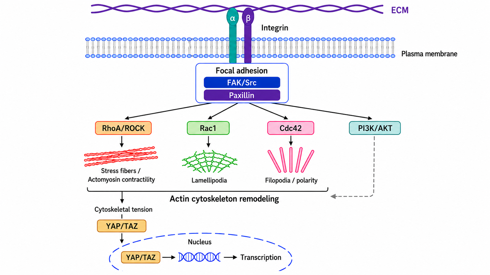
Figure 1 Schematic diagram of integrin-mediated cytoskeletal signaling
Table 3 Cytoskeleton-Related Signaling Nodes
Signaling Node | Main Cytoskeletal Association | Functional Output | Common Detection |
RhoA/ROCK | Actin stress fibers, myosin contraction | Cell contraction, enhanced adhesion, mechanical response | RhoA activity, p-MLC, stress fibers |
Rac1 | Branched actin network, lamellipodia | Leading-edge extension, migration | Rac1 activity, lamellipodia, F-actin |
Cdc42 | Filopodia, cell polarity | Directional migration, polarity establishment | Cdc42 activity, filopodia, cell polarity markers |
FAK/Src | Focal adhesions, actin linkage | Adhesion, migration, survival | p-FAK, p-Src, Paxillin |
Paxillin | Focal adhesion scaffold | Focal adhesion assembly and turnover | Paxillin, p-Paxillin, focal adhesion area |
PI3K/AKT | Leading edge, membrane lipid signaling | Cell migration, survival, polarity | p-AKT, PIP3 localization, migration assays |
YAP/TAZ | Actin tension, nuclear skeleton | Mechanotransduction, proliferation, differentiation | YAP/TAZ nuclear localization, CTGF, CYR61 |
4 Cytoskeleton and Cell Migration
Cell migration is a multistep process jointly driven by morphological changes, cell adhesion, cytoskeletal remodeling, and signal transduction. It participates in embryonic development, wound repair, immune cell patrol, angiogenesis, and tumor metastasis.
(1) Polarity establishment
Migrating cells first establish front-rear polarity. Rac1, Cdc42, PI3K, and actin polymerization signals are enriched at the leading edge, while RhoA/ROCK and myosin contraction signals are enriched at the rear.
(2) Leading-edge extension
Actin polymerizes at the leading edge to form lamellipodia or filopodia, allowing the cell membrane to extend toward the migration direction.
(3) Adhesion formation
Integrins mediate cell binding to the extracellular matrix and form focal adhesions through FAK, Paxillin, Talin, and Vinculin, transmitting external matrix signals to the cytoskeleton.
(4) Cell body translocation and rear retraction
Myosin II mediates actin contraction, moving the cell body forward while adhesions at the rear are released, completing one migration cycle.
4.2 Migration Modes
Cell migration is not a single mode. Depending on cell type, matrix environment, adhesion strength, and cytoskeletal state, cells can show mesenchymal migration, amoeboid migration, collective migration, or invasive migration.
Table 4 Cell Migration Modes and Cytoskeletal Features
Migration Mode | Cytoskeletal Features | Common Scenario | Key Detection |
Mesenchymal migration | Obvious actin stress fibers and focal adhesions; integrin-dependent | Fibroblasts, tumor cell invasion | FAK/Paxillin, MMP, Transwell |
Amoeboid migration | Weak adhesion; dependent on cortical actomyosin contraction | Immune cells, some tumor cells | p-MLC, RhoA/ROCK, cell morphology |
Collective migration | Cell-cell junctions are retained; leader cells provide traction | Epithelial repair, collective invasion of cancer cells | E-cadherin, F-actin, leader cell markers |
Neurite extension | Coordinated regulation by microtubules and actin in the growth cone | Neural development, axon regeneration | βIII-tubulin, GAP43, F-actin |
4.3 Interpretation of Migration Assays
Cell migration assays should avoid misinterpreting proliferation, cell death, or matrix degradation as enhanced migration. Wound healing assays are suitable for observing population migration trends, but are easily affected by cell proliferation. Transwell assays are more suitable for chemotaxis analysis. Live-cell imaging can reflect migration speed, directionality, and morphological dynamics.
Table 5 Common Experiments in Cytoskeleton and Migration Research
Experiment Type | Main Use | Recommended Indicators | Interpretation Focus |
Phalloidin staining | Observing F-actin structures | Stress fibers, pseudopodia, cortical actin | Should be combined with cell morphology and localization analysis |
α/β-Tubulin staining | Observing microtubule networks | Microtubule arrangement, spindle, axonal microtubules | Fixation methods can affect microtubule structure |
Vimentin/Keratin staining | Assessing intermediate filaments and cell state | Intermediate filament distribution, EMT status | Should be combined with cell type markers |
Wound healing assay | Population migration | Wound closure rate | Cell proliferation should be controlled |
Transwell assay | Chemotaxis and invasion | Number of migrated cells | In invasion assays, matrix degradation ability should be distinguished |
Focal adhesion staining | Adhesion structure analysis | FAK, Paxillin, Vinculin | Focus on focal adhesion size, number, and turnover |
Live-cell imaging | Dynamic migration analysis | Speed, directionality, trajectory | Can distinguish random migration from directional migration |
5 Cytoskeleton in Cell Division and Intracellular Transport
5.1 Mitosis
Microtubules are the core structures of the mitotic spindle and are responsible for chromosome alignment, spindle checkpoint regulation, and sister chromatid separation. Actin and myosin participate in cytokinetic contractile ring formation, while intermediate filaments and lamins are reorganized during cell division.
(1) Microtubules and the spindle
Microtubule dynamic stability directly affects spindle formation and chromosome segregation. Both microtubule stabilizers and destabilizers can block mitosis and induce cell cycle arrest and cell death.
(2) Actomyosin contractile ring
During cytokinesis, actin and myosin II form a contractile ring, causing membrane ingression and completing separation into two daughter cells.
5.2 Intracellular Transport
Microtubules and actin together form intracellular transport tracks. Microtubules are mainly responsible for long-distance transport, while actin more often participates in transport near the cell cortex, membrane-proximal regions, and short-distance transport. Motor proteins such as kinesin, dynein, and myosin transport organelles, vesicles, protein complexes, and RNA to specific regions.
Table 6 Cytoskeleton-Related Functions in Cell Division and Transport
Process | Main Cytoskeletal Components | Key Molecules | Functional Significance |
Spindle formation | Microtubules | α/β-Tubulin, Aurora, Kinesin | Chromosome alignment and segregation |
Cytokinesis | Actin, Myosin II | F-actin, MLC, RhoA | Formation of the cellular contractile ring |
Vesicle transport | Microtubules, Actin | Kinesin, Dynein, Myosin | Secretion, endocytosis, and membrane transport |
Mitochondrial positioning | Microtubules, Actin | Miro, Milton, Dynein, Kinesin | Energy supply and cellular stress response |
Axonal transport | Microtubules | Kinesin, Dynein, Tau | Neuronal function and neurodegenerative diseases |
6 Cytoskeletal Abnormalities and Disease Mechanisms
6.1 Tumor Invasion and Metastasis
Tumor cell migration, invasion, and metastasis depend on cytoskeletal remodeling, focal adhesion turnover, and extracellular matrix degradation. During EMT, decreased E-cadherin, increased Vimentin, enhanced actin stress fibers, and altered cell adhesion patterns can all increase migratory and invasive capacity.
(1) Recommended detection
F-actin, Vimentin, E-cadherin, N-cadherin, p-FAK, Paxillin, RhoA/Rac1/Cdc42 activity, MMPs, and Transwell invasion assay.
(2) Interpretation focus
Enhanced migration does not equal enhanced metastasis. To support a metastatic mechanism, invasion, matrix degradation, intravasation/extravasation, in vivo metastasis models, or clinical tissue localization evidence should be combined.
6.2 Neurodegenerative Diseases
Neurons are highly dependent on microtubule stability and axonal transport. Tau abnormalities, microtubule depolymerization, motor protein transport defects, and abnormal neurofilament aggregation can all lead to impaired axonal transport, reduced synaptic function, and neuronal degeneration.
(1) Recommended detection
Acetylated tubulin, βIII-tubulin, Tau, p-Tau, Neurofilament, Kinesin, Dynein, mitochondrial transport, and axon length.
(2) Interpretation focus
Microtubule changes in neurons should be analyzed together with axonal transport and synaptic function. Microtubule function should not be judged only by changes in tubulin expression.
6.3 Cardiomyopathy, Myopathy, and Skin Diseases
Mutations in intermediate filaments and actin-related proteins can reduce tissue mechanical stability. Desmin abnormalities are associated with myopathy and cardiomyopathy, keratin abnormalities can cause epithelial mechanical fragility and blistering skin diseases, and lamin abnormalities can affect nuclear structure and tissue mechanics.
6.4 Inflammation, Fibrosis, and Immune Cell Function
Immune cell migration, phagocytosis, immune synapse formation, and inflammatory factor release all depend on cytoskeletal remodeling. During fibroblast activation and fibrosis, actin stress fibers, α-SMA, FAK, and RhoA/ROCK signaling are often significantly enhanced.
Table 7 Disease Directions Related to Cytoskeletal Abnormalities
Disease Direction | Key Cytoskeletal Change | Main Mechanism | Common Indicators |
Tumor metastasis | Actin remodeling, focal adhesion turnover, increased Vimentin | Migration, invasion, EMT | F-actin, Vimentin, p-FAK, Transwell |
Neurodegenerative diseases | Microtubule instability, Tau abnormalities, axonal transport defects | Synaptic injury, axonal degeneration | p-Tau, Acetylated tubulin, Neurofilament |
Cardiomyopathy and myopathy | Abnormal Desmin or actin-related structures | Reduced mechanical support in muscle cells | Desmin, α-actinin, F-actin |
Skin fragility diseases | Abnormal Keratin network | Reduced epithelial mechanical stability | Keratin, cell junction markers |
Fibrosis | Enhanced actin stress fibers and α-SMA | Myofibroblast activation | α-SMA, F-actin, p-FAK, Collagen |
Kidney and nuclear lamina disorders | Abnormal Lamin or podocyte cytoskeleton | Nuclear mechanics and podocyte structural injury | Lamin, Synaptopodin, Actin |
7 Cytoskeleton-Targeted Intervention and Drug Research
7.1 Microtubule-Targeting Drugs
Microtubules are important targets of antitumor drugs. Microtubule stabilizers inhibit microtubule dynamics, while microtubule destabilizers promote microtubule depolymerization. Both types of drugs can interfere with spindle function and cause mitotic arrest.
(1) Microtubule stabilizers
Taxane drugs inhibit dynamic remodeling by stabilizing microtubule structures and are commonly used in studies of mitotic blockade and tumor cell death.
(2) Microtubule destabilizers
Colchicine, vinblastine, nocodazole, and related compounds can interfere with microtubule polymerization or promote microtubule depolymerization. They are commonly used to study microtubule dynamics, cell division, and intracellular transport.
7.2 Actin-Targeted Intervention
Inhibitors of actin polymerization or depolymerization can be used to analyze the roles of microfilaments in migration, phagocytosis, cytokinesis, and cell morphology maintenance. For example, cytochalasins, latrunculins, and jasplakinolide can interfere with actin network dynamics.
7.3 Targeting Cytoskeletal Regulatory Signaling
Signaling nodes such as RhoA/ROCK, FAK, Src, Paxillin, PI3K, and YAP/TAZ regulate cytoskeletal structure and cellular mechanical state. They are important intervention directions in studies of tumor invasion, fibrosis, inflammation, and vascular remodeling.
Table 8 Cytoskeleton-Targeted Intervention Directions
Intervention Direction | Representative Effect | Research Use | Key Interpretation |
Microtubule stabilization | Enhances microtubule stability and inhibits dynamic changes | Mitotic blockade and antitumor mechanisms | Spindle abnormalities and cell cycle arrest |
Microtubule destabilization | Inhibits microtubule polymerization or promotes depolymerization | Microtubule dynamics, intracellular transport, neural axon research | Microtubule network disruption and impaired transport |
Actin polymerization inhibition | Inhibits F-actin formation or membrane protrusions | Migration, phagocytosis, cytokinesis research | Reduced F-actin and decreased migration |
Actin stabilization | Excessively stabilizes F-actin | Validation of actin dynamics dependence | Cytoskeletal stiffening and abnormal migration |
ROCK inhibition | Reduces actomyosin contraction | Cellular mechanics, fibrosis, stem cell culture | Decreased p-MLC and reduced stress fibers |
FAK/Src inhibition | Blocks focal adhesion signaling | Tumor migration, invasion, and adhesion research | Reduced p-FAK/p-Src and inhibited migration |
Tubulin acetylation regulation | Alters the proportion of stable microtubules | Neural transport, cilia, and microtubule stability research | Changes in acetylated tubulin |
8 Representative Products Related to Cytoskeleton Research
Research Module | Representative Product | CAS No. | Mechanism or Detection Positioning | Applicable Research |
F-actin structure observation | Phalloidin | Binds F-actin and is used for actin filament labeling | F-actin staining, stress fibers, pseudopodia, and cell morphology observation | |
Actin polymerization inhibition | Cytochalasin D | Inhibits actin polymerization and disrupts F-actin dynamics | Cell migration, cytokinesis, phagocytosis, and microfilament function validation | |
Migration/invasion mechanism research | Cytochalasin B | Disrupts actin polymerization and cytoskeletal dynamics | Cell migration, phagocytosis, membrane protrusions, and glucose transport-related research | |
Actin polymerization inhibition | Latrunculin A | Binds G-actin and blocks actin polymerization | Microfilament depolymerization, cell morphology changes, and migration mechanism research | |
Actin polymerization inhibition | Latrunculin B | Interferes with G-actin incorporation into F-actin | Actin dynamics, cytoskeletal remodeling, and cell migration research | |
Actin stabilization intervention | Jasplakinolide | Stabilizes F-actin and promotes actin polymerization | Microfilament stability, cytoskeletal stiffening, and abnormal migration research | |
Myosin II contraction regulation | Blebbistatin | Myosin II ATPase inhibitor | Actomyosin contraction, cellular mechanics, rear retraction, and cytokinesis research | |
Microtubule stabilization intervention | Paclitaxel | Microtubule stabilizer that inhibits microtubule dynamics | Mitotic arrest, spindle abnormalities, and antitumor mechanism research | |
Microtubule destabilization intervention | Nocodazole | Interferes with microtubule polymerization and promotes microtubule network disruption | M-phase synchronization, microtubule dynamics, intracellular transport, and spindle research | |
Microtubule destabilization intervention | Colchicine | Binds tubulin and inhibits microtubule polymerization | Microtubule depolymerization, cell division arrest, and inflammation-related research | |
Microtubule destabilization intervention | Vinblastine sulfate | Inhibits microtubule polymerization | Spindle disruption, mitotic arrest, and anti-mitotic drug efficacy research | |
Microtubule destabilization intervention | Vincristine sulfate | Microtubule polymerization inhibitor | Microtubule dynamics, tumor cell mitotic arrest, and neurotoxicity research | |
Microtubule dynamics regulation | Demecolcine | Inhibits microtubule formation | Chromosome segregation, cell cycle arrest, and microtubule function research | |
ROCK signaling inhibition | Y-27632 | ROCK inhibitor that reduces actomyosin contraction | Cell tension, stress fibers, stem cell culture, and migration research | |
ROCK signaling inhibition | Fasudil hydrochloride | ROCK-related pathway inhibitor | RhoA/ROCK signaling, vascular response, fibrosis, and cell contraction research | |
FAK adhesion signaling research | PF-573228 | FAK inhibitor | Focal adhesion signaling, p-FAK, cell migration, and invasion research | |
FAK adhesion signaling research | PF-562271 | FAK/Pyk2 inhibitor | Integrin-FAK signaling, tumor migration, invasion, and adhesion research | |
Src/FAK crosstalk signaling research | PP2 | Src family kinase inhibitor | Src-FAK axis, focal adhesion turnover, migration, and invasion mechanism research | |
PI3K leading-edge signaling research | LY294002 | PI3K inhibitor | PI3K/AKT signaling, cell polarity, migration, and survival research | |
Microtubule acetylation regulation | Trichostatin A | HDAC inhibitor that can affect tubulin acetylation-related status | Acetylated tubulin, microtubule stability, neural axons, and cilia research |
9 Cytoskeleton-Related Product Lines and Application Directions
Product Line Direction | Research Positioning | Main Applications |
Proteins related to membrane-cytoskeleton linkage, membrane repair, and membrane structural remodeling | Cell membrane dynamics, vesicle transport, inflammatory responses, and membrane injury repair research | |
| Actin branch nucleation complex | Lamellipodia formation, cell migration, phagocytosis, and membrane protrusion research |
Dynamin-related membrane scission and endocytosis regulatory proteins | Receptor endocytosis, vesicle release, membrane transport, and cytoskeleton-membrane dynamics research | |
| Proteins related to intercellular communication and junction structures | Cell junctions, tissue synchronization, coordinated migration, and inflammatory signal propagation research |
| Cell-matrix adhesion receptors | Focal adhesion formation, FAK/Src signaling, migration, invasion, and mechanotransduction research |
| Microtubule motor proteins | Vesicle transport, chromosome movement, axonal transport, and organelle positioning research |
| Enzymes regulating extracellular matrix crosslinking and tissue mechanics | ECM stiffening, fibrosis, tumor invasion, and mechanical microenvironment research |
| Actin-binding and membrane phospholipid regulatory protein | Cell migration, membrane cytoskeleton regulation, secretion, and inflammation-related research |
| Mitotic regulatory kinase | Cell cycle, spindle regulation, mitotic progression, and tumor proliferation research |
| Core components of microtubule structure | Microtubule networks, spindle, intracellular transport, neural axons, and microtubule drug research |
Spindle assembly checkpoint kinase | Chromosome alignment, spindle checkpoint, mitotic errors, and antitumor mechanism research | |
| Actin-related motor proteins | Actomyosin contraction, cytokinesis, cell migration, and cellular mechanics research |
| Downstream effector kinase of Rho GTPases | Rac/Cdc42 signaling, cytoskeletal remodeling, cell migration, and tumor invasion research |
| RhoA downstream contraction-regulating kinase | Stress fibers, p-MLC, cell contraction, fibrosis, and mechanotransduction research |
10 Common Questions in Cytoskeleton Research
10.1 Does Increased F-actin Always Indicate Enhanced Migration?
Not necessarily. Increased F-actin may indicate enhanced stress fibers, increased cell contraction, or cytoskeletal stiffening, and does not necessarily mean increased migratory capacity. Enhanced migration should be judged together with leading-edge structures, focal adhesion turnover, migration speed, and Transwell results.
10.2 Does Increased Tubulin Expression Mean Microtubules Are More Stable?
Not necessarily. Total tubulin only reflects protein expression level. Microtubule stability is more appropriately evaluated by acetylated tubulin, detyrosinated tubulin, microtubule network morphology, and dynamic imaging.
10.3 Is Increased Vimentin Equivalent to EMT?
No. Vimentin is one of the markers of a mesenchymal phenotype, but EMT should be evaluated together with decreased E-cadherin, increased N-cadherin, transcription factor changes, morphological changes, and enhanced migration and invasion.
10.4 Can Cell Migration Be Studied Only with a Wound Healing Assay?
It is not recommended. Wound healing assays are easily affected by cell proliferation, cell death, and scratch width. To prove changes in chemotaxis or invasion ability, Transwell, matrix invasion, or live-cell imaging assays should be added.
10.5 Is Cell Death Induced by Microtubule Drugs Only Due to Microtubule Disruption?
Not necessarily. Microtubule drugs can induce mitotic arrest, DNA damage response, mitochondrial stress, and apoptosis. Interpretation should combine cell cycle analysis, spindle morphology, caspase activation, and cell death indicators.
Cytoskeleton research should comprehensively analyze structural components, dynamic regulation, the mechanical environment, and signaling networks. Microfilaments, microtubules, and intermediate filaments respectively participate in morphology changes, intracellular transport, cell division, and mechanical support, and connect cell migration, proliferation, differentiation, and disease progression through nodes such as Rho GTPases, FAK/Paxillin, PI3K, ROCK, and YAP/TAZ.