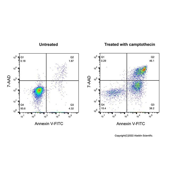
Annexins are a family of calcium-dependent phospholipid-binding proteins that preferentially bind phosphatidylserine (PS). Under normal physiologic conditions, PS is predominantly located in the inner leaflet of the plasma membrane. Upon initiation of apoptosis, PS loses its asymmetric distribution across the phospholipid bilayer and is translocated to the extracellular membrane leaflet marking cells as targets of phagocytosis. Once on the outer surface of the membrane, PS can be detected by fluorescently labeled Annexin V in a calcium-dependent manner.
In early-stage apoptosis, the plasma membrane excludes viability dyes such as propidium iodide (PI), 7-AAD. These cells will stain with Annexin V but not a viability dye, thus distinguishing cells in early apoptosis. However, in late stage apoptosis, the cell membrane loses integrity thereby allowing Annexin V to also access PS in the interior of the cell. A viability dye can be used to resolve these late-stage apoptotic and necrotic cells (Annexin V, viability dye-positive) from the early-stage apoptotic cells (Annexin V positive, viability dye-negative). This kit is suitable for the identification and enumeration of dead cells, such as apoptotic or necrotic cells, by flow cytometry.
A1456530 | Components | 20T | 50T | 100T | Storage | Quantity Per Test | A1456530A | 10X Annexin V Binding Buffer | 5 mL | 10 mL | 20 mL | 2-8℃ | 200 μL per 0.5-1.0x10⁵ cells | A1456530B | Annexin V-FITC | 40 μL | 100 μL | 200 μL | 2-8℃. Store in the dark. | 2 μL per 0.5-1.0x10⁵ cells | A1456530C | 7-AAD Staining Solution | 100 μL | 250 μL | 500 μL | 2-8℃. Store in the dark. | 5 μL/ 0.5-1.0x10⁵ cells |
Note: The recommended number of cells to stain per test is 0.5-1.0x10⁵ cells. |
Precautions
1. Please try to avoid light when using to slow down the quenching of fluorescence.
2. 7-AAD Solution is toxigenic and mutagenic; handle with care.
3. Since the binding of annexin V to phosphatidylserine (PS) is calcium-dependent, it is critical to avoid buffers containing EDTA or other calcium chelators during Annexin V experiments.
Instruction for use
1. Dilute 10x Binding Buffer to 1x using distilled water (1 mL 10x Binding Buffer + 9 mL ddH2O).
2. Wash cells twice with cold PBS and then resuspend the desired amount of cells in Annexin V Binding Buffer at a concentration of 0.5-1.0x10⁶ cells /mL .
3. Add 2 µL of FITC Annexin V and 5 µL 7-AAD to 100 µL of the cell suspension.
4. Add 100 µL of 1x Binding Buffer to each assay. Gently vortex the cells and incubate for 10 min at RT (25°C) in the dark.
5. Analyze by flow cytometry within 1 hr.