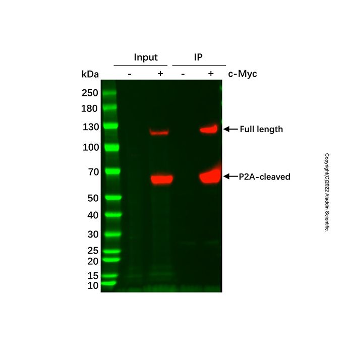
This product is designed for the detection of c-Myc tag fusion proteins expressed in both prokaryotic and eukaryotic systems. The c-Myc peptide is encoded by the c-Myc gene on human chromosome 8q24 and corresponds to amino acids 410-419 (EQKLISEEDL) of human P62. The Anti-c-Myc Agarose Resin can recognize C-terminal, N-terminal, or internal c-Myc tag fusion proteins. It is applicable in Western blotting, immunoprecipitation, and flow cytometry for detecting the expression of recombinant proteins in target cells.
Aladdin Anti-c-Myc Agarose Resin is stored in a solution containing 0.1% ProClin 300, with a settled gel to storage solution ratio of 1:1. The product specification refers to the actual volume of the settled gel.
Parameter
| Value / Description
|
Matrix
| 4% Agarose Microspheres
|
Ligand
| Anti-c-Myc Human Monoclonal Antibody
|
Particle Size Range
| 45~165 μm |
| Binding Capacity | ~1 mg c-Myc tagged protein / mL resin
|
Maximum Pressure
| 0.1 MPa, 1 bar |
Storage Conditions
| 0.1% ProClin 300, 2~8℃
|
Shelf Life
| 2 years |
Protocol
1. Sample Preparation
Prior to column loading, ensure the sample solution has appropriate ionic strength and pH. The sample or cell culture medium can be diluted with equilibration buffer or dialyzed against it.
It is recommended to centrifuge the sample or filter it through a 0.22 μm or 0.45 μm membrane before application to reduce impurities, improve protein purification efficiency, and prevent column clogging.
2. Buffer Preparation
It is recommended to filter all water and buffers through a 0.22 μm or 0.45 μm membrane before use.
Equilibration/Wash Buffer: 50 mM Tris, 0.15 M NaCl, pH 7.4
Acidic Elution Buffer: 0.1 M Glycine-HCl, pH 3.0
Competitive Elution Buffer: 50 mM Tris, 0.15 M NaCl, 100-500 μg c-Myc peptide/mL, pH 7.4
Neutralization Buffer: 1 M Tris-HCl, pH 8.0
3. Sample Purification
3.1 Column Chromatography
(1) Pack the Anti-c-Myc Agarose Resin into a suitable chromatography column. Equilibrate with 5 column volumes (CV) of equilibration buffer to bring the medium into the same buffer system as the target protein.
(2) Apply the sample to the equilibrated Anti-c-Myc Agarose Resin. Collect the flow-through. The sample can be reloaded to increase binding efficiency.
(3) Wash with 10-50 CV of wash buffer to remove non-specifically adsorbed impurities. Collect the wash fraction.
(4) Elution:
* A. Acidic Elution: Elute with 5 CV of acidic elution buffer. Add a volume of neutralization buffer equal to one-tenth of the elution volume to the collected fractions to adjust the pH to 7.0-8.0. Collect fractions separately.
*Note: After acidic elution, immediately re-equilibrate the medium with equilibration buffer. Do not leave the Anti-c-Myc Agarose Resin in the elution buffer for more than 20 minutes.*
* B. Competitive Elution: Elute with 5 CV of competitive elution buffer. Collect fractions separately.
(5) Regenerate the column with 3 CV of elution buffer, then re-equilibrate with equilibration buffer until neutral pH is reached.
(6) Store the medium in storage buffer at 2-8°C.
3.2 Batch/Binding Adsorption
(1) Medium Preparation: Transfer an appropriate amount of Anti-c-Myc Agarose Resin to a column or tube and allow the storage solution to drain. Wash with 5 CV of equilibration buffer.
(2) Add the sample solution and incubate with shaking or gentle mixing at 4°C or room temperature for at least 30 minutes (do not use a magnetic stirrer). Ensure thorough mixing of the medium and sample solution.
(3) After incubation, centrifuge the mixture (5000 × g, 1 min) or filter to collect the medium.
(4) Transfer the medium to a column. Wash with equilibration buffer until the UV baseline stabilizes.
(5) Elute using either the acidic or competitive elution buffer, as described in section 3.1 (4).
(6) Medium regeneration and storage are the same as described in sections 3.1 (5) and (6).
3.3 Immunoprecipitation (IP) Protocol
(1) Medium Preparation: Add 40 µL of the Anti-c-Myc Agarose Resin slurry (approx. 20 µL settled medium volume) to a 1.5 mL microcentrifuge tube. Centrifuge at 5000 × g for 1 min. Carefully remove and discard the supernatant.
(2) Medium Equilibration: Add 0.5 mL of equilibration buffer to resuspend the medium (this brings it into the same buffer system as the target protein, protecting the protein). Centrifuge at 5000 × g for 1 min. Discard the supernatant. Repeat this step once.
(3) Sample Binding: Add 200-1000 µL of sample lysate or cell extract to the prepared medium. Mix thoroughly. Incubate the tube at room temperature on a rotator/mixer for gentle inversion for at least 1 hour to ensure sufficient contact and adsorption. Centrifuge at 5000 × g for 1 min. Discard the supernatant.
(4) Washing: Add 0.5 mL of wash buffer to resuspend the medium. Mix gently. Centrifuge at 5000 × g for 1 min. Discard the supernatant. Repeat this washing step three more times to ensure removal of non-specifically bound material.
(5) Sample Elution: Choose the elution method based on downstream application needs.
* A. Acidic Elution: Add 100 µL of acidic elution buffer to resuspend the medium. Incubate at room temperature for 5 min. Centrifuge at 5000 × g for 1 min. Carefully collect the supernatant without disturbing the medium. Neutralize the eluate immediately using neutralization buffer. Store eluted samples at 4°C; for long-term storage, keep at -20°C.
* B. Competitive Elution: Add 100 µL of competitive elution buffer to resuspend the medium. Incubate at room temperature for 30 min. Centrifuge at 5000 × g for 1 min. Carefully collect the supernatant without disturbing the medium. Store eluted samples at 4°C; for long-term storage, keep at -20°C.
* C. Denaturing Elution: Conventional protein loading buffer contains β-mercaptoethanol or DTT, which reduces disulfide bonds, and SDS, which denatures proteins. This method will elute the target protein but also denature the Anti-c-Myc antibody, making the medium unsuitable for reuse.
Add 20 µL of 2× Loading Buffer to the medium. Heat at 95°C for 5 min. Centrifuge at 5000 × g for 1 min. Carefully collect the supernatant for SDS-PAGE analysis.
Troubleshooting Guide