
How Does Dynamic ECM Remodeling in the Tumor Microenvironment Regulate Tumor Immune Responses?
How Does Dynamic ECM Remodeling in the Tumor Microenvironment Regulate Tumor Immune Responses?
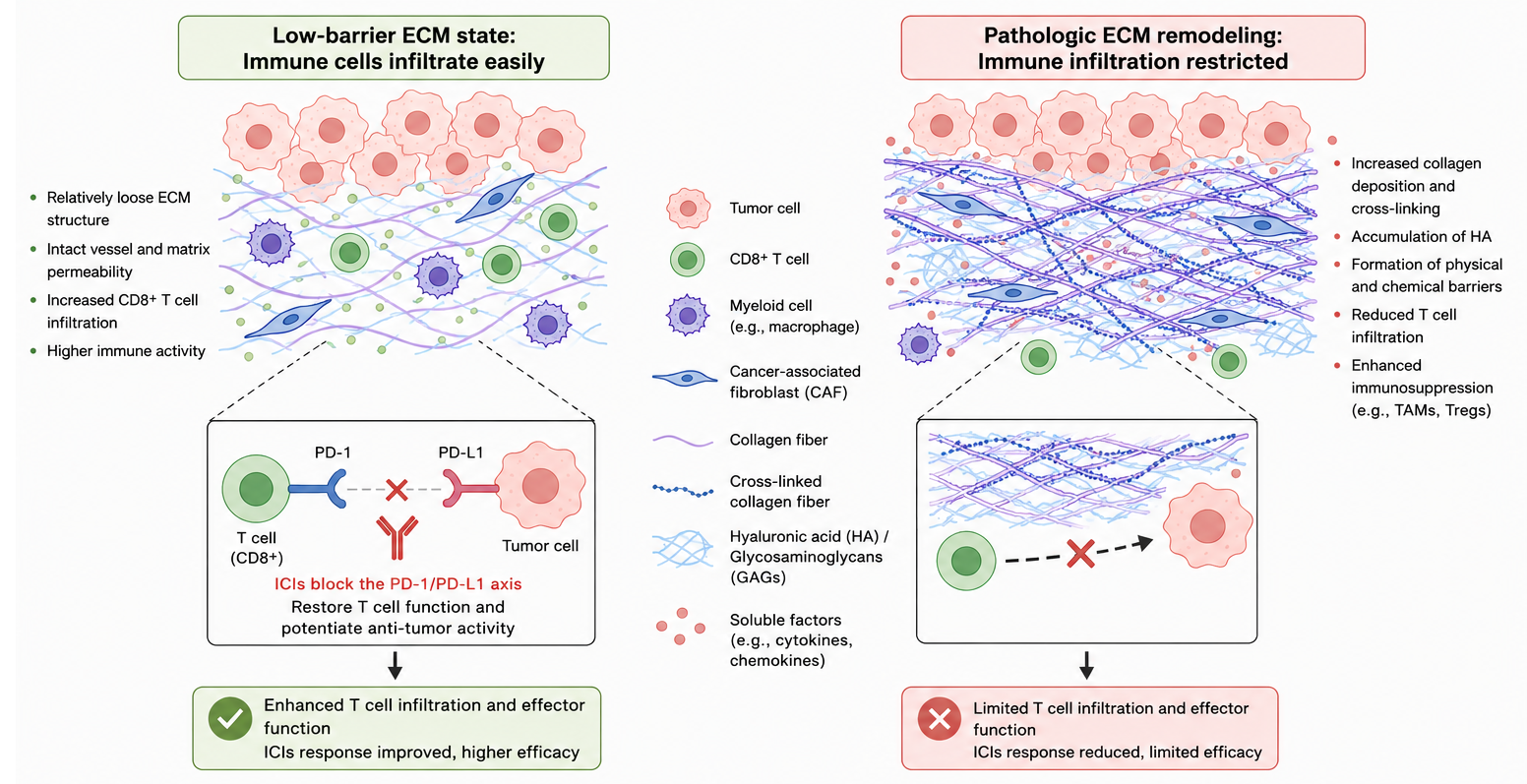
The extracellular matrix (ECM) in the tumor microenvironment not only supports tissue structure, but also continuously regulates immune cell infiltration, migration, activation, and effector functions through matrix deposition, crosslinking, degradation, and spatial reorganization. ECM remodeling can form physical barriers, alter tissue stiffness, store or release immunomodulatory factors, and work together with cancer-associated fibroblasts, TGF-β signaling, and myeloid immunosuppression to shape the immune response status of tumors.
Keywords: tumor microenvironment; extracellular matrix; ECM remodeling; tumor immunity; T cell infiltration; cancer-associated fibroblasts; TGF-β; immune exclusion
1 Basic Logic of Dynamic ECM Remodeling
1.1 ECM Is Not a Passive Scaffold
The ECM is composed of collagen, fibronectin, laminin, hyaluronic acid, proteoglycans, and matrix-bound factors. In normal tissues, the ECM maintains cell adhesion, tissue boundaries, mechanical stability, and wound repair. In tumor tissues, tumor cells, cancer-associated fibroblasts (CAFs), endothelial cells, macrophages, and neutrophils continuously alter ECM composition and structure, making it a dynamic regulatory interface that affects the spatial distribution and functional status of immune cells.
1.2 Major Forms of ECM Remodeling
(1) Matrix component reconstruction
Collagen I, collagen III, fibronectin, hyaluronic acid, tenascin-C, periostin, and various proteoglycans are often abnormally elevated in the tumor stroma. Different components correspond to different immune effects: collagen deposition is more likely to form a structural barrier, hyaluronic acid accumulation can increase interstitial pressure, and fibronectin and tenascin-C can alter cell adhesion, migration, and inflammatory signaling.
(2) Fiber crosslinking and matrix stiffening
After collagen crosslinking mediated by lysyl oxidase (LOX), tumor matrix stiffness increases, and mechanotransduction pathways such as integrin, FAK, Src, and YAP/TAZ are enhanced. A stiffened matrix not only promotes tumor cell invasion and epithelial-mesenchymal transition, but also affects T cell migration, immune synapse formation, and cytotoxic function.
(3) Proteolysis and fragment release
MMPs, ADAMTS, cysteine proteases, and hyaluronidases can degrade the ECM and release collagen fragments, hyaluronic acid fragments, and proteoglycan fragments. These fragments can act as matrikines and participate in chemotaxis, inflammation, and immune regulation. Therefore, matrix degradation is not merely “barrier removal”; it may also reshape myeloid cell recruitment and immunosuppressive states.
(4) Spatial topological alteration
ECM fiber orientation, density, pore structure, and distribution around tumor nests determine whether immune cells can enter tumor cell-enriched regions. Parallel collagen bundles can provide tracks for tumor cell migration, but may cause CD8+ T cells to remain at the stromal margin, forming an immune-excluded spatial pattern.
2 ECM Structural Barriers and Immune Cell Infiltration
2.1 Collagen Deposition and T Cell Exclusion
Massive deposition of collagen I and collagen III reduces tumor tissue porosity and increases the mechanical resistance against immune cell entry into tumor nests. In such tumors, CD8+ T cells are not necessarily absent; instead, they often accumulate in collagen-rich stromal areas, around blood vessels, or at tumor margins, making it difficult for them to establish effective contact with tumor cells.
(1) Spatial obstruction
Dense collagen bundles can retain T cells in the tumor stroma rather than allowing them to enter tumor cell-dense regions. During evaluation, the spatial relationship among Collagen I/III, α-SMA, CD8, and tumor cell markers should be analyzed together, avoiding reliance only on total CD8-positive area to assess immune infiltration.
(2) Reduced effector function
ECM stiffening can affect tumor cell survival through integrin-FAK and YAP/TAZ-related signaling, and can also alter T cell migration, adhesion, and immune synapse stability. In such samples, Granzyme B, Perforin, IFN-γ, and exhaustion markers should be detected simultaneously to distinguish “T cells present but without effective killing” from “true lack of T cell infiltration.”
(3) Resistance to immunotherapy
Tumors with marked collagen deposition often respond poorly to immune checkpoint inhibitors, partly because T cells are excluded from tumor nests. Simply releasing PD-1/PD-L1 inhibition may not restore cytotoxicity. Combination strategies involving CAF modulation, TGF-β blockade, LOX inhibition, or FAK inhibition are more suitable for validating ECM barrier-associated immune resistance.
2.2 Hyaluronic Acid Accumulation and Interstitial Pressure
Hyaluronic acid has strong water-retaining capacity. When abnormally accumulated, it can increase tumor interstitial pressure, compress blood vessels, and restrict molecular diffusion. A high-hyaluronic-acid environment is often accompanied by decreased vascular perfusion, local hypoxia, enhanced HIF-1α signaling, and myeloid cell recruitment, further limiting T cell infiltration and antitumor effects.
(1) Pressure barrier
Elevated hyaluronic acid can create a highly hydrated and high-pressure tumor interstitium, affecting the entry of T cells, NK cells, and therapeutic molecules into the tumor parenchyma. Such models can be analyzed by combining hyaluronic acid staining, vascular perfusion assessment, hypoxia markers, and immune cell localization.
(2) Hypoxia linkage
Vascular compression and insufficient perfusion enhance hypoxic responses and promote the expression of VEGF, CXCL12, and myeloid chemotactic signals. If hyaluronic acid accumulation is accompanied by increased HIF-1α, CD11b, or MDSC-related markers, this suggests that hyaluronic acid may aggravate immunosuppression through a hypoxia-myeloid axis.
2.3 Basement Membrane Remodeling and Immune Cell Entry
The basement membrane is composed of collagen IV, laminin, nidogen, and heparan sulfate proteoglycans, and determines epithelial boundaries and vascular barrier status. During tumor progression, the basement membrane may show local rupture, abnormal thickening, or component rearrangement. Local disruption may promote invasion and antigen leakage, whereas abnormal thickening may restrict lymphocyte transvascular migration and entry into the tumor parenchyma. Evaluation should jointly analyze Collagen IV, Laminin, CD31, CD8, and tumor cell boundaries.
3 ECM Biochemical Signaling and Immunosuppression
3.1 Integrin-FAK Signaling
The ECM connects with cells through integrins, converting matrix composition, adhesion status, and mechanical pressure into intracellular signaling. Tumor cells, CAFs, macrophages, and T cells can all sense ECM changes. Therefore, ECM remodeling can simultaneously affect tumor invasion, immune cell migration, and expression of immunosuppressive factors.
(1) Tumor cell immune escape
After collagen, fibronectin, and laminin bind to integrins, they can activate FAK, Src, PI3K-AKT, and YAP/TAZ signaling, enhancing tumor cell survival, migration, and anti-apoptotic capacity. In some tumor models, FAK activation is also associated with chemokine rearrangement, myeloid cell recruitment, and T cell exclusion.
(2) CAF activation
CAFs secrete collagen, fibronectin, LOX, MMPs, and TGF-β, continuously promoting ECM deposition and matrix contraction. Activated CAFs can also release CXCL12, IL-6, and TGF-β, causing T cells to remain in stromal regions and reducing their effector function.
(3) Altered immune cell function
T cells, macrophages, and dendritic cells can sense ECM stiffness, adhesive ligands, and degradation fragments. Abnormal adhesion and stiffened environments can alter immune cell migration speed, residence time, polarization state, and activation threshold.
3.2 TGF-β Storage and Activation
TGF-β can bind to latency-associated proteins and ECM structures and be stored in the matrix in a latent form. Proteolysis, integrin-mediated tension, and matrix stiffening in tumor tissues can promote TGF-β activation. Activated TGF-β further promotes CAF differentiation, collagen deposition, matrix crosslinking, and EMT, while suppressing CD8+ T cell effector function, NK cell cytotoxicity, and dendritic cell maturation. This creates a self-amplifying feedback loop between ECM remodeling and immunosuppression.
3.3 ECM-Bound Factors and Chemokine Gradients
The ECM can bind chemokines, growth factors, and inflammatory factors, forming local signaling reservoirs. Fibronectin, proteoglycans, and glycosaminoglycans can affect the local concentration and diffusion range of molecules such as CXCL12, CCL2, VEGF, and FGF, thereby altering immune cell migration direction and tissue localization.
(1) CXCL12-related immune exclusion
CXCL12 can form immune exclusion-related gradients in CAF-enriched and dense ECM regions, making it difficult for T cells to enter tumor cell areas. Detection should focus on the spatial relationship of CXCL12 with α-SMA, collagen, and CD8 distribution.
(2) CCL2 and myeloid cell recruitment
CCL2 promotes monocyte and macrophage recruitment and is associated with TAM and MDSC expansion. If increased CCL2 is accompanied by elevated CD68, CD163, CD206, or Arg1, this suggests that ECM remodeling may promote myeloid immunosuppression.
(3) VEGF and vascular-immune linkage
ECM remodeling and hypoxic environments can promote VEGF expression, altering vascular structure and immune cell entry routes. When VEGF is elevated, CD31, hypoxia markers, T cell localization, and myeloid cell infiltration should be evaluated simultaneously.
4 ECM Proteolysis and Immune Remodeling
4.1 MMP-Mediated Matrix Degradation
MMP-2, MMP-9, and other matrix proteases can degrade collagen, gelatin, fibronectin, laminin, and proteoglycans. Moderate matrix degradation may increase the space available for immune cell entry, whereas excessive or selective degradation may release pro-inflammatory, pro-angiogenic, or pro-invasive fragments. When MMP activity is increased, ECM structure, TAM/MDSC infiltration, and T cell spatial distribution should be measured simultaneously, rather than simply interpreting MMP upregulation as the removal of a matrix barrier.
4.2 Immune Effects of Matrix Fragments
Matrikines generated after ECM degradation can participate in inflammation, chemotaxis, angiogenesis, and immunosuppression. The function of different fragments depends on molecular size, receptor expression, local cellular composition, and tumor stage.
(1) Hyaluronic acid fragments
High-molecular-weight hyaluronic acid is generally associated with tissue stability, interstitial pressure, and immunosuppression, whereas low-molecular-weight hyaluronic acid fragments can enhance inflammatory responses and myeloid cell activation through pathways such as TLR signaling.
(2) Collagen degradation fragments
Collagen degradation fragments can affect immune cell chemotaxis, angiogenesis, and tissue repair responses. At the tumor invasion front, collagen degradation often appears together with MMP activity, TAM infiltration, and tumor cell migration.
(3) Proteoglycan fragments
Proteoglycans such as decorin and biglycan, as well as their fragments, can affect TLR, TGF-β, and inflammatory signaling. In different tumor models, their effects may be pro-inflammatory, antitumor, or protumor, and should be interpreted according to receptor status and cellular source.
5 Effects of ECM Remodeling on Different Immune Cells
5.1 CD8+ T Cells and NK Cells
CD8+ T cells and NK cells are the core effector cells of antitumor cytotoxic responses. ECM densification, CAF barriers, and TGF-β signaling can restrict their entry into tumor nests and weaken their killing function.
(1) Spatial exclusion
CD8+ T cells accumulating around collagen-rich regions while being insufficient within tumor cell areas is an important manifestation of ECM-related immune exclusion. Analysis should combine collagen staining, tumor cell regions, and CD8 localization.
(2) Reduced effector function
Stiffened ECM and TGF-β signaling can reduce the expression of Granzyme B, Perforin, and IFN-γ. NK cells may also be affected by hypoxia, high matrix pressure, and TGF-β, resulting in reduced cytotoxicity.
(3) Limited response to immunotherapy
When the ECM barrier is prominent, PD-1/PD-L1 blockade may not fully restore cytotoxicity because T cells have not effectively contacted tumor cells. ECM modulation or intervention in the CAF/TGF-β axis can serve as a combination direction for immunotherapy.
5.2 Macrophages, MDSCs, and Neutrophils
ECM remodeling often occurs together with myeloid immunosuppression. Collagen deposition, hyaluronic acid accumulation, MMP activation, and matrix fragment release can promote the recruitment of monocytes, neutrophils, and MDSCs, and drive TAMs toward an immunosuppressive phenotype.
(1) TAM polarization
M2-like TAMs can secrete MMPs, TGF-β, VEGF, and collagen-regulatory factors, further promoting ECM degradation, matrix deposition, and vascular abnormalities. Combined detection of CD68, CD163, CD206, and ECM markers can be used to evaluate the myeloid suppressive state.
(2) MDSC recruitment
MDSCs weaken T cell function through arginine metabolism, ROS, and immunosuppressive factors. If ECM remodeling is accompanied by increases in CD11b, Arg1, or Ly6G/Ly6C-related markers, this suggests that the tumor microenvironment may shift from cytotoxic immunity toward myeloid suppression.
(3) Neutrophils and proteases
Proteases released by tumor-associated neutrophils can participate in ECM degradation and remodeling of the invasion front. When neutrophils increase, MMP activity, collagen degradation, and T cell localization should be analyzed together to determine their immune impact.
5.3 Dendritic Cells and Tregs
Dendritic cells need to capture antigens in tumor tissues and migrate to lymphoid tissues to complete antigen presentation. Tregs are more likely to expand in environments enriched in TGF-β, IL-10, and CAFs. Dense ECM and high TGF-β activity restrict dendritic cell migration and maturation, while providing a stable niche for FOXP3+ Treg accumulation, shifting the local immune response from antitumor immunity toward immune tolerance.
6 Experimental Strategies for Studying ECM-Immune Interactions
6.1 Spatial Colocalization Analysis
The key to understanding how ECM remodeling affects immune responses is not only expression changes, but also altered spatial relationships. Collagen, hyaluronic acid, fibronectin, CAF markers, and immune cell markers should be detected together as much as possible to determine whether immune cells are blocked in stromal regions, around blood vessels, or at tumor margins.
(1) Tissue staining
Masson’s trichrome staining, Van Gieson staining, hyaluronic acid staining, Alcian blue staining, PASM staining, and reticular fiber staining can be used to evaluate collagen deposition, mucopolysaccharide accumulation, basement membrane structure, and reticular fiber scaffolds. Different staining results should be analyzed together with markers such as CD8, CD68, α-SMA, and CD31 to determine whether ECM structure truly affects immune cell entry routes.
(2) Multiplex immunofluorescence
Multiplex immunofluorescence is suitable for simultaneous detection of Collagen I, Fibronectin, α-SMA, CD8, CD68, FOXP3, and PD-L1, and can be used to analyze the spatial relationship between ECM structure and immune cells. DAPI or Hoechst nuclear staining can assist in cell localization and tissue structure interpretation.
(3) Second-harmonic generation imaging
Second-harmonic generation imaging can observe collagen fiber structure without labeling. It is suitable for analyzing fiber orientation, density, and tumor invasion fronts, and can be used to study the relationship between collagen alignment and T cell exclusion.
6.2 Three-Dimensional Matrix and Coculture Models
Two-dimensional culture cannot easily simulate the effects of ECM on immune cell migration and tumor cell mechanotransduction. Three-dimensional collagen gels, hyaluronic acid hydrogels, decellularized matrices, and organoid-immune cell coculture systems are more suitable for studying ECM-immune interactions.
(1) Three-dimensional collagen models
Three-dimensional collagen models allow adjustment of collagen concentration, fiber density, and stiffness, and can be used to analyze T cell migration, tumor cell invasion, and CAF contraction. Hydroxyproline quantification can supplement tissue staining results for collagen deposition.
(2) CAF-tumor-immune cell coculture
CAF, tumor cell, and T cell or macrophage coculture models are suitable for analyzing the relationship among CXCL12, TGF-β, collagen deposition, and T cell localization. MMP inhibitors can be used to verify whether matrix proteolysis participates in changes in immune cell migration.
(3) Organoid and matrix remodeling models
After combining tumor organoids with ECM materials and immune cells, the process by which immune cells enter organoids, recognize tumor cells, and execute killing can be observed. Hyaluronic acid- or collagen-enriched conditions can be used to simulate immune-excluded matrix barriers.
6.3 Causal Validation Strategies
The relationship between ECM remodeling and immunosuppression should not be analyzed only by correlation; intervention experiments are needed to verify causality. Common strategies include inhibiting MMPs to alter proteolysis, degrading hyaluronic acid to improve interstitial pressure, blocking TGF-β signaling to weaken the CAF-ECM axis, and inhibiting FAK or integrin signaling to improve immune exclusion.
7 Related Reagent and Material Selection
Table 1 Tissue staining and spatial localization materials for ECM remodeling and tumor immunity research
Cat. No. | Product Name | Specification/Purity | Application Module | Application Positioning |
Hyaluronic Acid Staining Solution | BioReagent,Biological Stain,for microscopy | Hyaluronic acid detection | Evaluates hyaluronic acid accumulation, interstitial pressure-related matrix changes, and barriers to immune cell entry | |
Alcian Blue Staining Solution (pH2.5) | BioReagent,for microscopy,Biological Stain | Acidic mucopolysaccharide staining | Detects acidic mucopolysaccharides and mucinous matrix, supporting evaluation of hyaluronic acid-enriched tissues | |
Alcian Blue Staining Solution (pH 1.0) | BioReagent, Biological Stain, for microscopy | Sulfated mucopolysaccharide staining | Analyzes sulfated glycosaminoglycans and proteoglycan-related matrix changes | |
Improved Hale Colloid Iron Polysaccharide Staining Solution | BioReagent,for microscopy,Biological Stain | Mucopolysaccharide detection | Supports analysis of hyaluronic acid, acidic mucopolysaccharides, and mucinous stromal changes in tumors | |
Modified Masson Trichrome Staining Solution | BioReagent, Biological Stain, for microscopy | Collagen fiber staining | Evaluates collagen fiber deposition, stromal fibrosis, and ECM barrier formation | |
Van Gieson Staining Solution | BioReagent, Biological Stain, for microscopy | Collagen fiber staining | Evaluates collagen deposition and tumor stromal fibrosis | |
Methen Amine Silver Staining Solution (PASM) | BioReagent, Biological Stain, for microscopy | Basement membrane staining | Evaluates basement membrane integrity, vascular basement membrane changes, and immune cell entry routes | |
Reticular Fibre Staining Solution (Gomori) | BioReagent, Biological Stain, for microscopy | Reticular fiber staining | Evaluates reticular fibers and matrix scaffold structural changes | |
Russell Modified Movat Pentachrome Staining Solution | BioReagent,Biological Stain,for microscopy | Multicomponent ECM staining | Simultaneously observes multiple stromal components, including collagen, elastic fibers, and mucinous matrix | |
Verhöeff Elastic Fiber Staining Solution (Eosin Counterstain) | BioReagent,Biological Stain,for microscopy | Elastic fiber staining | Analyzes changes in vascular wall and stromal elastic fibers, supporting evaluation of vascular-matrix remodeling | |
DAPI Staining Solution | BioReagent, for microscopy, sterile-filtered, Suitable for Immunofluorescence(IF), 1.0 mg/mL | Immunofluorescence nuclear staining | Used for spatial colocalization analysis of ECM markers, CAF markers, and immune cells | |
oechst 33342 Staining Solution | Suitable for Immunofluorescence(IF), BioReagent, ready-to-use, Biological Stain, for fluorescence analysis, Biological dye grade, for microscopy, for cell culture, 1.0 mg/ml in H₂O | Nuclear staining | Used for imaging analysis of ECM-immune cell coculture, organoids, and tissue sections |
Table 2 Products related to ECM proteolysis, collagen quantification, and MMP intervention
Cat. No. | Product Name | Specification/Purity | Application Module | Application Positioning |
Hydroxyproline (HYP) Content Assay Kit (Ehrlich, Micro Method) | BioReagent | Collagen quantification | Used to evaluate total collagen content and supplement tissue staining results such as Masson’s trichrome and Van Gieson staining | |
GM 6001 | Moligand™, ≥98% | MMP inhibition | Blocks MMP-mediated ECM degradation and evaluates the relationship between proteolysis and immune remodeling | |
MMP-2/MMP-9 Inhibitor I | Moligand™,≥99% | MMP-2/MMP-9 inhibition | Used to analyze the effect of gelatinase activity on basement membrane degradation and immune cell infiltration | |
SB-3CT | Moligand™, ≥98% | MMP-2/MMP-9 inhibition | Validates the roles of MMP-2/MMP-9 at the invasion front and in immune remodeling | |
JNJ 0966 | ≥98%(HPLC) | Pro-MMP9 activation inhibition | Analyzes the relationship between MMP-9 activation, tumor inflammation, invasion, and immune remodeling | |
MMP-9 inhibitor | ≥95% | MMP-9 inhibition | Used for studies on MMP-9-mediated matrix degradation, vascular remodeling, and myeloid cell recruitment | |
CL-82198 | Moligand™, ≥98% | MMP-13 inhibition | Used for research related to collagen degradation, fibrotic matrix, and the invasion front | |
MMP2 Mouse mAb | Carrier Free, ExactAb™, Validated, High Performance, See COA | MMP detection | Detects MMP-2 expression and evaluates gelatin/basement membrane degradation | |
MMP9 antibody | ExactAb™, Validated, Recombinant, See COA | MMP detection | Detects MMP-9 expression and analyzes ECM proteolysis, inflammation, and immune remodeling | |
Human Matrix MetalloProteinase 2 (MMP-2) ELISA Kit | BioReagent | MMP quantitative detection | Quantitatively detects MMP-2 levels in human samples and evaluates ECM degradation activity | |
Human Matrix MetalloProteinase 9 (MMP-9) ELISA Kit | BioReagent | MMP quantitative detection | Quantitatively detects MMP-9 levels in human samples and analyzes inflammatory matrix remodeling | |
Mouse Matrix MetalloProteinase 2 (MMP-2) ELISA Kit | BioReagent | Mouse MMP quantitative detection | Used for MMP-2 detection in mouse tumor models | |
Mouse Matrix MetalloProteinase 9 (MMP9) ELISA Kit | BioReagent | Mouse MMP quantitative detection | Used for MMP-9 detection in mouse tumor models | |
Recombinant Human MMP-2 Protein | Animal Free,Carrier Free,Bioactive,His Tag,≥90%(SDS-PAGE) | Recombinant MMP protein | Used for MMP-2 functional experiments, substrate validation, and positive controls | |
Recombinant Human MMP-9 Protein | Animal Free,Carrier Free,Bioactive,High Performance,His Tag,PBS Only,≥95%(SDS-PAGE) | Recombinant MMP protein | Used for MMP-9 activity research, substrate validation, and matrix degradation models |
8 Frequently Asked Questions
8.1 Why do ECM-rich tumors often show immune exclusion?
ECM enrichment does not simply mean more matrix; rather, dense collagen, CAF expansion, TGF-β activation, and chemokine rearrangement jointly form a spatial barrier. T cells may have already been recruited around the tumor but cannot enter tumor nests and contact tumor cells, resulting in immune exclusion rather than complete immune absence.
8.2 Does ECM remodeling always suppress antitumor immunity?
Not necessarily. Some ECM degradation can increase the space available for immune cell entry, and certain matrix fragments can also induce inflammatory responses. However, in most advanced solid tumors, persistent collagen deposition, matrix stiffening, CAF activation, and enhanced TGF-β signaling are more commonly associated with immunosuppression and therapeutic resistance.
8.3 Can collagen content alone determine the immunoregulatory role of ECM?
No. Collagen content reflects only part of ECM remodeling. Fiber alignment, crosslinking degree, CAF distribution, T cell spatial localization, and effector function also need to be considered. With the same collagen content, different fiber orientations and pore structures may generate completely different immune infiltration patterns.
8.4 Why are CAFs a key connection point between ECM remodeling and immunosuppression?
CAFs are both one of the main sources of ECM and a source of molecules such as TGF-β, CXCL12, IL-6, MMPs, and LOX. CAFs can form physical barriers through ECM deposition and contraction, and can also alter the function of T cells, macrophages, and dendritic cells through immunosuppressive factors.
8.5 Why does ECM regulation affect the efficacy of immune checkpoint therapy?
Immune checkpoint inhibitors require pre-existing or restorable antitumor T cell responses. If the ECM barrier prevents T cells from entering tumor nests, simply releasing PD-1/PD-L1 inhibition may be insufficient to restore killing. Reducing the matrix barrier, blocking TGF-β, or modulating CAF function can enhance T cell entry and immune checkpoint therapy response.
8.6 What is the most important experimental design in ECM-immune interaction research?
The core design is spatial colocalization combined with causal intervention. Spatial colocalization is used to determine whether the ECM truly restricts immune cell entry into tumor regions. Causal intervention is used to verify whether ECM remodeling drives immune exclusion. Only by observing ECM structural changes, altered immune cell localization, and restored effector function together can the regulatory role of ECM in tumor immune responses be more completely explained.
Dynamic ECM remodeling shapes the tumor immune microenvironment through structural barriers, mechanical signaling, proteolysis, TGF-β storage, and chemokine gradient rearrangement. Establishing an integrated evaluation system around ECM components, spatial structure, CAF activity, and immune cell localization helps determine the mechanisms underlying tumor immune exclusion and provides experimental evidence for ECM-targeted combination immunotherapy.