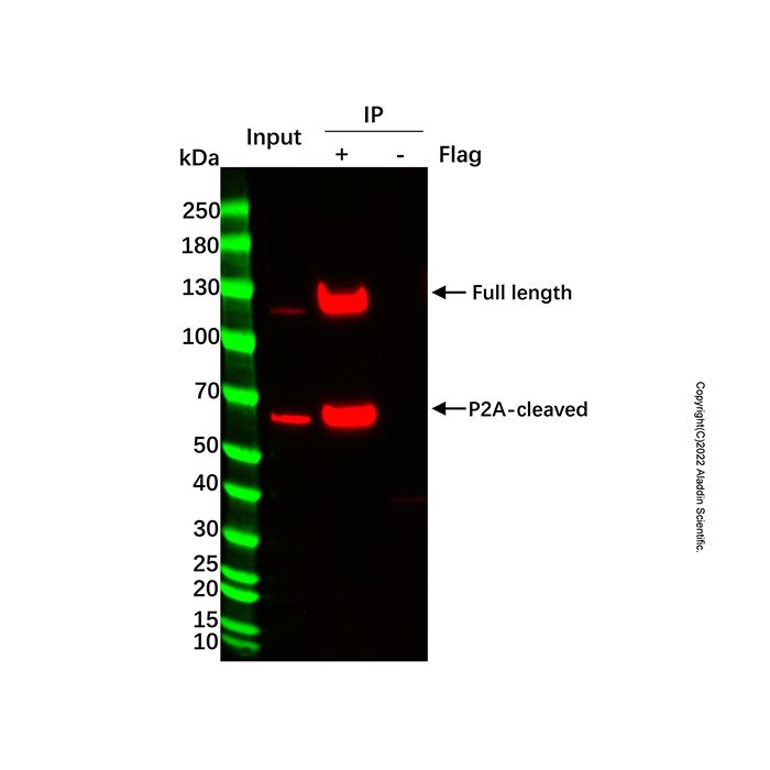
Background Introduction:
UltraBio™ Anti-Flag Magnetic Beads, also known as Anti-Flag magnetic beads, Anti-DYKDDDDK Magnetic Beads, Anti-Flag immunomagnetic beads, or Flag antibody magnetic beads, are conjugated with rigorously screened, optimized, and recombinantly expressed heavy/light chain-free Flag nanobodies. These beads are designed to capture Flag-tagged (DYKDDDDK) fusion proteins and their interacting proteins from cell or tissue extracts of various biological sources, including mammals, plants, bacteria, yeast, and insects.This kit includes high-quality Anti-Flag magnetic beads and optimized, validated essential reagents for immunoprecipitation, making Immunoprecipitation (IP) or Co-Immunoprecipitation (Co-IP) experiments simpler, more convenient, and efficient. It is widely used for the immunoprecipitation, co-immunoprecipitation, or purification of Flag-tagged fusion proteins or their protein complexes.
Tags such as His-tag, Myc-tag, Flag-tag, HA-tag, V5-tag, and GST-tag are among the most commonly used epitope tags in expression vectors. Fusion expression with these tags facilitates convenient detection of the target protein and its interacting partners, as well as efficient purification of the protein of interest.
Precautions
1.The IP Lysis Buffer included in this kit contains protease inhibitors. If specific experimental requirements exist, consider adding other appropriate inhibitor cocktails.
2.All sample lysis procedures should be performed on ice or at 4°C to minimize potential protein degradation. If cell lysis is insufficient, it can be supplemented with sonication after adding the lysis buffer.
3.The IP Lysis Buffer provided in this kit has been extensively tested and is suitable for sample lysis and subsequent washing in many immunoprecipitation scenarios. However, due to the complexity and specificity of immunoprecipitated protein samples, this lysis buffer may not be optimal for all IP sample types. If suboptimal performance is observed with the provided lysis or wash buffers during experimentation, optimization of lysis and wash conditions should be performed by the user.
4.The Magnetic Beads must be fully resuspended before use by inverting the vial several times to ensure a homogeneous mixture.
5.Protein samples should be purified as soon as possible after collection and must always be kept at 4°C or on ice to slow protein degradation or denaturation.
6.The lysis buffer provided in this kit is used for both sample lysis and the subsequent washing steps.
Instruction for use
1.Sample Preparation:(Note: Perform all sample lysis steps at 4°C or on ice)
* For Serum Samples:
If the target protein is abundant, dilute the serum sample with Lysis Buffer to a final target protein concentration of 50–150 µg/mL. Keep the diluted sample on ice for immediate use or store at -20°C for long-term preservation.
* For Adherent Cell Lysis and Preparation:
Aspirate the culture medium and wash the cells twice with PBS. Remove all residual liquid completely. Add 100-200 µL of Lysis Buffer with inhibitors per 0.5-1 million cells (equivalent to one well of a 6-well plate). Pipette gently to ensure thorough contact between the lysis buffer and cells. Animal cells are typically lysed within 1-2 seconds of contact with the buffer. For plant cells, lyse on ice for 2-10 minutes. After complete lysis, use a cell scraper to detach the cells and transfer the lysate to a 1.5 mL microcentrifuge tube. Centrifuge at 10,000-14,000 × g for 3-5 minutes at 4°C. Collect the supernatant for protein concentration determination before proceeding to subsequent immunoprecipitation or co-immunoprecipitation experiments.
Note: A small amount of insoluble material, primarily genomic DNA, may be present after lysis and will form a pellet upon centrifugation.
* For Suspension Cell Lysis and Preparation:
Collect cells by centrifugation at 250-1,000 × g for 5 minutes at room temperature. Wash the pellet twice with PBS and remove all residual liquid completely. Gently vortex or tap the tube to disperse the cells. Add 100-200 µL of Lysis Buffer with inhibitors per 0.5-1 million cells. Mix and incubate on ice for 5-20 minutes (mix several times during incubation). Tap the tube or pipette gently to ensure complete cell lysis; no significant cell pellet should remain after thorough lysis. If processing a large number of cells, it is recommended to aliquot them into tubes containing 0.5-1 million cells per tube before lysis. Large cell clumps are more difficult to lyse completely, whereas smaller numbers of cells allow better contact with the lysis buffer and lyse more efficiently. After complete lysis, centrifuge at 10,000-14,000 × g for 3-5 minutes at 4°C. Collect the supernatant for protein concentration determination before subsequent immunoprecipitation or co-immunoprecipitation experiments.
Note: A small amount of insoluble material, primarily genomic DNA, may be present after lysis and will form a pellet upon centrifugation.
* For Bacterial or Yeast Sample Lysis and Preparation:
For 1 mL of bacterial or yeast culture, centrifuge to pellet the cells and remove the supernatant. Wash the pellet twice with PBS and remove all residual liquid completely. Gently vortex or tap the tube to resuspend and disperse the bacterial or yeast cells. Add 100-200 µL of Lysis Buffer with inhibitors. Gently vortex or tap the tube to mix, and lyse on ice for 2-10 minutes. For improved lysis efficiency, bacteria and yeast can be pretreated with lysozyme and lyticase, respectively, before adding the Lysis Buffer with inhibitors. After complete lysis, centrifuge at 10,000-14,000 × g for 3-5 minutes at 4°C. Collect the supernatant for protein concentration determination before subsequent immunoprecipitation or co-immunoprecipitation experiments.
Note: A small amount of insoluble material, primarily genomic DNA, is likely present after lysis and will form a pellet upon centrifugation.
* For Tissue Sample Lysis and Preparation:
Mince the tissue into small fragments. Add approximately 100-200 µL of Lysis Buffer per 20 mg of tissue. Homogenize the mixture using a glass homogenizer or other suitable homogenization device. Thorough homogenization ensures complete tissue lysis. After lysis, centrifuge at 12,000 × g for 5 minutes at 4°C. Collect the supernatant for protein concentration determination before subsequent immunoprecipitation or co-immunoprecipitation experiments.
Note: A small amount of insoluble material, primarily genomic DNA, is likely present after lysis and will form a pellet upon centrifugation.
2. Anti-Flag Magnetic Beads Preparation:
a. Gently resuspend the Anti-Flag Magnetic Beads to form a uniform suspension. Transfer 20 μl of the bead suspension to a clean microcentrifuge tube and add pre-chilled 1X TBS to a final volume of approximately 0.5-1 mL.
b. Place the tube on a magnetic rack for 1 min to separate the beads, then carefully remove and discard the supernatant without disturbing the beads. Repeat this wash procedure two additional times.
3. Sample Binding
a. Combine the cell lysate with the pre-treated Anti-Flag Magnetic Beads in a microcentrifuge tube. The recommended total protein input per IP reaction is 500–1000 μg, as determined by a BCA protein assay kit.
b. Incubate the mixture on a rotator or rocking platform at 4°C for 4–6 hours or overnight (alternatively, incubation at room temperature for 1–2 hours is also acceptable).
c. Place the tube on a magnetic rack and let it stand for 1 minute. Carefully transfer the supernatant to a new microcentrifuge tube (the supernatant can be used to check for any residual DYKDDDDK-tagged protein). The remaining material in the original tube is the protein-bead complex.
d. Add 1 mL of 1X TBS buffer to the obtained protein-bead complex. Gently resuspend the beads by pipetting. Place the tube on the magnetic rack for 1 minute, then carefully aspirate and discard the supernatant. Repeat this wash procedure two additional times.
4. Elution:
This kit provides two elution protocols. The operator can choose the appropriate method based on downstream application requirements. Denaturing elution is suitable for Western blot analysis, while non-denaturing elution—after neutralization—is applicable for enzymatic assays or functional studies.
a. Denaturing Elution:
Samples eluted using this method are suitable for SDS-PAGE analysis. Add 20 µL of 1X SDS-PAGE loading buffer (prepared by diluting 5X loading buffer, if applicable) to the washed Anti-Flag Magnetic Beads containing the antigen-antibody complexes. Mix thoroughly and heat at 100°C for 10 minutes. After cooling, place the tube on a magnetic rack for 1 minute. Collect the supernatant for SDS-PAGE analysis.
b. Non-denaturing Elution:
This method preserves the native biological activity of the eluted samples for subsequent functional analysis. Add 50 µL of elution buffer to the washed Anti-Flag Magnetic Beads containing the antigen-antibody complexes. Mix and incubate on a rotator at room temperature for 5 minutes. After incubation, place the tube on a magnetic rack for 1 minute. Transfer the supernatant to a new microcentrifuge tube and immediately add 5 µL of neutralization buffer. Mix well to adjust the pH of the eluate to neutral for subsequent functional analysis.
Notes:
a. To maximize elution efficiency, repeat the elution step and pool the eluates.
b. Eluted and neutralized proteins/complexes can be stored at 4°C for immediate use or at -20°C/-80°C for long-term storage.
c. Although acidic elution is efficient, it may be less effective than denaturing elution.
d. Elution efficiency of the acidic method may vary depending on the target protein. If high elution efficiency is critical, optimize the pH of the elution buffer (with corresponding adjustments to the neutralization buffer). Alternatively, consider using potentially more efficient methods such as peptide competition elution or the highly efficient denaturing elution. Note that denaturing conditions may impact subsequent experiments requiring protein activity.