HPD Mouse mAb - Primärer Antikörper, specific to HPD, Mouse IgG1

Cat. No.: Ab108629
Zu bestellen verfügbar
QUALITÄT & REINHEIT Carrier Free ? Carrier-free — supplied without added carrier protein/stabilizer. Use when carriers (e.g. BSA) would interfere with conjugation or sensitive assays. ExactAb™ ? ExactAb™ — Aladdin's antibody line emphasizing precision and validated quality. Use for reliable, specificity-validated antibody applications. Azide Free ? Azide-free — without sodium azide preservative. Use in conjugations, cell work, or assays where azide is toxic or inhibitory. Validated ? Validated grade — performance confirmed in defined applications before release. Use when you want documented assurance the reagent works as stated. High Performance ? High-performance grade with optimized purity and performance characteristics. Use for sensitive analyses where ordinary grades fall short. See COA
Application
IHC, WB
Host species
Maus
Reactive species
Mensch, Rat, Schwein
Isotype
Mouse IgG1
Clone Number
E3
Conjugation
Unconjugated
★
Größe
Deutschland (EU)
USA*
Preis
Menge
10μl
Ab108629-10μl
—
Auf Lager ≥10
86,69€
50μl
Ab108629-50μl
—
4 Auf Lager
225,53€
100μl
Ab108629-100μl
—
2 Auf Lager
364,36€
1ml
Ab108629-1ml
Auf Bestellung · 8–12 Wochen
2.429,58€
Enter a quantity for the sizes you want to add.

Übersicht

Mouse anti Human HPD Antibody, Monoclonal (E3), could be used for WB, IHC and so on.

Application:

WB: 0.01-2 µg/mL

IHC: 5-20 µg/mL

Protein Function:

Key enzyme in the degradation of tyrosine.

Spezifikationen

Product Name
HPD Mouse mAb - Primärer Antikörper, specific to HPD, Mouse IgG1
Synonyme
HPD|HPPDase|4 HPPD antibody|4 hydroxyphenylpyruvate dioxygenase antibody|4 hydroxyphenylpyruvic acid oxidase antibody|4-hydroxyphenylpyruvate dioxygenase antibody|4-hydroxyphenylpyruvic acid oxidase antibody|4HPPD antibody|glyoxalase domain containing 3 antibody|HPD antibody|HPPDase antibody|PPD|HPPD|GLOD3|4-HPPD|HPPDASE
Spezifikationen & Reinheit
Carrier Free, ExactAb™, Azide Free, Validated, High Performance, See COA
Wirtsspezies
Maus
Spezifität
HPD
Immunogen
Recombinant HPD expressed in E.coli (AA 2-393).
Positive Kontrolle
WB: Hep G2 cell lysate. IHC: Rat liver tissue.
Konjugation
Unconjugated
Note
Azide Free, Carrier Free, ExactAb™, High Performance, Validated
Isotyp
Mouse IgG1
Lichterketten-Typ
kappa
SDS-PAGE
150 kDa
Verfahren zur Reinigung
Protein A purified + Protein G purified
Konzentration
See COA
Product Line
Biologische Produkte
Lagerung und Versand
Form
Liquid
Storage
Store at -20°C,Avoid repeated freezing and thawing
Verschickt in
Ice chest + Ice pads
Stabilität und Lagerung
Store at 4℃ short term (1-2 weeks). Store at -20℃ long term (24 months). Upon receipt, it is recommended to aliquot. Avoid freeze/thaw cycle.
Bilder

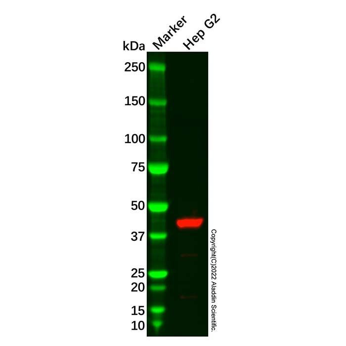
HPD Mouse mAb (Ab108629) - Western Blot
All lanes: HPD Mouse mAb (Ab108629) at 1/1000 dilution
Samples: Lysates at 20 µg per lane
Secondary: Goat Anti-Mouse IgG H&L (HRP) (Ab179001) at 1/20000 dilution

Predicted band size: 45 kDa
Observed band size: 42 kDa
Exposure time: 22.5 s

HPD Mouse mAb (Ab108629) - IHC
DAB staining on IHC
Sample: Rat Liver Tissue
Primary Ab: 20 µg/mL HPD Mouse mAb (Ab108629)
Second Ab: 2 µg/mL HRP-Linked Caprine Anti-Mouse IgG Polyclonal Antibody.

Anwendung
Anwendung
Dilution info
IHC

5-20 µg/mL

WB

0.01-2 µg/mL

Erweiterte Daten

Zugehörige Ziele (menschlich)
HPD Tclin 4-hydroxyphenylpyruvate dioxygenase (0 Activities)
Activity TypeActivity Value -log(M)Mechanism of ActionActivity ReferencePublications (PubMed IDs)
Wirkungsmechanismen
Zertifikate (CoA, COO, BSE/TSE und Analyse-Diagramm)
C of A & Other Certificates(BSE/TSE, COO):
Analytical Chart:

Find and download the COA for your product by matching the lot number on the packaging.

3 results found

Lot NumberCertificate TypeDatumArtikel
ZJ24F0404463ZJ24F0404463Certificate of AnalysisFeb 10, 2026 Ab108629
ZJ24F0404464ZJ24F0404464Certificate of AnalysisFeb 10, 2026 Ab108629
ZJ24F0404465ZJ24F0404465Certificate of AnalysisFeb 10, 2026 Ab108629
FAQs und Artikel
Lösungsrechner
Bewertungen

Kundenbewertungen

Application Protocols

Tested applications and recommended dilutions for this item are not specified; refer to CoA/Spec Sheet.

General starting protocols for monoclonal antibodies (literature/general; adjust empirically):

  • Western blot (WB):
    • Sample: Denatured/reduced lysates, 20–40 µg per lane.
    • Primary: 0.1–1 µg/mL in 5% BSA/TBST; 1 h RT or overnight 4°C.
    • Secondary: Anti‑mouse HRP 1:5,000–1:20,000.
    • Develop with ECL.
  • Immunofluorescence (IF/ICC):
    • Fix with 4% PFA or cold methanol (optimize).
    • Block/permeabilize: 3% BSA + 0.1% Triton X‑100 in PBS.
    • Primary: 1–10 µg/mL; 1 h RT or overnight 4°C.
    • Secondary: Anti‑mouse Alexa Fluor 1:500–1:1,000.
  • Immunohistochemistry (IHC):
    • Antigen retrieval as needed (citrate pH 6.0 or Tris‑EDTA pH 9.0).
    • Primary: 0.5–5 µg/mL; detect with HRP/DAB or fluorescence.
  • Immunoprecipitation (IP):
    • 1–5 µg Ab per 0.5–1 mg lysate; incubate 1–2 h, capture with Protein A/G beads (isotype dependent).
  • Flow cytometry (FC):
    • Start 0.1–1 µg per 10^6 cells in 100 µL; include Fc‑block where appropriate.

Always verify species compatibility and epitope accessibility for your system.

Biological Roles

Antibodies are affinity proteins used to detect, capture, or modulate specific biomolecular targets. The precise biological relevance depends on the antigen recognized; for this item, target details are not specified.

General roles of monoclonal antibodies in research (literature/general):

  • Molecular detection: High specificity recognition of epitopes on proteins, peptides, or modified residues enables mapping of signaling pathways, PTMs, and protein–protein interactions.
  • Quantitation: Sandwich ELISA calibrates analyte abundance in complex matrices with high sensitivity when paired with standards and orthogonal antibody pairs.
  • Localization: IF/ICC/IHC reveal subcellular or tissue distribution of targets. Epitope accessibility and fixation chemistry strongly influence signal.
  • Functional perturbation: Some mAbs can block ligand–receptor interactions or enzymatic sites in cell assays; suitability must be empirically confirmed.
  • Enrichment and proteomics: IP/Co‑IP coupled to MS enables interactome discovery; epitope tags or native proteins can be targeted.

Host species and applications for this specific HPD Mouse mAb are not specified for this item; refer to CoA/Spec Sheet. If the antigen is enzymatic or signaling in nature, confirm that the antibody recognizes the correct isoform, species ortholog(s), and modification state (e.g., phospho‑ vs total) before experimental deployment.

Buffer Applications

For monoclonal antibodies, buffer composition governs stability, background, and activity. Item‑specific buffer details are not specified; the product is Carrier Free and Azide Free.

General buffer guidance (literature/general):

  • Storage/handling: PBS or TBS, pH 7.2–7.5. Avoid low‑ionic‑strength buffers that promote aggregation/adsorption.
  • Labeling reactions: Use amine‑free buffers (e.g., 50 mM sodium phosphate, pH 7.2–7.4) for NHS‑ester chemistry; include 2–5 mM EDTA for maleimide conjugations to protect thiols.
  • Assay buffers: Add 0.05% Tween‑20 and 0.1–5% inert protein (BSA or casein) for blocking/wash in ELISA/WB; note that the primary stock here is Carrier Free—add these only at the assay stage as needed.
  • pH effects: Most IgG maintain stability near neutral pH; extremes (<pH 5 or >pH 9) can induce partial unfolding or aggregation.
  • Filtration/sterility: If sterility is required, filter through 0.22 µm low‑protein‑binding membranes (PES or PVDF).

Not applicable: Classical small‑molecule buffer systems and precise recipes are secondary to immunoassay‑specific formulations here. For lot‑specific recommendations, including any provided stabilizers, please refer to the CoA/Spec Sheet.

Green Alternatives

While “green chemistry” frameworks mostly target small‑molecule solvents and reagents, several sustainability considerations apply to antibodies:

  • Formulation aspects:
    • This item is Azide Free, reducing use of toxic sodium azide preservatives. Consider sterile technique, small aliquots, and low‑temperature storage to maintain stability without hazardous biocides.
    • Carrier Free minimizes protein additives (e.g., BSA), reducing animal‑derived inputs in your process.
  • Process efficiency:
    • High‑affinity/validated antibodies can lower required mass per assay, reducing upstream production and waste (general).
    • Use of reusable, protein A/G resins for capture and recycling of antibody in IP workflows can reduce consumables.
  • Cold‑chain optimization: Consolidate orders and select shipping options that minimize dry‑ice/ice‑pack usage while maintaining integrity; this item ships with ice packs (per Product Data).
  • Buffer choices: Employ phosphate or Good’s buffers with minimal environmental persistence; avoid heavy‑metal preservatives or azide when possible (general).

Comparison snapshot (general):

  • Azide‑preserved mAbs: Longer bench stability but introduce acute aquatic/toxicity hazards and waste restrictions.
  • Azide‑free mAbs (this item): Safer for live cells and drains (subject to local rules) but require stricter sterility and cold storage.

End‑of‑life: Deactivate residual biologicals with appropriate detergents and dispose via institutional aqueous waste protocols rather than organic solvent waste.

Pharmaceutical Uses

This product is supplied strictly for research use only (per Product Data). It is not intended for diagnostic, therapeutic, or in vivo applications.

Relevant formulation/bioprocess considerations for research antibodies (general/literature):

  • Excipient strategy: In drug‑product development, antibodies are often stabilized by sugars (trehalose/sucrose), amino acids (arginine), and surfactants (polysorbate‑20/80). This item is Carrier Free and Azide Free; if transitioning toward translational studies, appropriate excipients would need to be qualified separately.
  • Endotoxin and bioburden: Pharmaceutical‑grade specs typically require <0.1 EU/mg endotoxin and strict microbial limits. For this item, endotoxin/bioburden are not specified; refer to CoA/Spec Sheet.
  • Aggregation control: Size‑exclusion chromatography (SEC) is used to monitor aggregates; in research workflows, keep concentration and ionic strength within ranges that minimize self‑association.
  • Container/closure: Low‑binding polypropylene or siliconized glass vials to minimize adsorption.

Pharmacopeial status, GMP grade, and release tests are not specified for this item; refer to CoA/Spec Sheet. Any use outside basic research requires additional qualification and compliance steps not covered by this listing.

Physical Properties

Antibodies are aqueous macromolecular proteins; conventional small‑molecule constants (bp, mp) are not applicable.

  • Physical state/appearance: Not specified for this item; refer to CoA/Spec Sheet.
  • Concentration: Not specified for this item; refer to CoA/Spec Sheet.
  • Buffer composition: Carrier Free, Azide Free (per Product Data). Exact salts, pH, and excipients are not listed; refer to CoA/Spec Sheet.
  • pH: Not specified for this item; refer to CoA/Spec Sheet.
  • Solubility:
    • Antibodies are soluble in physiological aqueous buffers (PBS, TBS) (literature/general).
    • Insoluble in organic solvents (acetonitrile, methanol, DMSO) except at very low percentages used for conjugation protocols (literature/general).
  • Isoelectric point (pI) and extinction coefficient: Not specified for this item; refer to CoA/Spec Sheet.
    • Typical IgG pI ranges ~6–9 depending on sequence/glycoform (literature/general).
    • Typical A280 for 1 mg/mL IgG ~1.3–1.5 (pathlength 1 cm) (literature/general).
  • Stability: Store at −20°C; avoid repeated freeze–thaw (per Product Data). Stability at 2–8°C or room temperature is not specified for this item; refer to CoA/Spec Sheet.
  • Viscosity/osmolarity: Not specified for this item; refer to CoA/Spec Sheet.

Note: Boiling/dehydration denatures antibodies; never dry-down without appropriate protectants unless specified by protocol.

Quality and Grades

This item is designated: Carrier Free, ExactAb™, Azide Free, Validated, High Performance.

  • Carrier Free (item‑specific): Supplied without protein carriers (e.g., BSA, gelatin). Advantages include:
    • Unencumbered conjugation to enzymes, fluorophores, or beads.
    • Reduced background in immunoassays sensitive to carrier proteins.
    • Flexible custom buffer exchange.
  • Azide Free (item‑specific): Suitable for live‑cell compatible workflows and enzymatic conjugation systems that are inhibited by azide. Note: Without azide, microbial control depends on sterile technique and storage temperature.
  • Validated / High Performance (item‑specific descriptors): Indicates internal performance testing was performed; specific validation datasets, tested applications, host species reactivity, and lot‑to‑lot acceptance criteria are not specified for this item; refer to CoA/Spec Sheet.
  • ExactAb™ (brand descriptor): Proprietary designation from the supplier. In general, such branding implies a sequence‑defined, highly consistent monoclonal format and controlled production (general statement). The exact nature (hybridoma vs recombinant, subclass, kappa/lambda light chain) is not specified for this item; refer to CoA/Spec Sheet.
  • Purity/Grade: Listed collectively as above; numerical purity, endotoxin level, aggregate content, and residual host cell proteins/DNA are not specified for this item; refer to CoA/Spec Sheet.

Recommendation: For regulated workflows, request lot‑specific CoA detailing concentration (mg/mL), isotype, endotoxin (EU/mg), SEC‑HPLC monomer %, and residuals.

Reaction and Applications

This product is a biological affinity reagent, not a small‑molecule reagent for chemical synthesis. Classical organic reactions (e.g., Grignard, cross‑coupling) do not apply.

Relevant laboratory applications for monoclonal antibodies (general/literature):

  • Immunoassays: ELISA (capture/detection), Western blot, dot blot.
  • Imaging: Immunofluorescence (IF), immunocytochemistry (ICC), immunohistochemistry (IHC).
  • Enrichment: Immunoprecipitation (IP), chromatin IP (ChIP) where applicable.
  • Cell analysis: Flow cytometry (FC) when the epitope is cell‑surface accessible.
  • Conjugation: Enzyme, fluorophore, oligonucleotide, or bead conjugation facilitated by the Carrier Free format.

Manufacturer‑validated applications, species reactivity, and working dilutions are not specified for this item; refer to CoA/Spec Sheet. To expand utility:

  • Pair with an appropriate anti‑mouse secondary antibody (HRP, AP, Alexa Fluor dyes) when using unconjugated primary in WB/IF/ELISA.
  • For sandwich ELISA, orthogonal epitopes with a second anti‑target mAb are required; verify non‑overlapping binding sites experimentally.
  • For IP, pre‑clear lysates and optimize antibody:lysate protein ratio (typical starting 1–5 µg Ab per 500–1000 µg lysate; literature/general).

Note: If antigen is intracellular, fixation/permeabilization optimization is critical (paraformaldehyde vs methanol; detergent selection).

Reaction Conditions

Classical reaction condition guidance is not applicable to an antibody reagent. For bioconjugation and assay setup, typical conditions are provided as general literature guidance below; adjust empirically and consult the CoA.

General bioconjugation conditions (literature/general):

  • NHS‑ester labeling (amines): pH 7.4–8.3 in amine‑free buffer (50 mM phosphate or bicarbonate), 4–25°C, 30–90 min; quench with Tris or ethanolamine; target D/P 2–4 for imaging.
  • Maleimide–thiol labeling (sulfhydryls): pH 6.5–7.0 in PBS + 2–5 mM EDTA; 4–25°C, 30–120 min. For partial reduction, 1–5 mM TCEP for 15–30 min at room temp, then rapid desalting prior to labeling.
  • Click chemistry: Azide/alkyne cycloadditions require prior installation of clickable handles by the above methods or enzymatic tags. This item is azide‑free by formulation; it does not inherently contain click handles.

Assay working ranges (starting points; literature/general):

  • Western blot: 0.1–1 µg/mL primary; incubation 1–16 h at 4–25°C.
  • IF/ICC: 1–10 µg/mL; 0.1–2 µg per well on coverslips.
  • IP: 1–5 µg antibody per 0.5–1 mg lysate protein.

Item‑specific optimal conditions, tested applications, and dilutions are not specified for this item; refer to CoA/Spec Sheet.

Safety and Handling
  • GHS classification, pictograms, signal word, and H‑statements: Not specified for this item; refer to SDS.
  • General hazards (literature/general): Antibody preparations are aqueous protein solutions and typically present low acute hazards. However, they should be handled as potentially bioactive materials. Avoid inhalation, ingestion, and skin/eye contact. Follow institutional biosafety procedures.
  • Azide status: Azide Free (per Product Data). This reduces risks associated with sodium azide (e.g., acute toxicity, formation of heavy‑metal azides in plumbing). Nevertheless, use clean plastic/glassware and avoid metal drain disposal of proteinaceous materials per local regulations.
  • PPE: Lab coat, gloves (nitrile), and safety glasses. Use a biological safety cabinet if handling large volumes, aerosols, or cell culture applications (general guidance).
  • First aid (general guidance; defer to SDS):
    • Skin/eye contact: Rinse with water for several minutes; remove contaminated clothing; seek medical advice if irritation persists.
    • Ingestion/inhalation: Rinse mouth; move to fresh air; obtain medical attention if unwell.
  • Incompatibilities: Avoid strong oxidizers and extreme pH; proteases can degrade antibodies (general). Avoid repeated freeze–thaw cycles (per Product Data).
  • Spill/cleanup: Absorb with inert material, wash area with detergent; dispose of as laboratory biological/chemical waste per institutional policy.
  • Waste: Non‑hazardous biological/chemical waste stream unless local regulations specify otherwise; consult SDS and EHS.
Solvent Selection

As a protein biologic, this monoclonal antibody is intended for use in aqueous buffers, not organic solvents.

  • Polarity/miscibility: Antibodies are water‑soluble and structurally dependent on aqueous environments. They are not soluble or stable in neat organic solvents (literature/general).
  • Compatible media (general/literature):
    • PBS or TBS, pH 7.2–7.5 for storage/handling.
    • For conjugations, use carrier‑free buffers lacking primary amines (e.g., 50 mM sodium phosphate, pH ~7.2–7.4) for NHS ester labeling; avoid Tris/glycine.
    • For maleimide–thiol chemistry, maintain antibody in deoxygenated PBS with EDTA, pH 6.5–7.0.
    • For cell‑based work, isotonic sterile buffers; azide‑free status is advantageous.
  • Additives (general):
    • Stabilizers such as 2–5% trehalose or 0.1% inert proteins can enhance freeze–thaw resilience; not present here due to Carrier Free grade (item‑specific).
    • Low levels of nonionic surfactant (e.g., 0.01% Tween‑20) can minimize adsorption losses; evaluate assay compatibility.
  • When to choose alternatives: If your workflow demands low‑UV absorbance or special ionic strength, perform buffer exchange (spin columns, dialysis) into your assay buffer. For long‑term storage without carriers, consider aliquoting to avoid repeated freeze–thaw.

Item‑specific buffer composition and pH: Not specified for this item; refer to CoA/Spec Sheet.

Storage and Reconstitution
  • Storage conditions (item‑specific): Store at −20°C. Avoid repeated freezing and thawing. (per Product Data).
  • Shipping: Ice chest + ice pads (per Product Data).
  • Format: Carrier Free, Azide Free (per Product Data). Exact concentration, buffer salts, and pH are not specified; refer to CoA/Spec Sheet.
  • Aliquoting: Upon receipt, spin briefly to collect contents, then aliquot into low‑protein‑binding tubes sized for single use to prevent freeze–thaw cycles.
  • Freeze–thaw guidance: Multiple freeze–thaw events accelerate aggregation and activity loss; store working aliquots at −20°C or −80°C as appropriate. Avoid frost‑free freezers. Thaw on ice and mix gently; do not vortex.
  • Short‑term storage: If frequent use is expected within 1–2 weeks, 2–8°C storage can be used to avoid freeze–thaw, but microbial control must be ensured because the product is azide‑free (general guidance).
  • Reconstitution: If provided lyophilized (not specified), reconstitute with sterile, neutral pH buffer (e.g., PBS) to the desired concentration. For conjugation, reconstitute in amine‑free buffer. Exact reconstitution volume and target concentration are not specified; refer to CoA/Spec Sheet.
  • Adsorption losses: Minimize by using low‑bind plastics and adding 0.01% nonionic surfactant in assay buffers if compatible.
  • Stability/expiry: Lot‑specific shelf life and post‑thaw stability are not specified; refer to CoA/Spec Sheet.

Research Use Only: For research use only; not for diagnostic or therapeutic use (per Product Data).

Structure and Identity

HPD Mouse mAb is a mouse-derived monoclonal antibody supplied for research applications. As a biologic macromolecule, it does not have a conventional small‑molecule structure.

  • Product type: Monoclonal antibody (mouse mAb); clone/isotype: Not specified for this item; refer to CoA/Spec Sheet.
  • Antigen/target: Not specified for this item; refer to CoA/Spec Sheet.
  • Molecular formula: Not applicable to proteins in a fixed stoichiometric sense; sequence-defined macromolecule (Not specified for this item; refer to CoA/Spec Sheet).
  • Molecular weight: Not specified for this item; refer to CoA/Spec Sheet.
    • Typical IgG1 literature value: ~150 kDa (heavy+light chains, ~2x50 kDa + 2x25 kDa) (literature/general).
  • SMILES / InChI / InChIKey: Not applicable for antibodies; sequence-defined biopolymers lack standard small‑molecule line notations.
  • Structural features (general/literature):
    • Immunoglobulin G architecture with two identical heavy chains and two identical light chains linked by disulfide bonds.
    • Each arm contains a variable domain (VH/VL) forming the antigen‑binding site (paratope); Fc region mediates effector functions and enables Protein A/G binding (isotype-dependent).
    • Post‑translational modifications may include N‑glycosylation in Fc (N297 for many IgG subclasses); specific glycoform for this item is not specified.
  • ExactAb™ note: Branded designation provided; the underlying sequence/format is not detailed here (general description of brand; item‑specific attributes Not specified for this item; refer to CoA/Spec Sheet).
Synthetic Utility

Not typically applicable. This product is a monoclonal antibody used as an affinity reagent rather than a reagent or building block for organic synthesis.

For researchers performing antibody–conjugate synthesis (general/literature):

  • Functional handles: Native lysines (NHS‑ester chemistry) and reduced interchain disulfides or engineered cysteines (maleimide chemistry) allow bioconjugation to dyes, enzymes, chelators, or payloads. The Carrier Free format is advantageous for controlled conjugation without carrier protein interference.
  • Site‑selective strategies: Enzymatic methods (e.g., glycan remodeling with EndoS2 + GalT azido‑sugar, Sortase A LPXTG tagging) or Fc‑specific oxidation to generate aldehydes provide more homogeneous conjugates.
  • Characterization: Use UV–Vis (dye:protein ratio), SEC‑HPLC (aggregation), MS/CE‑SDS, and antigen‑binding assays to verify conjugate integrity.

Item‑specific sequence, isotype, and reactive cysteine content are not specified for this item; refer to CoA/Spec Sheet.

Target Specificity
  • Antigen/epitope: Not specified for this item; refer to CoA/Spec Sheet.
  • Clone ID and isotype: Not specified for this item; refer to CoA/Spec Sheet.
  • Species reactivity: Not specified for this item; refer to CoA/Spec Sheet.
  • Cross‑reactivity: Not specified for this item; refer to CoA/Spec Sheet.
  • Validation data: Not specified for this item; refer to CoA/Spec Sheet.

Note: “HPD Mouse mAb” suggests a mouse monoclonal antibody; however, specific target details are not provided in the Product Data and should be confirmed from the lot‑specific datasheet.

Benötigen Sie Hilfe bei der Auswahl der Qualität?

Unser Leitfaden zur Qualitätsauswahl behandelt Reinheit, Stabilisatorstatus und Anwendungseignung für alle Varianten in unserem Katalog.

Carrier Free Qualitätsleitfaden ansehen → Validated Qualitätsleitfaden ansehen → ExactAb™ Qualitätsleitfaden ansehen → Azide Free Qualitätsleitfaden ansehen → High Performance Qualitätsleitfaden ansehen →

Verwandte Produkte

Goat Anti-Mouse IgG H&L Antibody - Sekundärantikörper, for ELISA,LF,GICA various applications, from Mouse, generated in Goat
Goat Anti-Mouse IgG H&L (HRP) - Sekundärantikörper, for WB,ELISA,IHC-P various applications, from Mouse, generated in Goat, IgG
Rabbit Anti-Mouse IgG H&L (HRP) - Sekundärantikörper, IgG
Rabbit Anti-Mouse IgG antibody - Sekundärantikörper
Goat Anti-Mouse IgG H&L (FITC) - Sekundärantikörper
Goat Anti-Mouse IgG H&L (TRITC) - Sekundärantikörper
Goat Anti-Mouse IgG H&L Antibody - Sekundärantikörper, Goat IgG
Goat Anti-Mouse IgG H&L (Cy5) - Sekundärantikörper
Goat Anti-Mouse IgG H&L (HRP) - Sekundärantikörper
Goat Anti-Mouse IgG H&L (Biotin) - Sekundärantikörper
Rabbit Anti-Mouse IgG (HRP) - Sekundärantikörper
Rabbit Anti-Mouse IgG (Biotin) - Sekundärantikörper
Goat Anti-Mouse IgG H&L (HRP) - Sekundärantikörper, Goat IgG
Goat Anti-Mouse IgG H&L (TRITC) - Sekundärantikörper, Goat IgG
Goat Anti-Mouse IgG H&L (Biotin) - Sekundärantikörper, Goat IgG
Goat Anti-Mouse IgG H&L (FITC) - Sekundärantikörper, Goat IgG
Goat Anti-Mouse IgG H&L (Cy5) - Sekundärantikörper, Goat IgG
Goat Anti-Mouse IgG H&L (PE) - Sekundärantikörper, Goat IgG
Goat Anti-Mouse IgG H&L (APC) - Sekundärantikörper, Goat IgG
Goat Anti-Mouse IgG H&L (Alkaline Phosphatase) - Sekundärantikörper, Goat IgG
Goat Anti-Mouse IgG H&L (AF405) - Sekundärantikörper, Goat IgG
Goat Anti-Mouse IgG H&L (AF647) - Sekundärantikörper, Goat IgG
Anti-Maus-IgG H&L von der Ziege (AF555) - Sekundärantikörper, specific to Mouse IgG, Goat IgG
Goat Anti-Mouse IgG H&L (AF700) - Sekundärantikörper, specific to Mouse IgG, Goat IgG
Goat Anti-Mouse IgG H&L (PE-Cy5) - Sekundärantikörper, specific to Mouse IgG
Centrose patent anti-dysadherin (anti-Dysadherin) - Primärer Antikörper, specific to FXYD5
Goat Anti-Mouse IgG H&L (AF488) - Sekundärantikörper
Goat Anti-Mouse IgG H&L (PE-Cy7) - Sekundärantikörper
Goat Anti-Mouse IgG H&L (AF750) - Sekundärantikörper
Goat Anti-Mouse IgG H&L (AF546) - Sekundärantikörper
Goat Anti-Mouse IgG H&L (Oregon Green 488) - Sekundärantikörper
Goat Anti-Mouse IgG H&L (Cy3) - Sekundärantikörper

Dürfen wir Sie über Rabattaktionen informieren?

Später erinnern

Vielen Dank! Bitte überprüfen Sie Ihren E-Mail-Posteingang, um zu bestätigen.

Hoppla! Benachrichtigungen sind deaktiviert.