The His Pull‑Down Kit is designed for efficient co‑sedimentation of tagged bait proteins with target proteins. It enables the isolation and purification of unknown captured pro-teins that interact with His‑tagged bait proteins. The simple experimental principle is as follows: His residues are immobilized on agarose beads to form His‑agarose resin; the bait protein is expressed as a fusion with a His‑tag, and the resulting His‑bait fusion protein binds to the His‑agarose resin. If a captured protein that interacts with the bait protein exists in the system, an agarose‑His‑bait protein‑captured protein complex will be formed, allowing the interacting captured protein to be isolated and detected.
This product utilizes high‑quality His‑tag protein agarose resin. This resin is prepared by immobilizing IDA ligands onto a highly rigid agarose matrix via an ether linkage with a 9‑atom spacer arm, followed by chelation with Ni²⁺ metal ions.The highly rigid agarose matrix supports high flow rates and low backpressure. Its optimized pore structure provides higher binding capacity compared with conventional agarose matrices. The kit features easy operation, strong binding capacity, and wide applicability.
Important Notes:
1. This product is intended for scientific research use by professionals only.
2. Unless otherwise specified, all steps are recommended to be performed at 4°C to minimize potential protein degradation. If cell lysis is insufficient, sonication may be used after adding the lysis buffer.
3. Ni-IDA Agarose Resin should be stored in the storage solution to prevent drying. Before use, mix thoroughly by gently inverting the tube several times to ensure uniform suspension of the beads. Avoid vigorous vortex or shaking to prevent antibody denaturation.
4. Agarose Resin that has been boiled lose their binding capacity and should not be reused.
5. Reagent volumes can be adjusted according to experimental needs. It is recommended to perform preliminary experiments to validate immunoprecipitation conditions or optimize lysis parameters.
6. Please follow safety guidelines and adhere to laboratory reagent handling protocols.
7. The lysis buffer included in this kit already contains protease inhibitors. If specific needs arise, other appropriate inhibitor cocktails can be used.
8. After harvesting, protein samples should be purified as soon as possible and kept at 4°C or on ice at all times to minimize protein degradation and denaturation.
9. It is recommended to include positive and negative control groups during immunoprecipitation or purification procedures.
Procedure (Unless otherwise specified, all procedures should be performed at 4°C)
1. Reagent Preparation
Additional Required Materials:
(1) Reagents to be prepared by the user:
a) Primary Antibody: His tag antibody.
b) Secondary Antibodies: Goat Anti-Mouse IgG H&L (HRP), Goat Anti-Rabbit IgG H&L (HRP).
c) Other Reagents: BST, running buffer, transfer buffer, reducing SDS‑PAGE gel, as well as the bait protein and target protein used for protein‑protein interaction assays.
(2) Required Equipment:
Electrophoresis Apparatus, Transfer Apparatus, Imaging System
The above reagents, if required, can be ordered from Aladdin: His tag (C-terminal) Mouse mAb (Ab155835), Recombinant His tag Antibody (Ab086830), Goat Anti-Rabbit IgG H&L (HRP) (Ab176443), Goat Anti-Mouse IgG H&L (HRP) (Ab179001).
2. Solution Preparation
You may use the buffers provided in the kit, or prepare different buffer systems according to actual needs. It is recommended to filter all buffers through a 0.22 μm or 0.45 μm membrane filter before use. Buffers should be stored at 4°C. If any reagent appears cloudy, discard it immediately.
(1) Preparation of Elution Buffer: Prepare 1 mL of 250 mM imidazole solution. Specifically, add 62.5 µL of 4 M imidazole to 937.5 µL of 1× Wash Buffer.
(2) Prepare an appropriate amount of inhibitor-containing lysis buffer based on the proportion of using 100-200 μL of inhibitor-containing lysis buffer for lysis and 300-600 μL of inhibitor-containing lysis buffer for washing per 0.5-1 million cells. Mix Lysis Buffer and Protease Inhibitor Cocktail(100×) at a ratio of 100:1. For example, add 10 μL of Protease Inhibitor Cocktail (100×) to 1 mL of lysis Buffer to obtain 1 mL of inhibitor-containing lysis buffer (Lysis Buffer with Protease Inhibitor Cocktail). The prepared inhibitor-containing lysis buffer should be placed on ice or at 4°C.
Note 1: Lysis buffer containing inhibitors should be prepared fresh immediately before use and is not suitable for frozen storage and later use.
Note 2: This lysis buffer is suitable for mammalian cells.
(3) Preparation of 1×Wash Buffer: Dilute the 10×Wash Buffer with deionized water at a ratio of 9:1 before use. For example: add 9 mL of deionized water to 1 mL of 10× Wash Buffer, mix well to obtain 1×Wash Buffer.
(4) Ni-IDA Agarose Resin Washing: Gently resuspend the Ni-IDA Agarose Resin to form a homogeneous gel suspension. Using 40 μL of well-mixed gel suspension per sample (all subsequent affinity precipitation steps use 40 μL gel suspension per sample as an example), transfer an appropriate amount of Ni-IDA agarose resin to a clean centrifuge tube. Add 0.5 mL of 1×wash buffer, centrifuge at 1250×g for 30 seconds at 4°C, discard the supernatant, and repeat this step twice.
Note: It is more convenient to aspirate the gel suspension using a wide-bore tip (e.g., by cutting off the end of a pipette tip with scissors).
3. Preparation of Bait Protein Samples (Note: Perform all sample lysis steps at 4°C or on ice)
(1) For His-tagged fusion protein expressed in Escherichia coli:
Grow and transform E. coli according to standard protocols. Transfer 5 mL of IPTG-induced E. coli culture to a sterile centrifuge tube. Centrifuge at 5000×g for 5 minutes and discard the culture supernatant. Resuspend the pellet in 1 mL of PBS buffer per 5 mL of original culture volume, and mix by pipetting or vortex. Transfer 1 mL of cell suspension to a 1.5 mL microcentrifuge tube. Centrifuge at 5000×g for 5 minutes and discard the supernatant. Resuspend the pellet in 200 μL of ice-cold PBS buffer per 5 mL of original culture volume, and mix by pipetting or vortex. Protease inhibitors may be added as desired; for optimal results, use a protease inhibitor cocktail during cell lysate preparation. Add 200 μL of pull-down lysis buffer per 5 mL of original culture volume, and immediately invert the tube until thoroughly mixed. Incubate on ice for approximately 30 minutes, inverting the tube periodically. Centrifuge at 12000×g for 5 minutes to clarify the crude E. coli lysate. Transfer the supernatant to a new microcentrifuge tube, store on ice, and label as “bait lysate”.
(2) For secreted His-tagged fusion protein:
Collect the culture supernatant and determine the bait protein concentration. If the bait protein concentration is high, dilute it to a final concentration of 500 μg/mL with 1×PBS for subsequent experiments.
(3) For pre-purified His-tagged fusion protein
Determine the bait protein concentration. If the bait protein concentration is high, dilute it to a final concentration of 500 μg/mL with 1×PBS for subsequent experiments.
4. Pull-Down Assay
(1) Immobilization of Bait Protein.
1) Add the bait protein obtained in Step 3 to the pre-washed gel, mix gently, and incubate on a shaker at 4°C for 3 h. Reserve a portion of the bait protein sample as an input control for later detection.
2) Separation by centrifugation. After incubation, centrifuge at 1250×g for 30 seconds at 4 °C, transfer the supernatant to a new centrifuge tube, and store for analysis.
3) Transfer the complex in the centrifuge tube to a spin column, centrifuge at 1250×g for 30 seconds at 4°C, transfer the flow-through to a new centrifuge tube, and label as “bait flow-through” for later detection.
4) Add 500 μL of 1×PBS to the spin column, invert gently several times to wash the gel, centrifuge at 1250×g for 30 seconds at 4°C, and discard the eluate. Repeat 4 times to obtain the bait protein-gel complex.
(2) Capture of Target Protein.
1) Add 800 μL of the prepared prey protein sample to the bait protein–gel complex. Incubate with gentle shaking on a shaker at 4°C for at least 3 hours or overnight. Reserve a portion of the prey protein sample as a prey input for later detection.
Note: Longer incubation times may be required to ensure sufficient binding between bait and prey proteins, and can be adjusted according to experimental conditions.
2) After incubation, centrifuge at 1250×g for 30 seconds at 4°C, transfer the flow-through to a new centrifuge tube, and label as “prey flow-through” for later detection.
3) Add 500 μL of 1×PBS to the spin column, invert gently several times to wash the gel, centrifuge at 1250×g for 30 seconds at 4°C, and discard the eluate. Repeat 4 times to obtain the bait-prey protein-gel complex.
5. Elution of Bait-Prey Protein Complex: Elution can be performed using one of the following three methods, depending on the characteristics of the tagged protein and requirements of subsequent experiments.
(1) Non-Denaturing Elution
Pipette 100 μL of the prepared elution buffer into the washed resin, mix the resin and elution buffer thoroughly, then centrifuge and collect the eluate. If elution is inefficient, increase the glutathione concentration or perform multiple elution.
Note: If elution efficiency is unsatisfactory, the glutathione concentration may be increased or multiple elution steps may be performed.
(2) Peptide competitive elution
Add 100 µL of His‑peptide elution buffer to every 40 µL of the original gel volume. Incubate the mixture on a vertical rotator for 5-10 minutes, then centrifuge at 1250×g and 4 °C for 30 seconds. Aspirate the supernatant to obtain the eluted fraction, which contains the His‑tagged protein and its complexes.
Note: If elution efficiency is unsatisfactory, the His‑peptide concentration can be increased or multiple rounds of elution can be performed. Once the gel beads are fully settled, transfer the supernatant to a new centrifuge tube for Western Blot analysis.
(3) Elution with SDS-PAGE Loading Buffer
For every 40 μL of original beads volume, add 50 μL of 1×PBS to resuspend the beads, followed by 50 μL of 2×SDS Loading Buffer. Mix gently by flicking the tube, then heat at 95-100°C for 5-10 minutes. Centrifuge at 1250×g for 30 seconds. Collect the supernatant for SDS-PAGE electrophoresis or Western Blot analysis.
Note: The recommended loading volume for WB is 20 μL or less. The remaining sample can be stored at -20°C.

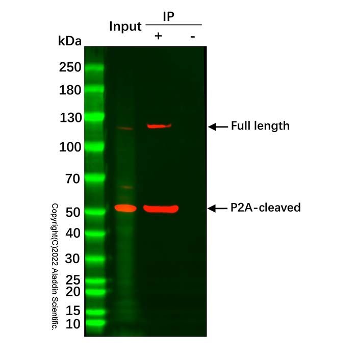
 ZJ26F0535413