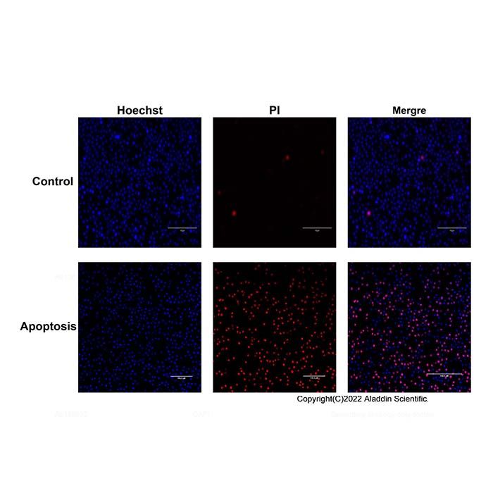
The Hoechst 33342/PI Double Staining Apoptosis Detection Kit provides a classic, rapid, and straightforward method for distinguishing between apoptotic and necrotic cells. This kit utilizes a dual-staining approach with Hoechst 33342 and propidium iodide (PI). The nuclear dye Hoechst 33342 permeates cell membranes and emits blue fluorescence upon binding to double-stranded DNA. In normal cells, limited dye entry results in weak blue fluorescence. In apoptotic cells, however, increased membrane permeability allows significantly more Hoechst 33342 to enter, leading to higher fluorescence intensity. Additionally, chromatin condensation during apoptosis enhances the efficiency and concentration of DNA binding by the dye. Impaired function of the P-glycoprotein pump on the membrane of apoptotic cells further prevents effective efflux of Hoechst 33342, resulting in its intracellular accumulation. These characteristics collectively contribute to a markedly stronger fluorescence in apoptotic cells compared to normal cells. In contrast, propidium iodide (PI), another nuclear dye, cannot penetrate the intact membranes of normal or apoptotic cells, making viable cells impermeable to PI staining. Necrotic cells, which lose membrane integrity at an early stage, are readily stained by PI.
When dual-stained with these dyes, normal cells exhibit weak blue fluorescence primarily from Hoechst 33342. Apoptotic cells display intense blue fluorescence due to enhanced Hoechst 33342 staining, while necrotic cells show strong red fluorescence from PI.
Component
H1373483 | Component | 100T | 500T | Storage | Quantity Per Test |
H1373483A | Hoechst 33342 Staining Solution | 1 mL | 5 mL | -20℃, Store in the dark. | 10 μL per 0.5–1.0×10⁶ cells/mL. |
H1373483B | Propidium iodide Staining Solution (PI) | 1 mL | 5 mL | -20℃, Store in the dark. | 10 μL per 0.5–1.0×10⁶ cells/mL. |
Note: The recommended number of cells to stain per test 0.5–1.0×10⁶ cells.
Usage method:
1. Cell Preparation
Adherent cells: Grow cells in 6-well plates to the logarithmic growth phase. Wash twice with PBS and add 1 mL of PBS.
Suspension cells: Wash cells twice with pre-cooled PBS and resuspend in PBS at a density of 0.5–1.0×10⁶ cells/mL.
2. Staining
Adherent cells: Add 10 µL of Hoechst 33342 and 10 µL of PI directly to each well of the 6-well plate. Incubate at 4 °C in the dark for 20–30 minutes.
Suspension cells: Take 1 mL of the cell suspension, add 10 µL of Hoechst 33342 and 10 µL of PI, and incubate at 4 °C in the dark for 20–30 minutes.
Note: After staining, proceed with fluorescence detection as soon as possible. The number of cells for each detection should not exceed 1×10⁶.
3. Fluorescence Microscopy Detection and Analysis
Adherent cells: Remove the staining solution, wash twice with PBS, add an appropriate amount of PBS, and observe under a fluorescence microscope.
Suspension cells: Wash twice with PBS, resuspend the cells in PBS, transfer to a culture dish, and observe for red and blue fluorescence.
Matters needing attention:
1. After the staining process is completed, the detection should be carried out as soon as possible.
Hoechst 33342 and PI are harmful to the human body. Please take protective measures when using them.
2. For your safety and health, please wear a laboratory coat and put on disposable gloves when operating.
3. This product is exclusively for scientific research purposes and must not be used for clinical diagnosis or treatment.

 ZJ26F0636640