
ERAD, ER-phagy, and Cell-Fate Regulation of Misfolded Proteins Under Endoplasmic Reticulum Stress
ERAD, ER-phagy, and Cell-Fate Regulation of Misfolded Proteins Under Endoplasmic Reticulum Stress
The endoplasmic reticulum maintains protein homeostasis through molecular chaperones, endoplasmic reticulum-associated degradation, and endoplasmic reticulum-selective autophagy. Abnormal proteins that can be unfolded are mainly retrotranslocated through ERAD and degraded by the proteasome, whereas large aggregates and abnormal endoplasmic reticulum fragments are more frequently delivered to lysosomes through ER-phagy. When the burden of misfolded proteins persistently exceeds cellular clearance capacity, the unfolded protein response may shift from adaptive regulation toward inflammation and cell death.
Keywords: endoplasmic reticulum stress; unfolded protein response; endoplasmic reticulum-associated degradation; ERAD; ER-phagy; HRD1; SEL1L; VCP; proteasome; cell death
1 Endoplasmic Reticulum Protein Quality Control and Stress Responses
1.1 Protein-Folding Environment of the Endoplasmic Reticulum
Proteins entering the secretory pathway undergo N-glycosylation, disulfide-bond formation, subunit assembly, and conformational maturation in the endoplasmic reticulum. BiP, calnexin, calreticulin, and members of the protein disulfide isomerase family jointly recognize hydrophobic regions, unpaired cysteine residues, and abnormal glycan structures in nascent polypeptide chains. The redox state, calcium concentration, and molecular-chaperone capacity of the endoplasmic reticulum lumen all influence protein-folding efficiency. Hypoxia, nutrient deprivation, disruption of calcium homeostasis, oxidative stress, and increased secretory demand can all cause accumulation of misfolded proteins.
1.2 Unfolded Protein Response
(1) PERK Signaling Pathway
PERK phosphorylates eIF2α and temporarily reduces global protein translation, thereby limiting the entry of newly synthesized polypeptides into the endoplasmic reticulum while promoting expression of ATF4-associated stress-response genes. Under short-term stress, this pathway primarily reduces the protein-folding burden and improves cellular adaptability.
(2) IRE1α Signaling Pathway
IRE1α splices XBP1 mRNA to generate XBP1s, which promotes expression of molecular chaperones, lipid-synthesis enzymes, and ERAD-associated genes, thereby expanding the folding and degradation capacities of the endoplasmic reticulum. IRE1α can also alter the protein load entering the endoplasmic reticulum through regulated IRE1-dependent mRNA decay.
(3) ATF6 Signaling Pathway
Under stress conditions, ATF6 is transported from the endoplasmic reticulum to the Golgi apparatus, where proteolysis releases a transcriptionally active cytoplasmic fragment. Activated ATF6 enters the nucleus and induces expression of molecular chaperones, folding enzymes, and certain ERAD components, cooperating with PERK and IRE1α to restore protein homeostasis.
1.3 Major Clearance Pathways for Misfolded Proteins
Endoplasmic reticulum-associated degradation (ERAD) mainly processes monomeric proteins or small complexes that can be unfolded and transported across the endoplasmic reticulum membrane, with the cytosolic ubiquitin-proteasome system as the final degradation machinery. Endoplasmic reticulum-selective autophagy (ER-phagy) delivers specific endoplasmic reticulum membrane regions and associated protein aggregates to lysosomes and is better suited for removing large aggregates, abnormal membrane structures, and excess endoplasmic reticulum that cannot be efficiently retrotranslocated. These pathways are not interchangeable but divide substrates according to conformation, aggregation state, and localization within specific endoplasmic reticulum subdomains.
Table 1 Major Clearance Pathways in Endoplasmic Reticulum Protein Quality Control
Clearance Pathway | Major Substrates | Core Process | Major Degradation System |
ERAD | Misfolded luminal proteins that can be unfolded and retrotranslocated, abnormal membrane proteins, and proteins with abnormal cytosolic domains | Substrate recognition, retrotranslocation, ubiquitination, and membrane extraction | 26S proteasome |
ER-phagy | Endoplasmic reticulum fragments, membrane-protein aggregates, and large substrates that are difficult to translocate | Receptor recognition, endoplasmic reticulum membrane remodeling, and autophagosome sequestration | Lysosome |
ER-to-lysosome-associated degradation | Aggregated secretory proteins and specific endoplasmic reticulum luminal substrates | Formation of ER-derived vesicles and fusion with endosomes or lysosomes | Lysosome |
Proteolysis within the endoplasmic reticulum lumen | Certain abnormal proteins or short peptides | Luminal proteolysis and local processing | Endoplasmic reticulum luminal proteases |
2 Recognition and Selection of ERAD Substrates
2.1 Glycoprotein Folding Surveillance
Oligosaccharyltransferase transfers the Glc₃Man₉GlcNAc₂ oligosaccharide to Asn-X-Ser/Thr sites in nascent polypeptides, where X is generally not proline. After sequential trimming by glucosidases, monoglucosylated glycoproteins enter the calnexin-calreticulin cycle. If a protein has not folded correctly, UDP-glucose:glycoprotein glucosyltransferase can re-add a glucose residue, allowing the substrate to re-enter the chaperone-assisted folding cycle. Correctly folded proteins leave the endoplasmic reticulum and enter the downstream secretory pathway.
2.2 Mannose Trimming and Degradation Signals
Glycoproteins that fail to fold over prolonged periods can undergo mannose trimming by MAN1B1 and EDEM-family proteins. Exposure of specific mannose residues generates a glycan signal for ERAD entry. OS-9 and XTP3-B recognize these trimmed glycans through mannose-6-phosphate receptor homology domains and deliver substrates to the SEL1L-HRD1 complex. Mannose trimming is not merely a deglycosylation process but an important timing mechanism that transfers repeatedly misfolded glycoproteins from the calnexin-calreticulin cycle into the degradation pathway.
2.3 Recognition of Nonglycosylated Substrates
Nonglycosylated proteins cannot rely on glycan-based degradation signals and are generally recognized by BiP and other chaperones through exposed hydrophobic regions, abnormal assembly interfaces, or prolonged unfolded states. OS-9 and XTP3-B can also influence degradation of certain nonglycosylated substrates, but their functions in recognizing glycan-dependent and glycan-independent degradation signals are not identical. Therefore, all nonglycosylated ERAD substrates should not be assigned to a single chaperone pathway.
2.4 Disulfide-Bond Reduction and Substrate Unfolding
Many secretory proteins depend on disulfide bonds to maintain stable conformations. Misfolded proteins retaining complex disulfide-bond structures are difficult to pass through retrotranslocation channels. ERdj5 cooperates with EDEM and BiP to reduce incorrect or nonnative disulfide bonds, converting substrates from compact conformations into states suitable for translocation. The J domain of ERdj5 also promotes the ATPase cycle of BiP, allowing BiP to bind reduced substrates stably and prevent their reaggregation.
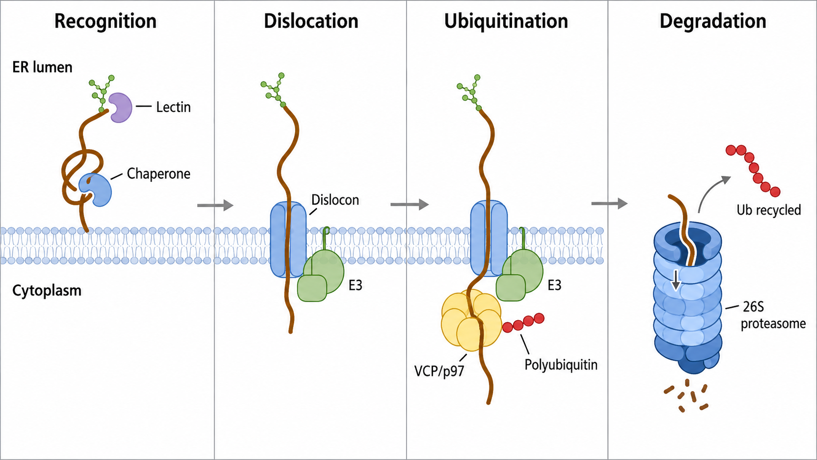
Figure 1 ER-associated degradation (ERAD) pathway for clearance of misfolded proteins
3 Retrotranslocation of ERAD Substrates
3.1 SEL1L-HRD1 Complex
SEL1L is a core scaffold that connects the luminal substrate-recognition system with the membrane-associated E3 ubiquitin ligase HRD1. Its luminal region forms complexes with OS-9, XTP3-B, and other chaperones, whereas its membrane-associated region cooperates with HRD1 and Derlin proteins to deliver substrates judged to be irreparable to the retrotranslocation machinery. Loss of SEL1L destabilizes the HRD1 complex and markedly reduces clearance of multiple classes of luminal ERAD substrates.
3.2 Retrotranslocation Channels
(1) HRD1-Mediated Membrane Translocation
HRD1 functions both as an E3 ubiquitin ligase and as an important membrane component of the retrotranslocation complex. Its transmembrane region can form a channel with a hydrophilic internal environment, providing a pathway through which certain luminal or intramembrane misfolded substrates exit the endoplasmic reticulum.
(2) Cooperation of Derlins and Other Membrane Components
Derlin-family proteins may participate in recognition of abnormal transmembrane regions, membrane insertion, substrate transfer, or assembly of retrotranslocation complexes, depending on substrate type. Distinct ERAD complexes process substrates with luminal abnormalities, transmembrane-domain defects, or abnormal cytosolic domains.
(3) Limitations of the Role of Sec61
Early models proposed that Sec61 might mediate retrotranslocation of certain ERAD substrates. However, it cannot be regarded as a universal retrotranslocation channel for all mammalian ERAD substrates. Whether Sec61 participates in degradation of a specific substrate depends on substrate type, cellular context, and the composition of the associated ERAD complex.
3.3 Substrate Conformation and Translocation Efficiency
Retrotranslocation generally requires substrates to be at least partially unfolded. Large oligomers, stable aggregates, and highly cross-linked glycoproteins cannot readily pass through ERAD channels and may remain in the endoplasmic reticulum before being transferred to ER-phagy or other lysosomal clearance pathways. Membrane proteins may first undergo recognition of abnormal transmembrane regions and then be ubiquitinated and extracted from the membrane by HRD1, gp78, or other E3 ligases.
4 Ubiquitination, Extraction, and Proteasomal Degradation of ERAD Substrates
4.1 Ubiquitination Cascade
(1) Ubiquitin Activation
E1 ubiquitin-activating enzymes use ATP to activate ubiquitin and form a high-energy thioester bond between the catalytic cysteine of E1 and the C terminus of ubiquitin.
(2) Ubiquitin Transfer
Activated ubiquitin is subsequently transferred from E1 to the catalytic cysteine of an E2 ubiquitin-conjugating enzyme. Different E2 enzymes cooperate with specific endoplasmic reticulum membrane E3 ligases and influence ubiquitin-chain type and elongation efficiency.
(3) Substrate Ubiquitination
E3 ubiquitin ligases recognize both E2 and the substrate and promote formation of an isopeptide bond between the C-terminal glycine of ubiquitin and a substrate lysine residue. ERAD substrates generally carry K48-linked polyubiquitin chains, although some substrates may also contain mixed or branched ubiquitin chains.
4.2 ERAD-Associated E3 Ligases
HRD1 is a core E3 ligase involved in degradation of luminal and certain intramembrane abnormal substrates. Other E3 ligases participating in endoplasmic reticulum protein quality control include gp78/AMFR, MARCHF6/TEB4, RNF5, RNF170, RNF185, and TRC8/RNF139. Different E3 ligases have distinct substrate localizations, tissue distributions, and stress-related functions. Ubiquitination of all endoplasmic reticulum substrates should therefore not be attributed exclusively to HRD1.
4.3 VCP/p97-Mediated Substrate Extraction
VCP/p97 is an AAA+ ATPase that binds the UFD1-NPL4 complex and recognizes polyubiquitinated substrates. Through the mechanical force generated by ATP hydrolysis, p97 separates substrates from the endoplasmic reticulum membrane or membrane-protein complexes and pulls them into the cytosol. UBXD2, UBXD8, VIMP, and other cofactors recruit p97 to specific ERAD complexes, whereas deubiquitinases such as YOD1 and Ataxin-3 regulate ubiquitin-chain length and the efficiency with which substrates pass through the central channel of p97.
4.4 Deglycosylation and Proteasomal Degradation
Retrotranslocated glycoproteins can be deglycosylated in the cytosol by NGLY1, accompanied by conversion of glycosylated asparagine residues into aspartate. Deglycosylation reduces substrate size and facilitates proteasomal processing, although NGLY1 is not absolutely required for degradation of all ERAD substrates. After the necessary unfolding, deglycosylation, and ubiquitin-chain remodeling steps, substrates are recognized by the 19S regulatory particle of the 26S proteasome and delivered into the 20S core particle for proteolysis.
Table 2 Major Steps and Core Molecules of ERAD
ERAD Stage | Core Molecules | Major Function |
Folding surveillance | BiP, calnexin, calreticulin, UGGT | Recognize unfolded regions and promote refolding |
Glycan editing | MAN1B1, EDEM1-3 | Trim mannose residues and generate degradation signals |
Substrate delivery | OS-9, XTP3-B, SEL1L | Recognize degradation signals and deliver substrates to membrane complexes |
Disulfide-bond reduction and substrate delivery | ERdj5, ERFAD, ERp90 | ERdj5 directly reduces aberrant disulfide bonds and promotes substrate unfolding; ERFAD and ERp90 participate in ERAD-complex organization, substrate delivery, or retrotranslocation, but ERp90 has not been established as a direct disulfide reductase |
Retrotranslocation | HRD1, Derlin family, and associated complexes | Move substrates across or out of the endoplasmic reticulum membrane |
Ubiquitination | E1, E2, HRD1, gp78, and other E3 ligases | Generate ubiquitin chains required for proteasomal recognition |
Membrane extraction | VCP/p97, UFD1, NPL4, UBXD proteins | Use ATP to extract ubiquitinated substrates into the cytosol |
Cytosolic processing | NGLY1, deubiquitinases, and ubiquitin-chain elongation factors | Remove glycans and remodel ubiquitination states |
Final degradation | 26S proteasome | Hydrolyze substrates into short peptides |
5 Endoplasmic Reticulum Clearance Mediated by ER-phagy
5.1 Substrate Range of ER-phagy
ER-phagy is a form of selective autophagy that delivers specific endoplasmic reticulum membrane fragments and associated luminal proteins, membrane proteins, and aggregates to lysosomes. ER-phagy serves as an important complement to ERAD for substrates that cannot be sufficiently unfolded, retrotranslocated, or resolved from large aggregated structures. Misfolded procollagen, for example, can be delivered to lysosomes through a quality-control complex formed by calnexin and FAM134B.
5.2 ER-phagy Receptors
(1) Sheet-Associated Endoplasmic Reticulum Receptors
FAM134B/RETREG1 is mainly localized to the edges of endoplasmic reticulum sheets. Its reticulon homology domain promotes membrane curvature, whereas its LC3-interacting region recruits autophagic membranes. CCPG1 also participates in the clearance of endoplasmic reticulum sheets and misfolded proteins in secretory cells.
(2) Tubular Endoplasmic Reticulum-Associated Receptors
RTN3L, ATL3, and TEX264 participate predominantly in selective clearance of tubular endoplasmic reticulum. RTN3L and ATL3 are associated with membrane curvature and network remodeling, whereas TEX264 is an important regulator of ER-phagy flux during nutrient deprivation.
(3) Stress-Recovery-Associated Receptors
SEC62 is closely associated with endoplasmic reticulum remodeling after stress resolution. Through its LC3-interacting region, SEC62 promotes lysosomal clearance of expanded endoplasmic reticulum and helps restore basal endoplasmic reticulum capacity.
5.3 Endoplasmic Reticulum Membrane Scission and Lysosomal Delivery
ER-phagy requires not only substrate recognition but also local bending, scission, and separation of the continuous endoplasmic reticulum network. The reticulon homology domains of FAM134B and RTN3L increase membrane curvature, whereas Atlastin-family GTPases participate in endoplasmic reticulum membrane remodeling and fragment separation. The resulting endoplasmic reticulum fragments are enclosed by isolation membranes, forming autophagosomes that subsequently fuse with lysosomes, allowing simultaneous degradation of membranes and luminal substrates.
5.4 Recovery ER-phagy
The unfolded protein response can cause expansion of the endoplasmic reticulum to increase protein-folding and lipid-synthesis capacity. After stress resolution, excess endoplasmic reticulum must be removed to restore normal structure. This process is commonly referred to as recovery ER-phagy or recovER-phagy. During the recovery phase, SEC62 promotes entry of excess endoplasmic reticulum into lysosomes through its LC3-interacting motif, allowing cells to return from an expanded stress state to basal endoplasmic reticulum levels.
Table 3 Functional Comparison of ERAD and ER-phagy
Comparison Item | ERAD | ER-phagy |
Major substrates | Monomeric proteins and small complexes that can be unfolded and retrotranslocated, as well as abnormal membrane proteins | Large aggregates, endoplasmic reticulum fragments, and substrates that are difficult to translocate |
Substrate-processing mode | Retrotranslocation from the endoplasmic reticulum into the cytosol | Sequestration together with the endoplasmic reticulum membrane |
Major recognition signals | Misfolded structures, trimmed glycans, and polyubiquitin chains | ER-phagy receptors and LC3/GABARAP-binding motifs |
Energy-dependent stages | p97 ATP hydrolysis, ubiquitin activation, and proteasomal unfolding | Autophagosome formation, membrane transport, and lysosomal acidification |
Final degradation system | 26S proteasome | Lysosome |
Major function | Precise removal of individual abnormal proteins | Endoplasmic reticulum remodeling and large-scale quality control |
6 Persistent Endoplasmic Reticulum Stress and Cell Death
6.1 PERK-eIF2α-ATF4-CHOP Pathway
(1) Short-Term Adaptive Regulation
Transient PERK activation reduces the entry of newly synthesized proteins into the endoplasmic reticulum by suppressing global translation while promoting expression of genes involved in amino acid metabolism, antioxidant responses, and stress recovery. These effects support maintenance of protein homeostasis.
(2) Death Signaling Under Persistent Stress
Prolonged PERK-eIF2α-ATF4 signaling can continuously induce CHOP. CHOP regulates BCL-2-family proteins, redox homeostasis, and death receptor 5 expression, shifting cells from a protective load-reduction state toward mitochondrial apoptosis and death receptor-associated apoptosis. The biological outcome of PERK signaling depends on signal duration, intensity, and cell type.
6.2 Dual Functions of IRE1α Signaling
(1) XBP1s-Mediated Adaptive Response
IRE1α-mediated XBP1 splicing generally enhances endoplasmic reticulum folding, lipid synthesis, and ERAD capacity, enabling cells to manage short-term increases in secretory demand and misfolded protein burden.
(2) Persistent IRE1α Signaling and Cellular Injury
Persistent or high-intensity IRE1α oligomerization can enhance regulated IRE1-dependent mRNA decay and promote inflammation and cell death through pathways involving TRAF2, ASK1, and JNK. STF-083010 and 4μ8C directly inhibit IRE1α RNase activity, whereas KIRA compounds allosterically reduce RNase output by binding the kinase domain. Because 4μ8C also has reactive oxygen species-scavenging activity, effects observed at high concentrations cannot be attributed entirely to IRE1α inhibition.
Depletion of endoplasmic reticulum calcium reduces the folding capacity of calcium-dependent molecular chaperones and increases abnormal calcium transfer from the endoplasmic reticulum to mitochondria. Mitochondrial calcium overload can promote reactive oxygen species generation, loss of membrane potential, and cytochrome c release, leading to activation of caspase-9 and caspase-3. Human caspase-4 and rodent caspase-12 are also frequently measured in studies of endoplasmic reticulum stress-associated inflammation and apoptosis, but they should not be considered directly equivalent across species.
6.4 Protective and Damaging Effects of Autophagy
Moderate autophagy can improve cell survival by removing aggregated proteins, damaged endoplasmic reticulum, and dysfunctional mitochondria. When stress persists, lysosomal capacity is insufficient, or autophagic flux is blocked, autophagosome accumulation may occur together with cell death. An increase in LC3-II does not directly demonstrate enhanced autophagic degradation and may instead indicate impaired autophagosome-lysosome fusion. Lysosomal inhibitors and flux assays are therefore required for interpretation.
7 Products for Research on Endoplasmic Reticulum Stress, ERAD, and ER-phagy
7.1 Products for Endoplasmic Reticulum Stress Induction and UPR Signaling Regulation
Product Name | CAS No. | Product Type | Major Target or Site of Action | Main Research Application |
Tunicamycin | N-glycosylation inhibitor | N-glycan precursor synthesis | Inhibition of N-glycosylation, induction of glycoprotein misfolding, and establishment of classic endoplasmic reticulum stress models | |
Thapsigargin | Endoplasmic reticulum stress inducer | SERCA | Depletion of endoplasmic reticulum calcium stores and activation of PERK, IRE1α, and ATF6 pathways | |
Dithiothreitol (DTT) | Thiol-reducing agent | Protein disulfide bonds and the endoplasmic reticulum redox environment | Disruption of disulfide-bond formation and induction of reductive endoplasmic reticulum stress | |
Brefeldin A | ER-Golgi transport inhibitor | ARF guanine nucleotide exchange factors and secretory transport | Inhibition of ER-Golgi transport, induction of protein retention, and investigation of ATF6 trafficking | |
GSK2656157 | ATP-competitive kinase inhibitor | PERK/EIF2AK3 | Studies of PERK, eIF2α, ATF4, CHOP signaling, and stress-induced cell death | |
GSK2606414 | ATP-competitive kinase inhibitor | PERK/EIF2AK3 | Inhibition of PERK-dependent translational regulation and endoplasmic reticulum stress responses | |
AMG PERK 44 | Highly selective PERK inhibitor | PERK/EIF2AK3 | Studies of PERK-dependent UPR, autophagy, and cell-death mechanisms | |
CCT020312 | PERK-eIF2α pathway modulator | eIF2α phosphorylation-associated signaling | Investigation of eIF2α phosphorylation, ATF4 expression, and stress adaptation | |
Salubrinal | eIF2α dephosphorylation inhibitor | eIF2α phosphatase complex-associated processes | Maintenance of eIF2α phosphorylation and investigation of translational suppression and stress protection | |
ISRIB | Integrated stress response inhibitor | eIF2B | Restoration of eIF2B activity and suppression of phosphorylated eIF2α-dependent translational reprogramming | |
STF-083010 | IRE1α RNase inhibitor | IRE1α endoribonuclease | Studies of XBP1 splicing, IRE1α RNase output, and tumor-cell dependence on stress signaling | |
4μ8C | IRE1α RNase inhibitor | IRE1α RNase, XBP1 splicing, and RIDD | Studies of IRE1α output and inflammation; reactive oxygen species-scavenging interference should be controlled | |
KIRA-7 | Kinase-inhibiting RNase attenuator | IRE1α kinase domain | Allosteric inhibition of IRE1α RNase, XBP1 splicing, and fibrosis research | |
KIRA8 (AMG-18) | IRE1α allosteric inhibitor | IRE1α kinase domain and RNase output | Inhibition of XBP1 splicing and IRE1α-dependent stress responses | |
Tauroursodeoxycholic Acid (TUDCA) | Chemical chaperone and hydrophilic bile acid | Protein folding and endoplasmic reticulum stress | Reduction of misfolded protein accumulation, excessive UPR activation, and stress-induced apoptosis | |
4-Phenylbutyric Acid (4-PBA) | Chemical chaperone and HDAC modulator | Protein folding and chromatin regulation | Alleviation of endoplasmic reticulum stress, improvement of misfolded protein processing, and use as a protective control | |
Melatonin | Pleiotropic indoleamine | Oxidative stress and UPR-associated signaling | Endoplasmic reticulum stress, oxidative stress, and cytoprotection studies; should not be defined as an ATF6-specific inhibitor | |
Oleandrin | Cardiac glycoside | Na⁺/K⁺-ATPase and stress-associated signaling | Studies of Na⁺/K⁺-ATPase, endoplasmic reticulum stress, and cell death; not a PDI-specific inhibitor |
7.2 Products for ERAD, Proteasome, and ER-phagy Research
Product Name | CAS No. | Product Type | Major Target or Site of Action | Main Research Application |
Kifunensine | Class I α-mannosidase inhibitor | Endoplasmic reticulum α-mannosidases and glycan trimming | Inhibition of mannose trimming, delayed glycoprotein entry into ERAD, and investigation of glycan-dependent substrate recognition | |
3-Methyltoxoflavin | PDI-family inhibitor | Protein disulfide isomerase | Studies of oxidative folding, disulfide-bond formation, and PDI-dependent protein homeostasis | |
LOC14 | PDI/PDIA3 allosteric modulator | PDI and PDIA3 | Investigation of PDI oxidative conformations, misfolded protein stress, and viral glycoprotein maturation | |
Eeyarestatin I | ERAD inhibitor | ERAD retrotranslocation and associated complexes | Inhibition of endoplasmic reticulum substrate retrotranslocation and investigation of ERAD substrate retention | |
DBeQ | Reversible ATP-competitive p97 inhibitor | VCP/p97 | Blockade of ubiquitinated-substrate extraction from the endoplasmic reticulum membrane and investigation of p97-dependent ERAD | |
NMS-873 | p97 allosteric inhibitor | VCP/p97 AAA+ ATPase | Studies of ERAD substrate extraction, polyubiquitinated protein processing, and proteostasis collapse | |
CB-5083 | p97 ATPase inhibitor | VCP/p97 D2 ATPase domain | Inhibition of ubiquitin-dependent protein degradation and induction of proteotoxic stress | |
MG132 | Peptide-aldehyde proteasome inhibitor | 26S proteasome | Detection of ERAD substrate and polyubiquitinated protein accumulation; inhibition of other proteases should be considered | |
Epoxomicin | Irreversible proteasome inhibitor | Proteasome catalytic subunits | Relatively selective inhibition of proteasomal activity and validation of proteasome dependence in abnormal protein degradation | |
Bortezomib | Reversible boronic acid proteasome inhibitor | 26S proteasome | Studies of ubiquitinated protein accumulation, UPR activation, and stress-induced cell death | |
Bafilomycin A1 | Vacuolar H⁺-ATPase inhibitor | V-ATPase and lysosomal acidification | Inhibition of lysosomal acidification and autophagosome degradation and evaluation of ER-phagy and total autophagic flux | |
Chloroquine Diphosphate | Lysosomal-function inhibitor | Lysosomal acidic environment and autophagic degradation | Inhibition of lysosomal degradation of autophagic substrates and auxiliary analysis of ER-phagy flux | |
Cycloheximide | Eukaryotic translation inhibitor | Ribosomal elongation | Cycloheximide-chase experiments and measurement of ERAD-substrate degradation half-lives |
ERAD mainly removes abnormal proteins that can undergo retrotranslocation, whereas ER-phagy processes large aggregates and abnormal endoplasmic reticulum fragments. Together with the UPR, these pathways determine whether cells can restore protein homeostasis. When persistent stress exceeds their adaptive and clearance capacities, cells progressively shift toward inflammation and irreversible cell death.
References
[1] Iurlaro R, et al. Cell Death Induced by Endoplasmic Reticulum Stress. FEBS J. 2016;283(14):2640–2652.
[2] Faitova J, et al. Endoplasmic Reticulum Stress and Apoptosis. Cell Mol Biol Lett. 2006;11(4):488–505. Epub 2006 Sep 5.
[3] Tepedelen BE, et al. Endoplasmic Reticulum-Associated Degradation (ERAD). In: Endoplasmic Reticulum. 2019 Feb.
[4] Christianson JC, et al. OS-9 and GRP94 Deliver Mutant Alpha1-Antitrypsin to the Hrd1-SEL1L Ubiquitin Ligase Complex for ERAD. Nat Cell Biol. 2008;10(3):272–282.
For more related articles, please see below:
[1] Structural Coding of the Ubiquitin System and Mechanisms of Protein Homeostasis Regulation