Hemagglutinin (HA), also known as erythroagglutinin, has good immunogenicity, and the anti-hemagglutinin antibody induced by it can specifically neutralize hemagglutinin activity. The HA tag is a short peptide sequence derived from amino acids 98–106 of the hemagglutinin protein of human influenza virus, with the amino acid sequence YPYDVPDYA and a molecular weight of approximately 1.1 kDa. It is one of the most widely used epitope tags in current biological experiments. Through recombinant DNA technology, fusing the HA tag to the N-terminus or C-terminus of the target protein can effectively assist in the affinity purification and expression detection of the target protein, with minimal interference to the natural function of the target protein.
Anti-HA antibody can specifically recognize target proteins fused with the HA tag, and is widely used for the expression verification, intracellular localization analysis of such fusion proteins, as well as the affinity purification, qualitative and quantitative detection of fusion proteins. The Anti-HA antibody developed by our company has high affinity and strong specificity, and is compatible with protein expression detection and protein-protein interaction related experiments such as Western blot (WB) and Immunoprecipitation (IP).
The polymer magnetic beads used in this product perfectly combine superparamagnetic materials and polymer matrices through advanced polymer polymerization technology. They have faster magnetic responsiveness while maintaining excellent microsphere dispersibility, extremely low non-specific adsorption, and more abundant ligand binding sites. These beads can be efficiently adapted for the conjugation of Anti-HA antibody and subsequent capture experiments of HA-tagged fusion proteins. For detailed performance parameters of the magnetic beads, see Table below.
Technical Parameters
Parameter
| Specification
|
Bead Matrix
| Polymer magnetic beads
|
Ligand
| Anti-HA recombinant tag antibody (mouse anti)
|
Particle Size
| 1 μm
|
Bead Concentration
| 10 mg/mL
|
Storage Buffer
| Tris (pH 8.0), 0.01% Tween 20, 0.1% ProClin 300. |
Usage Instructions
This protocol is primarily designed for protein interaction experiments. Each reaction uses 20 µL of UltraBio™ Anti-HA Magnetic Beads. The workflow can be referenced in Figure 1.

Figure 1: Workflow for using UltraBio™ Anti-HA Magnetic Beads
1. Recommended Buffers (Self-prepared)
Buffer Name
| Preparation
|
Wash Buffer
| 20 mM Na₂HPO₄, 0.15 M NaCl, 0.05% (v/v) Tween-20, pH 7.4
|
Elution Buffer
| 0.1 M Glycine, 0.15 M NaCl, pH 3.0
|
Neutralization Buffer
| 1 M Tris-HCl, pH 8.0 |
2. Bead Washing
2.1 Transfer 20 μL of thoroughly resuspended magnetic beads to a 1.5 mL microcentrifuge tube. Add 500 μL of Wash Buffer and gently invert the tube 5–10 times to mix (avoid vortexing).
2.2 Place the tube on a magnetic stand. Once the beads are fully captured (approximately 30 seconds to 1 minute), carefully aspirate and discard all supernatant (avoid disturbing the bead pellet).
2.3 Repeat steps 2.1 and 2.2 four more times, for a total of five washes. After the final wash, remove as much supernatant as possible, leaving the beads ready for use.
3. Sample Incubation
3.1 Add 500–1000 μL of your cell lysate sample to the washed beads. Incubate at room temperature or 4°C for 60 minutes (incubation on a rotary mixer is recommended for thorough mixing).
3.2 After incubation, place the tube on the magnetic stand. Once all beads are captured, transfer the supernatant to a new tube (keep for later analysis if needed).
3.3 Add 500 μL of Wash Buffer to the tube with the beads, gently invert 5–10 times, place on the magnetic stand, and discard the supernatant once beads are captured.
3.4 Repeat step 3.3 five times.
Note: Recommended incubation temperature and duration: Room temperature for 0.5–2 hours or 4°C for 1–4 hours. Optimize incubation time based on actual binding efficiency. Overnight incubation is generally not recommended as it may increase non-specific binding. During each wash step, after adding Wash Buffer, the beads can be left on ice for 3–5 minutes.
4. Protein Elution
4.1 Elution Buffer Method
Add 40 μL of Elution Buffer per 20 μL of original bead volume. Incubate at room temperature or 4°C for 5–10 minutes, gently mixing 3–5 times during incubation. Place the tube on the magnetic stand. Once beads are captured, transfer the supernatant to a new tube. Add a small amount of Neutralization Buffer to adjust the pH to neutral, then proceed to Western blot analysis.
4.2 SDS-PAGE Loading Buffer Method
Add 60 μL of 1× SDS Loading Buffer per 20 μL of original bead volume. Mix gently and heat at 95–100°C for 5–10 minutes. Place the tube on the magnetic stand for 10 seconds, then collect the supernatant for SDS-PAGE electrophoresis or Western blot analysis.
Precautions
1. Storage: Store this product at 2–8°C. Do not freeze.
2. Appearance: Minor aggregation is normal and does not affect performance.
3. Reducing Agents: Do not use cell lysis samples containing high concentrations of reducing agents (e.g., DTT), as these may cause antibody ligand detachment.
4. Urea Tolerance: This product can tolerate urea concentrations ≤ 2 M. Higher concentrations may cause antibody ligand detachment.
5. Handling: Avoid aspirating beads during washing and elution steps. Aspirating beads during washing reduces final capacity; aspirating beads during elution affects protein quality and electrophoresis results. After the first elution, you can transfer the eluate to a new tube and place it on the magnetic stand again to capture any beads accidentally transferred.
Frequently Asked Questions (FAQ)
Q: What is the difference between polymer magnetic beads and agarose magnetic beads?
A:
Agarose beads are porous microspheres with a particle size of 30–100 μm. During coupling, some ligands enter the pores, resulting in relatively high binding capacity, making them suitable for rapid protein purification.
Polymer beads typically have a particle size below 1–3 μm, with ligands mainly distributed on the surface. This type of bead allows rapid protein binding with minimal steric hindrance, making them suitable for protein interaction experiments like IP, Co-IP, and Pull-down, although their binding capacity is not as high.
We recommend using agarose beads for protein purification experiments and polymer beads for protein interaction experiments such as IP, Co-IP, and Pull-down.
Q: Why is the target protein not binding?
A:
No or low expression of the target protein: It is recommended to check the original expression level by Western blot (WB) before purification. If there is no WB signal, capturing the protein will be difficult.
Sample contains high concentrations of reducing agents (e.g., DTT, β-ME): These can break antibody disulfide bonds, causing ligand detachment. Use mild lysis buffers without reducing agents.
Flag tag is sterically hindered: Protein folding may hide the tag. Try moving the tag to the N-terminus/C-terminus or adding a flexible linker peptide (e.g., (GGS)ₙ).
Insufficient incubation conditions: Temperature/time not optimized (e.g., room temperature incubation < 30 min, 4°C incubation < 1 h), or lack of rotation/mixing leading to incomplete binding.
Insufficient bead amount: Adjust the bead volume according to the amount of target protein in the sample (typically 20 μL beads for 1–5 mg total protein).
Excessively vigorous washing: Vortexing can cause bound protein to detach. Gently invert the tube instead.
Application Example
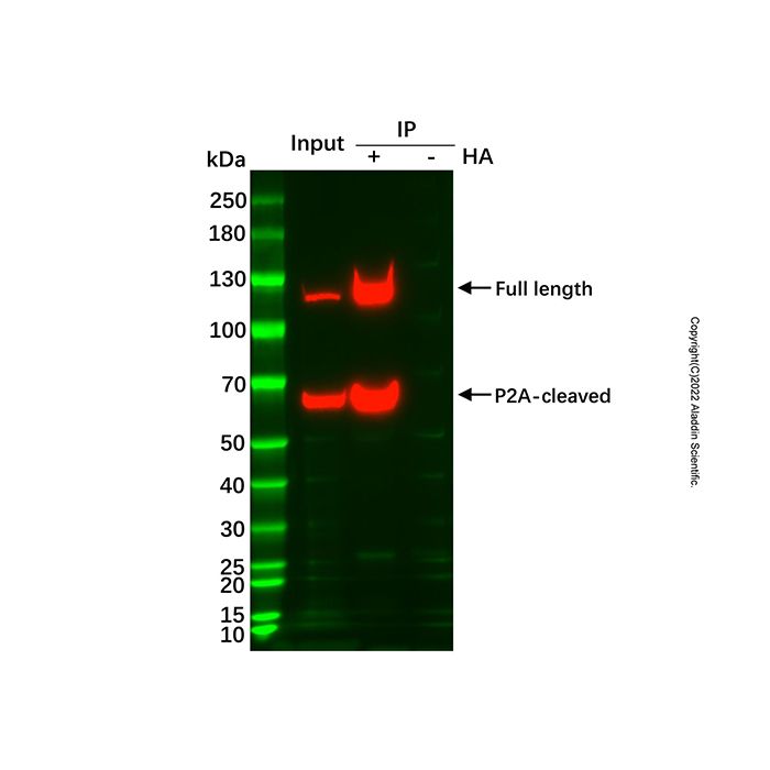
1. HA tag IP Experiment: The sample is an intracellularly expressed protein containing both His and HA tags.
2. Take 0.5 mL of suspension cells (≈1.5×10⁶ cells), wash three times with 1×PBS (pH 7.4) at room temperature, then quickly perform lysis and centrifugation.
3. Add 20 μL of pre-washed magnetic beads to the post-centrifugation supernatant and incubate with shaking for 30 minutes.
4. Transfer the tube containing the beads to a magnetic stand to separate beads from liquid. Aspirate the supernatant, leaving the beads.
5. Wash with Wash Buffer five times, then add 1× SDS Loading Buffer and heat at 99°C for 10 minutes before proceeding to WB.
6. The detection antibody used was Anti-His-HRP mAb. The negative control (-) used blank polymer magnetic beads (not conjugated with Anti-HA antibody) incubated with the sample. Results are shown in Figure 2.

Figure 2: Western blot after IP using UltraBio™ Anti-HA Magnetic Beads.
Figure 2 demonstrates that UltraBio™ Anti-HA Magnetic Beads effectively capture the HA-tagged fusion protein from the sample, with no non-specific protein binding.

 ZJ26F0434482