Lipids are a general term for neutral fats, lipoids and their derivatives, which are insoluble in water but soluble in organic solvents. There are two major categories of lipids in the human body. The first is storage lipids, represented by neutral fats (triglycerides), which are mainly distributed in subcutaneous tissue, kidneys, pancreas and other tissues. The second category is structural lipids, namely lipoids including phospholipids, glycolipids, cholesterol and more, predominantly located inside cells.
Lipid droplets serve as the primary intracellular storage compartment for neutral fats. Under physiological conditions, lipid droplets are barely visible under light microscopy in all cell types except adipocytes. Massive accumulation of lipid droplets in the cytoplasm indicates fatty degeneration, which is commonly observed in hepatocytes, cardiomyocytes, renal tubular epithelial cells and other cell types.
Oil Red O is a lipophilic azo dye with powerful liposolvent and lipid-staining properties. It specifically stains intracellular neutral triglycerides, lipoids, lipoproteins and other lipid components in tissues and cells, delivering superior staining performance compared with classic dyes such as Sudan II, Sudan III, Sudan IV and Sudan Black B. During Oil Red O staining, the dye exhibits higher solubility in cellular lipids than in its original organic solvent, driving the migration of Oil Red O from the solvent into lipid compartments to produce distinct staining.
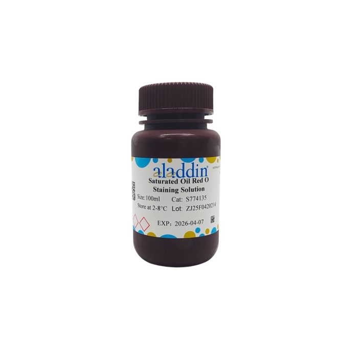
Saturated Oil Red O Staining Solution detects fatty degeneration and abnormal deposition of lipoids in tissues and organs by visualizing intracellular lipid droplets. It can also identify tumors arising from adipose tissue and determine their pathological properties. This product is compatible with both tissue section staining and lipid profiling analysis of cellular samples. Lipid-positive staining ranges from orange-yellow to deep red depending on lipid abundance. Manufactured with high-quality imported raw materials, this reagent delivers clear, stable staining results with simple operating procedures. Approximately 33 mL of working solution can be prepared from 20 mL saturated Oil Red O stock solution, and 100 mL saturated stock solution yields roughly 166 mL working solution.
Instructions for Use
I. Preparation of Working Solution
Mix saturated Oil Red O staining solution with distilled water at a volume ratio of 3:2 to prepare Oil Red O working solution. Homogenize the mixture, let it stand at room temperature for 5–10 minutes, and filter before use.
Note: Oil Red O working solution is unstable and shall not be prepared in advance.
II. Staining Protocols
1. Frozen Sections
1.1 Fix prepared frozen sections for 10 minutes with 10% neutral formalin, or skip fixation entirely; rinse thoroughly with distilled water.
1.2 Immerse slides in 60% isopropanol for 20–30 seconds.
1.3 Stain with Oil Red O working solution for 10–15 minutes. Adjust staining time according to expected staining intensity and experimental requirements.
1.4 Differentiate with 60% isopropanol until stroma is clearly visible, then briefly rinse with distilled water.
1.5 Counterstain nuclei with Mayer’s hematoxylin staining solution for 1–5 minutes. Adjust staining time as needed.
1.6 (Optional) Briefly differentiate slides with 1% hydrochloric acid solution.
1.7 (Optional) Rinse with running tap water for 10 minutes or bluing in dilute lithium carbonate solution.
1.8 Briefly rinse with distilled water.
1.9 Blot excess liquid around sections with filter paper, mount slides with glycerin gelatin, and observe under a microscope.
2. Cultured Cells
2.1 Aspirate cell culture medium, wash cells twice with PBS, and fix cells with 10% neutral formalin fixative for 20–30 minutes.
2.2 Discard fixative and rinse cells twice with distilled water.
2.3 Immerse cells in 60% isopropanol for 5 minutes.
2.4 Remove 60% isopropanol, add sufficient Oil Red O working solution to fully cover the cell substrate, and incubate for 10–20 minutes. Adjust staining time according to expected staining intensity and experimental requirements.
2.5 Discard staining solution, rinse cells with distilled water 2–5 times to remove residual dye.
2.6 Counterstain nuclei with Mayer’s hematoxylin staining solution for 1–2 minutes. Adjust staining time as needed; rinse cells 2–5 times with distilled water after discarding dye solution.
2.7 Differentiate with 1% hydrochloric acid solution for 1 minute, then discard the solution.
2.8 Add distilled water to cover cells and observe under a microscope.
3. Cell Smears
3.1 Prepare fresh bone marrow or blood smears, and fix smears in 10% neutral formalin for 10–15 minutes.
3.2 Take out smears and air-dry in circulating air for 10–15 minutes.
3.3 Immerse smears in Oil Red O working solution for 15 minutes. Adjust staining time according to expected staining intensity and experimental requirements.
3.4 Differentiate with 60% isopropanol for 20–30 seconds, flush with running water, then briefly rinse with distilled water.
3.5 Counterstain nuclei with Mayer’s hematoxylin staining solution for 1–2 minutes. Adjust staining time as needed.
3.6 Differentiate with 1% hydrochloric acid solution for 1 minute, then briefly rinse with distilled water.
3.7 Blot excess liquid around smears with filter paper, mount with glycerin gelatin, and observe under a microscope.
III. Staining Results
| Target Component | Staining Color | | Neutral fats | Orange-yellow to red | | Phospholipids | Pink | | Cell nuclei | Blue |
|
Precautions
1.Oil Red O working solution is unstable and prone to precipitate; it must not be prepared in advance. The working solution can be reused, but any leftover solution must be discarded at the end of the day.
2.Organic solvents dissolve intracellular lipids. Therefore, fixatives containing ethanol are prohibited for sample fixation. Use 10% neutral formalin or 10% formalin if fixation is required. Paraffin sections are not applicable; frozen sections or carbowax sections should be adopted instead.
3.For frozen sections of adipose tissue, a lower freezing temperature is required compared with regular specimens. Sections cannot be cut too thin, as excessive thinness will cause lipid loss. The recommended section thickness ranges from 6 to 10 µm.
4.Fully mature and plump adipocytes are fragile and prone to rupture, resulting in lipid droplet leakage. Extra care should be taken during slide pressing and sectioning.
5.Specimens mounted with glycerin gelatin have a short shelf life. For long-term preservation, seal the edges between the coverslip and slide with neutral balsam.
6.Excessive evaporation of staining reagent during incubation will lead to background precipitates. Ensure tissues and cells are fully submerged in staining solution throughout the staining process.
7.Differentiate with 60% isopropanol under microscopic observation until adipose tissue turns bright red and the stroma appears colorless. Counterstaining with hematoxylin is recommended to improve staining contrast.
8.A pilot test with 1–2 specimens is recommended for first-time use of this reagent.
9.This product is a saturated Oil Red O stock solution, so a small amount of precipitate is normal. Filtration is mandatory after preparing the working solution before use.
10.For personal safety and health protection, wear a lab coat and disposable gloves during all operations.

 ZJ26F0838327