
FeRhoNox-1 (Fe²⁺ Indicator) Fluorescent Probe Instruction Manual — Practical Guide for Specific Detection of Intracellular Fe²⁺ in Live Cells
FeRhoNox-1 (Fe²⁺ Indicator) Fluorescent Probe Instruction Manual — Practical Guide for Specific Detection of Intracellular Fe²⁺ in Live Cells
Product Description
FeRhoNox-1 (also known as RhoNox-1) is a fluorescent probe designed for detecting labile ferrous ions (Fe²⁺) in live cells. The probe exhibits good cell membrane permeability and high metal ion selectivity, making it suitable for fluorescence imaging of intracellular Fe²⁺. It preferentially localizes to the Golgi apparatus.
FeRhoNox-1 shows very weak fluorescence in the absence of Fe²⁺. Upon specific reaction with Fe²⁺, it irreversibly generates an orange-red fluorescent product with the following typical spectral characteristics:
(a) Maximum absorption wavelength (Abs max): approximately 540 nm
(b) Maximum emission wavelength (FL max): approximately 575 nm
At 37°C, FeRhoNox-1 reacts with Fe²⁺ for about 1 h, resulting in a pronounced increase in fluorescence intensity (see Figure 1). Within physiologically relevant concentration ranges, ferric ions (Fe³⁺) and other common divalent metal ions generally do not significantly enhance its fluorescence signal, indicating high selectivity toward Fe²⁺ (see Figure 2: Selectivity and reaction specificity of FeRhoNox-1).
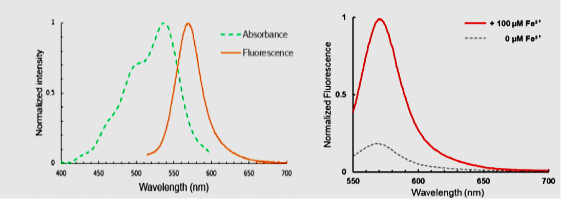
Figure 1. Spectral characteristics of FeRhoNox-1 Schematic absorption and emission spectra of the product formed after FeRhoNox-1 reacts with Fe²⁺. The maximum fluorescence emission peak is around 575 nm.
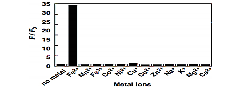
Figure 2. Selectivity of FeRhoNox-1 Comparison of fluorescence responses of FeRhoNox-1 in the presence of common metal ions. A marked fluorescence increase is observed only in the presence of Fe²⁺.
Workflow for Fluorescent Staining and Detection of Intracellular Fe²⁺ in Live Cells
I. Reagents and Materials Required
1. High-quality DMSO
(1) Cell culture grade or ultrapure DMSO.
(2) It is strongly recommended to aliquot high-purity DMSO into single-use vials and store at −80°C. Thaw immediately before use.
(3) DMSO is highly hygroscopic and may slowly degrade over time. Degraded DMSO may increase the background signal of FeRhoNox-1.
2. Cell-compatible neutral buffer (phenol red-free)
Examples:
(1) PBS (pH 7.2–7.4, without Ca²⁺/Mg²⁺ or with ions as needed).
(2) HBSS (Hank's Balanced Salt Solution).
(3) Other neutral buffers suitable for cell handling.
Notes:
(4) The buffer should be phenol red-free to avoid interference with fluorescence detection.
(5) It is recommended to use serum-free buffer for staining and washing steps to minimize the impact of serum proteins on iron distribution and signal readout.
II. Probe Preparation (FeRhoNox-1 Stock and Working Solutions)
Reconstitution of Powder to 1 mM Stock Solution
1. Remove FeRhoNox-1 from a −20°C freezer and allow it to equilibrate to room temperature for at least 30 min.
2. Place the vial in a microcentrifuge and spin briefly at low speed to collect all powder at the bottom of the tube.
3. Add 109 μL of high-quality DMSO to one vial of FeRhoNox-1 (50 μg).
4. Pipette up and down at least 5 times until the powder is completely dissolved to obtain a 1 mM FeRhoNox-1 stock solution in DMSO.
5. If the stock solution cannot be used up in a single experiment:
(a) Aliquot the stock into light-protected microtubes according to single-use volume.
(b) Store the aliquots at −80°C, protected from light.
(c) Avoid repeated freeze–thaw cycles.
It is recommended to consume a single aliquot in one experiment to ensure optimal signal-to-noise ratio and reproducibility.
III. Preparation of Working Solution
Before starting the actual experiment, dilute the 1 mM FeRhoNox-1 stock solution with HBSS or another neutral buffer to the desired working concentration, for example:
(1) 5 μM FeRhoNox-1 working solution
(a) Example preparation: Add 5 μL of 1 mM stock solution to 1 mL of buffer to obtain a 5 μM working solution.
Important Notes:
(2) The FeRhoNox-1 working solution should be prepared freshly and used within a short period of time.
(3) Avoid prolonged exposure of the working solution to room temperature or strong light to prevent probe degradation and increased background fluorescence.
IV. Cell Staining Procedure
The following procedure uses adherent cells cultured in glass-bottom dishes as an example. Adjust as needed according to the actual experimental system.
1. Removal of culture medium
(a) Retrieve the cell culture dish from a 37°C incubator and carefully aspirate the culture medium.
2. Cell washing
(a) Add an appropriate volume of pre-warmed HBSS or PBS (phenol red-free, serum-free) to gently cover the cells.
(b) Gently rock the dish and aspirate the buffer. Repeat the wash twice to remove residual culture medium and serum components.
3. Addition of staining working solution
(a) Add the FeRhoNox-1 staining working solution (e.g., 5 μM) to the dish, ensuring complete coverage of the cells.
4. Incubation
(a) Incubate the cells at 37°C in a 5% CO₂ incubator for 30–60 min.
(b) Different cell types may vary in sensitivity to incubation time. Optimize based on actual signal intensity and cell health.
5. Post-incubation washing
(a) After incubation, aspirate the staining solution and wash the cells 2–3 times with HBSS or PBS to remove free probe and unbound Fe²⁺.
6. Observation or downstream detection
(a) After washing, add a small volume of HBSS or an imaging-compatible buffer to cover the cells.
(b) Immediately observe the cells under a fluorescence microscope, or proceed to flow cytometry or other detection methods as required by the experimental design.
V. Fluorescence Detection Conditions
Fluorescence microscopy
(1) Excitation filter: Use a general green excitation filter set, such as those commonly used for Cy3 or tetramethylrhodamine (TMR).
(2) Emission range: The detection channel is recommended to be centered around 570–580 nm.
Laser confocal microscopy
(1) Laser excitation wavelength: Recommended lasers are 532 nm or 543 nm.
(2) Emission detection: Set the emission collection window around 570 nm. The bandwidth can be adjusted according to instrument configuration.
In practice, parameters may be fine-tuned according to the configuration of the fluorescence or confocal microscope in your laboratory to achieve the best signal-to-noise ratio.
VI. Precautions and Safety
1. Intracellular localization and signal interpretation
(a) FeRhoNox-1 preferentially localizes to the Golgi apparatus, but it may also partially reflect changes in the cytosolic labile Fe²⁺ pool.
(b) Detailed, cell-type–specific evaluation of its subcellular localization selectivity is still limited. Signal interpretation should therefore be made in conjunction with other markers and the overall experimental design.
2. Avoid acidic environments
(a) Acidic solutions can oxidize FeRhoNox-1, severely compromising the specificity and efficiency of its reaction with Fe²⁺.
(b) Whenever possible, use neutral buffers with pH 7.2–7.4 during the experiment and avoid handling the probe or samples under strongly acidic conditions.
3. Light protection to reduce photobleaching
(a) Similar to most fluorescent dyes, FeRhoNox-1 and its fluorescent product can undergo varying degrees of photobleaching under light exposure.
(b) During solution preparation, incubation, and imaging, work under low light and protect solutions and samples from strong light to slow photobleaching and improve signal stability.
4. DMSO and cytotoxicity
(a) Excessive DMSO concentrations may be cytotoxic. Ensure that the final DMSO concentration in the working solution is within a range tolerated by the cells (typically ≤0.1%–0.5%, depending on cell type).
5. Safety precautions
(a) For your safety and health, please always:
Wear a lab coat.
Wear disposable gloves.
Wear safety goggles when necessary.
(b) Handle waste liquids and solid waste in accordance with your institution’s chemical and biosafety regulations.
Aladdin: https://www.aladdinsci.com/