Introduction
Calcein-AM (acetoxymethyl ester) is a highly lipophilic and cell membrane-permeant compound. Calcein-AM itself is non-fluorescent. However, within live cells, intracellular esterases hydrolyze Calcein-AM into Calcein, which emits intense green fluorescence (Ex/Em=494nm/517nm). Therefore, Calcein-AM specifically stains live cells and reflects the state of cell viability. Propidium Iodide (PI) is a red fluorescent dye that cannot penetrate the intact membranes of live cells. Upon cell death, PI enters the cell and specifically binds to double-stranded DNA in the nucleus, emitting red fluorescence (Ex/Em=535nm/617nm).
This Live/Dead Cell Viability/Toxicity Assay Kit (Calcein AM/PI) enables simultaneous dual-fluorescence staining of live and dead cells for the detection of cell viability and cytotoxicity. The hydrolysis product of Calcein-AM, Calcein, has maximum excitation and emission wavelengths of 494 nm and 517 nm, respectively. The PI-DNA complex has maximum excitation and emission wavelengths of 535 nm and 617 nm, respectively.
Product Information
V1373482 | Components | 100T | 500T | Storage | Quantity Per Test | V1373482A | Calcein AM Staining Solution | 100μL | 500μL | -20℃. Store in the dark. | 1μL per 0.5-1.0 × 10⁶ cells | V1373482B | PI Staining Solution | 500μL | 2500μL | -20℃. Store in the dark. | 5μL per 0.5-1.0 × 10⁶ cells | V1373482C | Calcein AM/PI Dilution buffer | 10mL | 50mL | 2-8℃ | 0.1mL per 0.5-1.0 × 10⁶ cells |
Note: The recommended number of cells to stain per test is 0.5-1.0 × 10⁶ cells. |
|
Procedure
I. Pre-experiment to Determine Optimal Staining Concentration (Optional)
As optimal staining conditions vary depending on cell type and density, a pre-experiment is recommended to determine the best concentrations for Calcein-AM and PI.
(1) Take out the reagents and allow them to reach room temperature. Dilute the Dilution Buffer 10-fold with double-distilled water before use.
(2) Prepare cell samples: Prepare live and dead cell samples on coverslips.
Note: Dead cells can be prepared by treating with 0.1% saponin for 10 min, 0.1-0.5% digitonin for 10 min, or 70% methanol for 30 min.
(3) Determine PI working concentration: Test PI concentrations ranging from 0.1-10 μM on dead cell samples. Select the concentration that results in bright red nuclear staining of dead cells with minimal cytoplasmic staining.
(4) Determine Calcein-AM working concentration: Test Calcein-AM concentrations ranging from 0.1-10 μM on dead cell samples. Choose the concentration that does not produce significant fluorescence in the cytoplasm of dead cells. Then verify on live cells that this concentration produces sufficient fluorescence; increase the concentration appropriately if fluorescence is insufficient.
II. Example Dilution Protocol (for preparing 10 mL of Staining Working Solution)
1. Fluorescence Microscopy Detection
(1) Take out the reagents and allow them to reach room temperature. Dilute the Dilution Buffer 10-fold with double-distilled water before use.
(2) Prepare Calcein AM/PI Staining Working Solution: The concentration of Calcein-AM stock solution in this product is 2 mM, and the PI stock solution is 1.5 mM. Add 50 μL of PI stock solution and 10 μL of Calcein-AM stock solution to 10 mL of Dilution Buffer. Vortex to mix thoroughly to obtain 10 mL of Calcein AM/PI Staining Working Solution.
(3) For adherent cells: Wash 2-3 times with 1x PBS before staining.
For suspension cells: Centrifuge at 500 x g for 5 min at room temperature, collect the cell pellet for staining.
(4) Wash cells thoroughly 2-3 times with 1x PBS to sufficiently remove residual esterase activity.
(5) For adherent cells: Add a sufficient amount of Calcein AM/PI Staining Working Solution. For example, for adherent cells cultured in a 6-well plate with >80% confluency, it is recommended to add 1 mL/well of staining working solution. Optimize according to your specific experimental system.
For suspension cells: Add an appropriate amount of staining working solution to resuspend the cells, adjusting the cell density to 10^6 cells/mL.
(6) Incubate at room temperature protected from light for 10-45 minutes.
Note: The optimal incubation time varies for different cell types. Start with 10 min as the initial incubation time, and subsequently adjust and optimize based on the actual staining results.
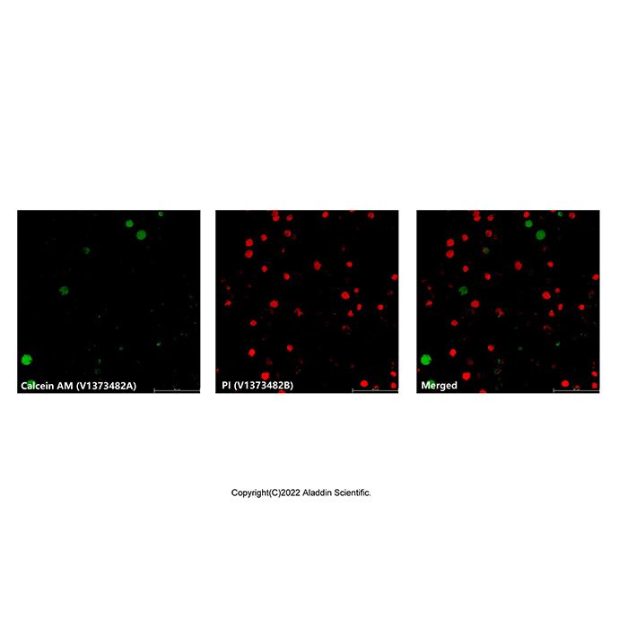
(7) Observe cells under a fluorescence microscope using Ex/Em=494/517nm and Ex/Em=535/617nm. Green fluorescence indicates live cells, red fluorescence indicates dead cells.
2. Fluorescence Microplate Reader Detection
(1) Take out the reagents and allow them to reach room temperature. Dilute the Dilution Buffer 10-fold with double-distilled water before use.
(2) Prepare Calcein AM/PI Staining Working Solution as described in step 1.(2).
(3) Culture an appropriate number of adherent or suspension cells in a black 96-well microplate.
(4) Wash cells thoroughly with 1x PBS to remove residual esterase activity.
(5) For adherent cells: Add 100 μL PBS per well to wash. For suspension cells: Resuspend in 100 μL PBS, centrifuge, and aspirate supernatant. Repeat this wash step.
(6) Add 100 μL of Staining Working Solution to each well. Gently shake the plate to ensure even coverage.
(7) Incubate at room temperature protected from light for 10-45 minutes.
Note: As above, optimize incubation time based on cell type.
(8) Read the plate using a microplate reader:
Detect green fluorescence intensity at Ex/Em=494/517nm.
Detect red fluorescence intensity at Ex/Em=535/617nm.
Calculating Live/Dead Cell Ratio Using Controls
For more precise calculation of the ratio of dead to live cells, set up controls.
(1) Culture and treat cells as before.
(2) In addition to the Calcein AM/PI Staining Working Solution, prepare separate Calcein-AM working solution and PI working solution for control measurements, using the same dilution method.
(3) Sample and control group setup:The live cell control group consists of cells without drug treatment.The dead cell control group can be prepared using methods like 0.1% saponin treatment for 10 min, etc. Ensure all groups have the same cell number, working solution concentration, incubation time, and temperature.
No. | Group | Staining Solution | Excitation | Emission | Result Designation | (1) | Sample Group | Calcein AM/PI | 494 nm | 517 nm | F(517)sam | (2) | Sample Group | Calcein AM/PI | 535 nm | 617 nm | F(617)sam | (3) | Live Cell Control | PI only | 494 nm | 517 nm | F(517)min | (4) | Live Cell Control | Calcein AM only | 494 nm | 517 nm | F(517)max | (5) | Dead Cell Control | PI only | 535 nm | 617 nm | F(617)max | (6) | Dead Cell Control | Calcein AM only | 535 nm | 617 nm | F(617)min | (7) | Cell-free Blank | Calcein AM/PI | 494 nm | 517 nm | F(517)₀ | (8) | Cell-free Blank | Calcein AM/PI | 535 nm | 617 nm | F(617)₀ |
|
(4) Calculate the percentage of live cells and dead cells based on the detection data:
% Live cell=(F(517)sam-F(517)max)/(F(517)max-F(517)min)×100%
% Dead cell=(F(617)sam-F(617)max)/(F(617)max-F(617)min)×100%
Note: All F(517) and F(617) values in the formulas should be subtracted by their corresponding background values (F(517)₀ and F(617)₀) before calculation.
(5) To determine the absolute number of live and dead cells, create standard curves using different numbers of pure live and dead cells, measuring fluorescence at 517 nm and 617 nm. Using the linear standard curves and the sample fluorescence intensities, calculate the numbers of live and dead cells.
Flow Cytometry Detection
(1) Take out the reagents and allow them to reach room temperature. Dilute the Dilution Buffer 10-fold with double-distilled water before use.
(2) Prepare Calcein AM/PI Staining Working Solution as described in step 1.(2).
(3) Wash cells thoroughly 2-3 times with 1x PBS.
(4) Resuspend cells in 1 mL of Staining Working Solution, adjusting the cell density to 0.5-1.0x10^6 cells/mL.
Note: It is recommended to prepare two additional cell samples, each stained with only one dye (Calcein-AM only or PI only), for compensation adjustment in flow cytometry. Also prepare a cell sample with buffer only as a negative control.
(5) Incubate at room temperature protected from light for 15-20 minutes.
(6) Analyze cell viability by flow cytometry within 1-2 hours. Calcein-AM can be excited by the 488 nm laser, with emission detected around 530 nm. PI emission is detected around 617 nm.
Note: When gating cells, exclude debris. Use the single-stained controls to adjust compensation. The double-stained sample should show two distinct populations: live cells (green fluorescence) and dead cells (red fluorescence).
Precautions
1. For your safety and health, wear a lab coat and disposable gloves.
2. Fluorescent dyes are susceptible to quenching. It is recommended to complete detection on the same day after staining.

 G2601310