
Antibody Fluorescent Labeling Techniques and Dye Selection Guide
Antibody Fluorescent Labeling Techniques and Dye Selection Guide
Antibody fluorescent labeling introduces fluorophores or reporter systems into antibody molecules via covalent conjugation or other stable linkage strategies, enabling target antigens to yield imageable and quantitatively measurable signals. It is a foundational technology for platforms such as flow cytometry, immunofluorescence, and confocal imaging. Because samples differ in autofluorescence and instruments differ in optical configurations, and because multicolor panel strategies vary in their sensitivity to excitation/emission channel matching, background interference, photostability, and spectral crosstalk, the choice of dyes and labeling systems directly determines signal-to-noise ratio, compensation burden, and interpretability of results. Using rapid labeling of 100 μg antibody as an example workflow, standardized kits combined with matched purification tools can reduce operational complexity and improve labeling consistency, thereby supporting rapid panel iteration and cross-lot comparable analyses.
Keywords: antibody fluorescent labeling; DOL; multicolor flow cytometry; immunofluorescence; far-red to near-infrared channels; purification and desalting
I. Technical Background of Antibody Fluorescent Labeling
1.1 Technical positioning and typical applications
Antibody fluorescent labeling is a key detection and imaging technology in life science research. By introducing fluorescent dyes or reporter systems into antibody molecules, it enables highly sensitive, visual, and quantitative detection of target antigens and is widely used in:
(1) Flow cytometry
(2) Immunofluorescence (IF/ICC/IHC)
(3) Confocal microscopy and super-resolution imaging
(4) Protein interaction studies and spatial-omics–assisted validation
(5) Multicolor detection and high-throughput analysis
1.2 Why selection determines experimental quality
Different experimental systems have markedly different requirements for excitation/emission channels, brightness, photostability, background noise, multicolor compatibility, and tissue penetration. Dye or reporter selection often determines signal-to-noise ratio, compensation burden, and interpretability. Common risks of suboptimal selection include:
(1) Signal–background mismatch
- Insufficient signal for low-abundance targets.
- Tissue autofluorescence masking true signal.
(2) Inadequate multicolor compatibility
- Spectral overlap leading to channel spillover and difficult compensation.
- Spillover spreading increasing resolution loss and reducing population separability.
(3) Insufficient data stability
- Photobleaching or system variability reducing reproducibility.
- Improper labeling conditions impairing antibody functionality or increasing background.
II. Design Rationale and Core Features of Aladdin 100 μg Antibody Labeling Kits
2.1 Coverage and design objectives
(1) Small-molecule fluorescent antibody labeling kits: designed to label antibodies with small-molecule fluorophores, covering UV/violet through far-red and near-infrared extended channels.
(2) Fluorescent protein/tandem labeling kits: designed for PE, APC, PerCP, and related tandem systems.
(3) HRP labeling kits: designed for enzyme conjugation to antibodies, typically distinguished by SMCC-based methods versus sodium periodate–based methods.
Because the chemical routes and QC criteria differ across these systems, selection should be driven by the target platform and channel plan, and parameters should be confirmed against the relevant instructions for use and certificates of analysis.
2.2 General characteristics of small-molecule fluorescent labeling
In small-molecule fluorophore antibody labeling, kit design typically emphasizes:
(1) Mild reaction conditions and stable linkage (typically covalent conjugation)
- Compatible with common IgG labeling requirements.
- Produces stable fluorescent conjugates suitable for flow cytometry and imaging.
(2) Controllability of degree of labeling (DOL)
- DOL that is too low may yield insufficient signal.
- DOL that is too high may cause self-quenching, increased hydrophobicity, and elevated background.
(3) Streamlined workflow and reproducibility
- Integrates labeling and purification into a continuous process.
- Reduces dependence on specialized instruments and improves laboratory-to-laboratory reproducibility.
2.3 Advantages of Aladdin Products
(1) High dose matching efficiency
- Fits common research antibody usage and supports rapid multicolor panel iteration.
- Reduces reagent waste and improves overall cost-effectiveness.
(2) Broad applicability
- Monoclonal and polyclonal antibodies.
- Common IgG subclasses (mouse, human, rabbit, etc.).
(3) Supports R&D workflows and rapid iteration
- Enables parallel multi-channel labeling of the same antibody clone for panel optimization and channel replacement.
(4) Performance Validation and Experimental Data
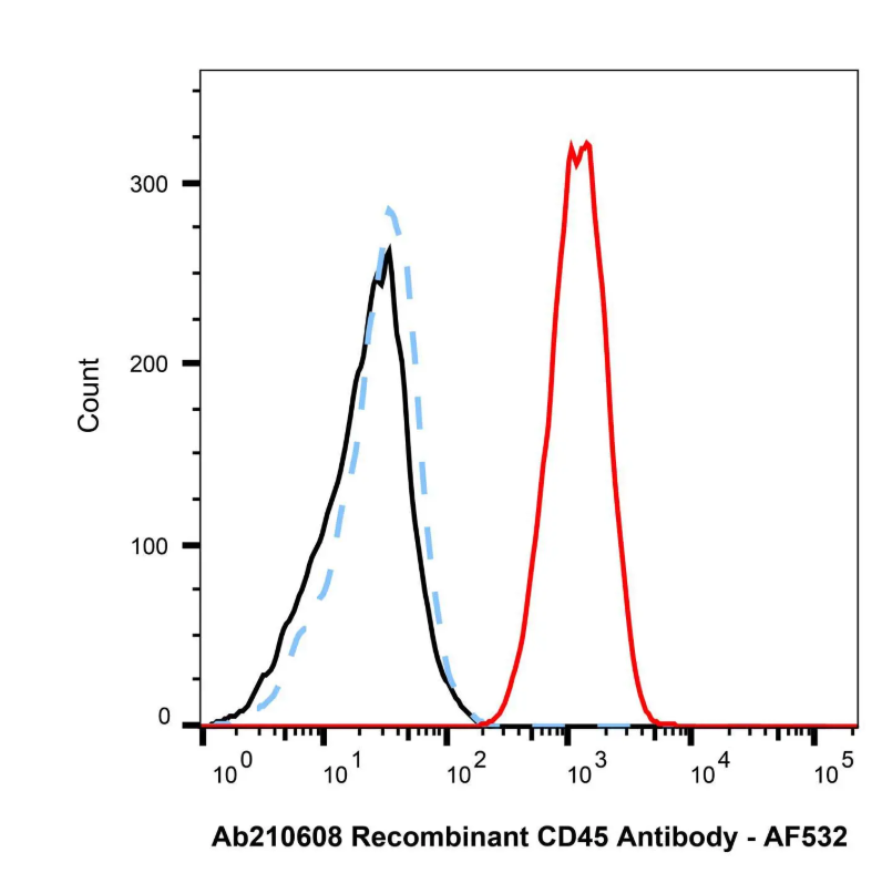
Flow Cytometry: AF532 Antibody Labeling Kit (A1491935)
Cytometry analysis of Jurkat cells stained with 0.5 μg/mL Recombinant CD45 Antibody (Ab210608). Followed by a goat anti-mouse IgG conjugated to AF532 using the AF532 Antibody Labeling Kit (A1491935) at 0.1 μg/mL for 1 hour at room temperature in the dark. Blue - Isotype control, Mouse IgG (Ab170221). Black - Unlabelled control, cells without incubation with primary antibody.
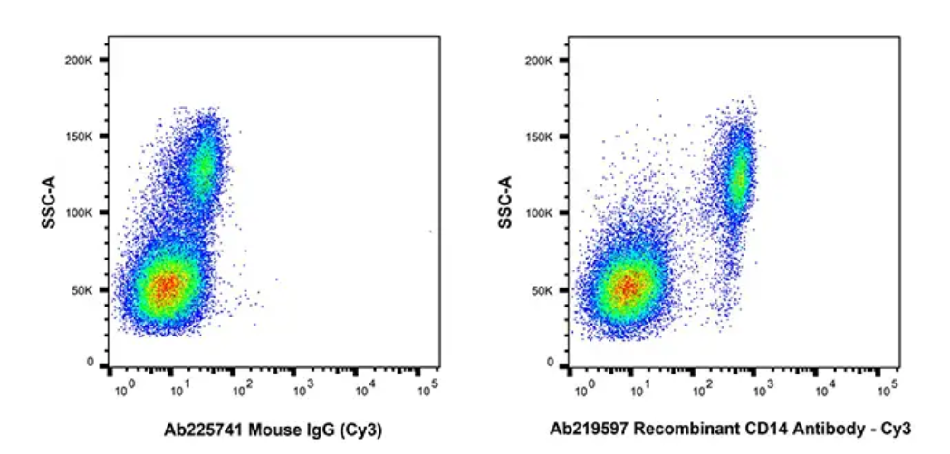
Flow Cytometry: Cy3 Antibody Labeling Kit (C1491949)
Flow cytometry analysis of CD14 expression by human peripheral blood cells (right panel) compared to an isotype control (left panel). Cells were preincubated with Human Fc Receptor Blocking Solution (H1374078). The cells were then stained with Recombinant CD14 Antibody-Cy3 prepared using the Cy3 Antibody Labeling Kit (C1491949) at 0.1μg/test for 1 hour at 4°C.
III. Spectral Properties and Typical Use Scenarios of Different Fluorophore Series
3.1 UV/violet channels (low background; entry channels for multicolor expansion)
(1) Representative dyes/systems
- AF350 (Ex/Em ≈ 346/442 nm; typically requires UV/355 nm excitation or can be detected in DAPI filter sets).
- Pacific Blue (Ex/Em ≈ 410/455 nm; compatible with 405 nm violet excitation).
(2) Typical applications
- Entry channels and channel expansion in multicolor designs.
- Flow cytometry or immunofluorescence under low-autofluorescence conditions.
3.2 Green channel (broad compatibility; mature ecosystems)
(1) Representative dyes
- AF488, Oregon Green 488, FITC.
(2) Typical applications
- Primary workhorse channels in flow cytometry.
- Routine imaging channels for immunofluorescence and immunohistochemistry.
(3) Risk note
- If green autofluorescence is high in certain tissues or cell types, background elevation risk should be evaluated.
3.3 Yellow–orange channels (panel “filler” channels for multicolor designs)
(1) Representative dyes
- AF532, AF555, Cy3.
(2) Typical applications
- Filling the spectral gap between green and red channels.
- Multichannel imaging and high-parameter flow cytometry expansion.
3.4 Red/far-red channels (high contrast; trend toward lower background)
(1) Representative dyes
- AF594, AF647, Cy5.
(2) Typical applications
- Preferred channels for tissue imaging to mitigate autofluorescence.
- High-contrast channels commonly used in flow cytometry and confocal microscopy.
3.5 Far-red to near-infrared extended channels (panel expansion and low-background options)
(1) Representative dyes
- AF660, AF680 (far-red to near-infrared transition); AF700, AF750 (near-infrared extension).
(2) Typical applications
- High-parameter flow cytometry panel expansion.
- Additional channels for thick samples or high-background conditions (instrument configuration must be evaluated).
3.6 Related product overview
(1) 100 μg antibody labeling kits
Catalog No. | Product Name | Category | Spectral Channel | Typical Platforms |
AF350 Antibody Labeling Kit | Small-molecule fluorescence (antibody fluorescent labeling) | UV channel (UV/355 or DAPI filter set) | IF/ICC; multicolor entry channel | |
Pacific Blue Antibody Labeling Kit | Small-molecule fluorescence (antibody fluorescent labeling) | Violet channel (405 nm) | Flow cytometry; low-autofluorescence systems | |
AF488 Antibody Labeling Kit | Small-molecule fluorescence (antibody fluorescent labeling) | Green channel | Flow cytometry; IF/ICC/IHC | |
Oregon Green 488 Antibody Labeling Kit | Small-molecule fluorescence (antibody fluorescent labeling) | Green channel | Flow cytometry; IF/ICC | |
FITC Antibody Labeling Kit | Small-molecule fluorescence (antibody fluorescent labeling) | Green channel | Routine microscopy; basic flow cytometry panels | |
AF532 Antibody Labeling Kit | Small-molecule fluorescence (antibody fluorescent labeling) | Yellow–orange channel | Multicolor flow; multichannel imaging | |
AF555 Antibody Labeling Kit | Small-molecule fluorescence (antibody fluorescent labeling) | Yellow–orange channel | Multicolor flow; IF/ICC | |
Cy3 Antibody Labeling Kit | Small-molecule fluorescence (antibody fluorescent labeling) | Yellow–orange channel | Traditional filter sets; imaging | |
AF594 Antibody Labeling Kit | Small-molecule fluorescence (antibody fluorescent labeling) | Red channel | Tissue imaging; stringent background control | |
AF647 Antibody Labeling Kit | Small-molecule fluorescence (antibody fluorescent labeling) | Far-red channel | Core flow channel; confocal imaging | |
Cy5 Antibody Labeling Kit | Small-molecule fluorescence (antibody fluorescent labeling) | Far-red channel | Traditional far-red; flow/imaging | |
AF660 Antibody Labeling Kit | Small-molecule fluorescence (antibody fluorescent labeling) | Far-red/NIR extension | High-parameter flow expansion; low background | |
AF680 Antibody Labeling Kit | Small-molecule fluorescence (antibody fluorescent labeling) | Far-red/NIR extension | High-parameter flow expansion; low background | |
AF700 Antibody Labeling Kit | Small-molecule fluorescence (antibody fluorescent labeling) | NIR extension | High-parameter flow expansion; panel iteration | |
AF750 Antibody Labeling Kit | Small-molecule fluorescence (antibody fluorescent labeling) | NIR extension | High-parameter flow expansion; low background | |
P1491951 | PE Labeling Kit | Fluorescent protein/tandem system (antibody labeling) | Phycobiliprotein channel (PE) | Flow cytometry high-brightness channel |
P1491952 | PE-Cy5 Labeling Kit | Fluorescent protein/tandem system (antibody labeling) | Tandem system (PE-Cy5) | Flow cytometry tandem expansion |
P1491953 | PE-Cy7 Labeling Kit | Fluorescent protein/tandem system (antibody labeling) | Tandem system (PE-Cy7) | Flow cytometry tandem expansion |
A1491946 | APC Labeling Kit | Fluorescent protein/tandem system (antibody labeling) | Phycobiliprotein channel (APC) | Flow cytometry; panel core channel |
P1491954 | PerCP Labeling Kit | Fluorescent protein/tandem system (antibody labeling) | PerCP complex channel (Ex ≈ 482/488 nm; Em ≈ 675–677 nm) | Flow cytometry; classic long-emission channel |
H1506714 | HRP Labeling Kit (SMCC method) | Enzyme labeling (HRP conjugation) | Enzymatic readout | IHC/ELISA and related assays |
M1508746 | HRP Labeling Kit (SMCC method) | Enzyme labeling (HRP conjugation) | Enzymatic readout | IHC/ELISA and related assays |
P1506712 | HRP Labeling Kit (Sodium periodate method) | Enzyme labeling (HRP conjugation) | Enzymatic readout | IHC/ELISA and related assays |
P1508744 | HRP Labeling Kit (Sodium periodate method) | Enzyme labeling (HRP conjugation) | Enzymatic readout | IHC/ELISA and related assays |
(2) Core reagents/coupling components for labeling
Catalog No. | Product Name | Category (Use classification) |
LumiDye™ AF488 NHS Ester | NHS ester dye | |
LumiDye™ AF532 NHS Ester | NHS ester dye | |
LumiDye™ AF546 NHS Ester | NHS ester dye | |
LumiDye™ AF555 NHS Ester | NHS ester dye | |
LumiDye™ AF568 NHS Ester | NHS ester dye | |
LumiDye™ AF594 NHS Ester | NHS ester dye | |
LumiDye™ AF647 NHS Ester | NHS ester dye | |
LumiDye™ AF660 NHS Ester | NHS ester dye | |
LumiDye™ AF680 NHS Ester | NHS ester dye | |
LumiDye™ AF700 NHS Ester | NHS ester dye | |
LumiDye™ AF750 NHS Ester | NHS ester dye | |
M1491723 | LumiDye™ SMCC Activated Phycoerythrin (SMCC-PE) | SMCC-activated component |
M1491727 | LumiDye™ SMCC Activated PE-Cy5 (SMCC-PE-Cy5) | SMCC-activated component |
M1491729 | LumiDye™ SMCC Activated PE-Cy7 (SMCC-PE-Cy7) | SMCC-activated component |
M1491730 | LumiDye™ SMCC Activated APC (SMCC-APC) | SMCC-activated component |
M1491733 | LumiDye™ SMCC Activated PerCP (SMCC-PerCP) | SMCC-activated component |
M1491737 | LumiDye™ SMCC Activated HRP (SMCC-HRP) | SMCC-activated component |
LumiDye™ PE-Cy5 | Tandem component | |
LumiDye™ PE-Cy7 | Tandem component | |
LumiDye™ APC-Cy7 | Tandem component | |
R-Phycoerythrin | Fluorescent protein/supporting component | |
Cross-linked Allophycocyanin | Fluorescent protein/supporting component | |
PerCP | Fluorescent protein/supporting component | |
PerCP-Cy5.5 | Tandem component | |
Biotinamidohexanoic acid 3-sulfo-N-hydroxysuccinimide ester sodium salt | Biotinylation reagent (NHS ester) |
(3) Supporting purification/desalting consumables
Catalog No. | Product Name | Typical Use | Selection Notes |
G1506506 | G25 Mini Desalting Column | Buffer exchange before labeling; removal of free dye after labeling | Better for small-volume samples and scenarios requiring minimal dilution |
G1506505 | G25 Spin Desalting Column | Buffer exchange before labeling; removal of free dye after labeling | Better for routine workflows and fast desalting/purification with efficiency priority |
G1506507 | G25 Max Desalting Column | Buffer exchange before labeling; removal of free dye after labeling | Better for larger processing volumes or higher-throughput needs |
IV. How to Select an Appropriate Labeling Strategy and Channel Combination
4.1 Selection by experimental platform
(1) Flow cytometry
① Recommended channel-allocation principles
Assign low-abundance targets preferentially to high-brightness, low-background channels.
Assign high-abundance targets to far-red or far-red/NIR extension channels to balance dynamic range.
② QC recommendations
Use single-color compensation for high-spillover combinations and evaluate resolution changes with FMO controls.
(2) Immunofluorescence/confocal microscopy
① Recommended channel-allocation principles
For tissue sections or high-autofluorescence samples, prioritize red, far-red, or far-red/NIR channels to reduce background interference.
For long acquisition sessions or time-lapse experiments, prioritize channel combinations with superior photostability.
② Acquisition strategy recommendations
Use sequential scanning and properly defined excitation/detection windows to reduce spectral bleed-through risk.
4.2 Selection by sample background and target abundance
(1) High autofluorescence (e.g., tissue sections, tumor samples)
- Prioritize red/far-red or far-red/NIR extension channels.
- Combine with optimized blocking systems and imaging exposure strategies.
(2) Low-abundance targets (weak signals)
- Prioritize high-brightness channels.
- Optimize staining conditions and perform small-scale parameter screening.
4.3 Selection by multicolor combination and channel planning
(1) Spectral and compensation principles
- Avoid severe overlap from closely spaced emission peaks.
- Evaluate filter bandwidth and spillover-driven resolution loss during panel design.
- Consider brightness, antigen abundance, and sample background simultaneously during channel allocation.
(2) Practical combination examples
- UV/violet: AF350, Pacific Blue
- Green: AF488, Oregon Green 488, FITC
- Yellow–orange: AF532, AF555, Cy3
- Red/far-red: AF594, AF647, Cy5
- Far-red/NIR extension: AF660, AF680, AF700, AF750
V. Technical Principles: Linkage Chemistry and DOL Logic Using Small-Molecule Fluorescent Labeling as an Example
5.1 Core chemical logic of small-molecule fluorescent antibody labeling
(1) Overview of linkage mechanisms
- In small-molecule fluorescent labeling, a common strategy is to use reactive functional groups to react with accessible sites on the antibody molecule, forming stable linkages that introduce fluorophores into the antibody.
(2) Key factors influencing reaction conditions
- Reactive groups may undergo hydrolysis in aqueous environments; reaction time should be controlled and conditions kept stable.
- Reaction efficiency depends on buffer composition, pH window, antibody concentration, and related factors.
(3) Buffer-compatibility essentials
- Avoid primary-amine–containing buffers and additives that can interfere with reactions.
- Avoid protein additives that introduce additional reaction sites and increase purification burden.
5.2 Definition, significance, and common QC logic for DOL
(1) Significance of DOL
- DOL represents the average number of fluorophore molecules introduced per antibody molecule and is a key determinant of signal intensity and background.
(2) Interpretation recommendations
- Low DOL: prioritize checks on buffer exchange adequacy, antibody concentration, and reaction condition matching.
- High DOL: focus on risks of self-quenching, aggregation, and functional loss; optimize labeling conditions when necessary.
5.3 Purpose and acceptance criteria of post-labeling purification
(1) Purification objectives
- Remove free dye and other small molecules to reduce non-specific background and improve data stability.
- Improve reproducibility for compensation and quantitative analysis.
(2) Acceptance criteria
- Background reduction in blank controls and decreased non-specific signals.
- Verifiable labeled-antibody concentration and QC metrics to support cross-lot comparability.
VI. FAQ
6.1 Applicable antibodies and buffer systems
Q: Which antibodies are suitable for labeling?
A: Most IgG antibodies can be labeled provided that buffer systems and additives meet kit requirements. If antibodies are supplied in primary-amine buffers or contain protein stabilizers, buffer exchange is recommended before labeling. Details should follow the relevant kit instructions for use.
6.2 Antibody concentration and sample quality requirements
Q: What happens if the antibody concentration is too low?
A: Low antibody concentration can reduce labeling efficiency, resulting in insufficient signal or increased lot-to-lot variability. Ensure the antibody concentration falls within an appropriate range for the reaction and minimize unnecessary dilution.
6.3 Fixation, permeabilization, and downstream compatibility
Q: Can labeled antibodies be used on fixed/permeabilized samples?
A: Typically yes, but evaluation should consider antigen localization and experimental conditions. Permeabilization conditions may alter background and antigen accessibility; small-scale condition screening is recommended.
6.4 Storage and stability
Q: How should labeled antibodies be stored?
A: Store protected from light and aliquot to reduce repeated freeze–thaw cycles. Specific storage conditions and shelf life should be determined based on intrinsic antibody stability and kit guidance.
6.5 Whether additional purification is required
Q: Is additional purification needed after labeling?
A: If the kit includes a built-in step to remove free small molecules, the conjugate can typically be used directly. For applications requiring extremely low background, an additional mild purification step may be applied provided it does not cause substantial dilution.
6.6 Practical recommendations for multicolor experiments
Q: How can compensation burden be reduced in multicolor experiments?
A: During panel design, evaluate spectral overlap and filter bandwidth, quantify spillover-driven resolution loss using single-color compensation and FMO controls, allocate weak targets to high-brightness/low-background channels, and allocate strong targets to far-red or far-red/NIR extension channels.
Antibody labeling introduces detectable fluorescent or enzymatic reporter systems into antibody molecules, enabling quantitative readouts in flow cytometry and imaging platforms. In practice, selection should begin with instrument configuration and available excitation/detection channels, followed by channel allocation that accounts for spectral overlap and compensation cost in multicolor designs. Standardized purification and QC workflows should be used to control free-dye residues and labeling consistency, thereby delivering stable signal-to-noise ratios and robust interpretability for multicolor panel iteration, tissue imaging, and high-parameter analyses.
Aladdin: https://www.aladdinsci.com/