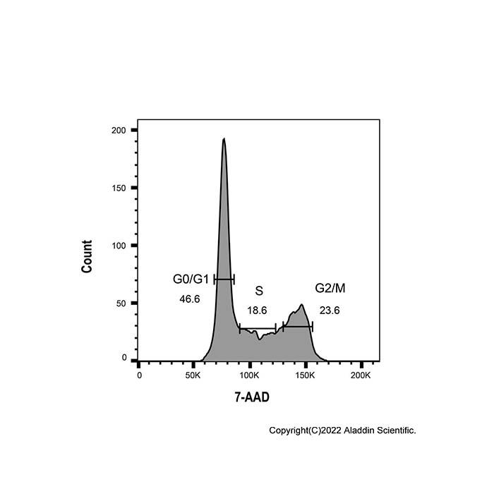
The cell cycle refers to the complete process from the completion of one cell division to the end of the next division. The cell cycle can be divided into four phases: G1 (pre-DNA synthesis phase), S phase (DNA synthesis phase), G2 phase (post-DNA synthesis phase), and M phase (mitotic phase). Based on chromosome ploidy, it is classified into three phases: G1 phase, S phase, and G2/M phase.
7-AAD (7-Aminoactinomycin D) is a fluorescent dye that binds to double-stranded DNA. When 7-AAD binds to double-stranded DNA, it produces fluorescence, and the fluorescence intensity is proportional to the amount of double-stranded DNA present. After staining intracellular DNA, the DNA content of cells can be measured using flow cytometry. Cell cycle analysis can then be performed based on the distribution of DNA content. Assuming the fluorescence intensity of G0/G1 phase cells is 1, the theoretical fluorescence intensity of G2/M phase cells, which contain twice the genomic DNA, is 2, while the fluorescence intensity of S phase cells undergoing DNA replication ranges between 1 and 2.
This kit is commonly used for cell cycle and apoptosis detection in cultured adherent or suspension cells. If it is used for cell cycle and apoptosis detection in tissues, the tissue must first be digested into a single-cell suspension before analysis can be performed.
Instructions for use
1.Collect cells and wash twice with ice-cold PBS; resuspend in ice-cold PBS at a density of 0.1–1.0 × 10⁷ cells/mL.
2.In the cell suspension, anhydrous ethanol was added dropwise while shaking until the final ethanol concentration reached 70%.
3.Mix gently and fix overnight at 4 °C.
4.Wash cells twice with ice-cold PBS, then resuspend in dyeing buffer solution of 2 × 10⁶ cells/mL.
5.Add 5 µL of 7-AAD staining solution and 20 µL RNase A to 500 µL of the cell suspension.
6.Resuspend cells gently and thoroughly, then incubate at room temperature for 30 min in the dark.
7.Analyze samples by flow cytometry.
Note: Please bring your own PBS and absolute ethanol.
Precautions
1. Fluorescent dyes all have the problem of quenching. Please try to avoid light during storage and use to slow down fluorescence quenching.
2. Iodophor is irritating to the human body. Please take appropriate protective measures.
3. When fixing cells with 70% ethanol, make sure it is thorough; otherwise, uneven staining will result in unclear or inaccurate results. It is recommended to fix the cells overnight at -4℃ with 70% ethanol.
4. After cell fixation, ensure that the cells are in a single-cell suspension; cell adhesion may affect our results.
5. For your safety and health, please wear laboratory coats and disposable gloves during operation.
6. This product is only for scientific research and cannot be used for clinical diagnosis or treatment, nor for food or medicine. It must not be stored in ordinary residences.
7. For your safety and health, please wear laboratory coats and disposable gloves during operation.