ATP is the core molecule of cellular energy metabolism, and its concentration directly reflects the energy status of an organism and its organs. As a crucial energy substance, ATP participates in and regulates various physiological and pathological processes within cells; fluctuations in its levels significantly impact cellular function. Typically, ATP levels decrease during cell apoptosis, necrosis, or under toxic conditions, while specific stimuli like high glucose can upregulate ATP levels in certain cells. A decline in ATP often indicates impaired mitochondrial function and, during apoptosis, frequently occurs concurrently with a decrease in mitochondrial membrane potential.
This kit is designed based on the catalytic principle of firefly luciferase: this enzyme requires ATP as an energy source to catalyze the light emission from luciferin. When both the luciferase and luciferin are in excess, the luminescence intensity is directly proportional to the ATP concentration within a certain range, enabling highly sensitive quantitative detection of ATP content in solutions, cells, or tissue samples.
This kit can also be used to estimate cell concentration by measuring the amount of ATP released from living cell samples. This method specifically counts viable cells, as ATP is rapidly degraded upon cell death. Typically, each viable cell contains approximately 1 pg (10⁻¹² g) or about 2 fmol (2 × 10⁻¹⁵ mol) of ATP. For a specific cell line and growth medium, a more accurate ATP-to-cell ratio can be obtained by consulting the literature or by staining cells and counting viable cells under a microscope before measuring ATP content. Using the described method, this kit can measure ATP released from up to 5 × 10⁵ viable cells (sample density of 2 × 10⁷ cells/mL). Its sensitivity is significantly superior to that of microscopy-based counting using a hemocytometer, which typically detects only about 2 × 10⁵ cells/mL.
R1375244 | Components | 100T | 200 T | Storage | Quantity Per Test |
R1375244A | ATP Assay Lysis Buffer | 50 mL | 100 mL | -20℃ | 200μL |
R1375244B | ATP Standard Solution, 0.5mM | 0.1 mL | 0.2 mL | -20℃, Store in the dark. | 20μL |
R1375244C | ATP Assay Reagent | 10 mL | 20 mL | -20℃, Store in the dark. | 100μL |
Precautions
1. The detection reagent in this kit contains luciferase. Repeated freeze-thaw cycles will lead to gradual loss of its activity. For optimal results, after the first thaw, it can be appropriately aliquoted for storage. However, ensure the aliquoting containers are free from ATP contamination.
2. Luciferase activity is sensitive to temperature. Therefore, both cells and the ATP detection reagent must be equilibrated to room temperature before the assay. Do not store the reagent at room temperature long-term.
3. ATP, especially in lysed samples, is relatively unstable at room temperature. Operations should be performed on ice or at 4°C.
4. Use white- or black-walled 96-well or 384-well plates suitable for cell culture for detection. Using standard clear plates may cause signal crosstalk between adjacent wells.
5. Use 0.2 µm filtered ultrapure water (resistivity 17 MΩ·cm or equivalent specification).
6. If dilution of sample solutions or dissolution of solid samples is required, the use of 0.2 µm filtered ultrapure water or a dilute buffer with a pH of approximately 7.8 is recommended. The use of arsenate as a sample buffer is not recommended, as it tends to reduce sensitivity through quenching. Additionally, high salt concentrations in samples can inhibit luciferase and decrease sensitivity. The Kₘ value for ATP increases with ionic strength.
7. The addition of cell culture medium does not affect the detection reagent of this kit compared to simple ATP detection. If the medium contains unusually complex additional components, a preliminary test may be performed.
8. The ATP Assay Lysis Buffer provided effectively lyses common cultured cells and tissues to release ATP. For certain specific tissues or samples, if the detected ATP level is significantly lower than expected, a portion of the lysate can be boiled for 2 minutes after lysis but before centrifugation to fully release ATP. Boiling will denature proteins, which will precipitate during subsequent centrifugation. Therefore, boiled samples cannot be used for protein concentration determination, SDS-PAGE, or Western blotting. Use the remaining, unboiled portion of the sample for these analyses.
9. The final experimental results are closely related to factors such as reagent validity, the operator's technique, and the experimental environment. It is essential to pay close attention to these factors.
Instructions for Use 1 (Standard Curve Method)
1. Sample Preparation (Note: Perform cell/tissue lysis on ice or at 4°C):
(1) For Adherent Cells:
Aspirate the culture medium. Add lysis buffer proportional to 200 μL per well of a 6-well plate (i.e., approximately 1/10 of the 2 mL culture medium volume). Lyse the cells by pipetting up and down repeatedly or gently rocking the plate to ensure complete contact. Cells typically lyse immediately upon contact with the buffer. Centrifuge the lysate at 12,000× g for 5 minutes at 4°C. Collect the supernatant for subsequent assay.
(2) For Suspension Cells:
Centrifuge to pellet the cells, discard the supernatant, and gently resuspend the pellet. Add lysis buffer proportional to 200 μL per well of a 6-well plate (based on cell count). Lyse the cells by flicking the tube or brief vortexing to ensure complete contact. Cells typically lyse immediately. Centrifuge the lysate at 12,000× g for 5 minutes at 4°C. Collect the supernatant for subsequent assay.
(3) For Tissue Samples:
Add approximately 100-200 μL of lysis buffer per 20 mg of tissue. Homogenize using a glass homogenizer or other appropriate device. Thorough homogenization ensures complete lysis. Centrifuge the homogenate at 12,000× g for 5 minutes at 4°C. Collect the supernatant for subsequent assay.
2. Standard Curve Preparation:
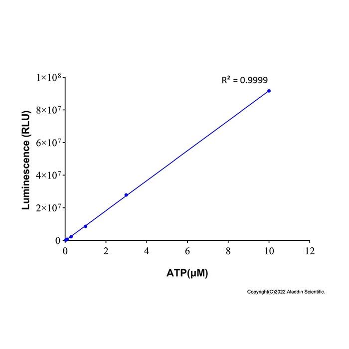
Thaw the necessary reagents on ice. Dilute the ATP Standard Solution with the ATP Assay Lysis Buffer to create an appropriate concentration gradient. The specific concentrations depend on the expected ATP levels in your samples. For initial experiments, concentrations like 0.01, 0.03, 0.1, 0.3, 1, 3, and 10 µM can be tested. Adjust the range in subsequent experiments based on sample ATP concentrations.
3. ATP Concentration Measurement:
(1) Add 100 μL of the ATP Detection Reagent to each well of the assay plate. Incubate at room temperature for 3-5 minutes.
(2) Add 20 μL of the sample or the diluted ATP standard solution to the wells containing the reagent.
(3) Measure the Relative Light Units (RLU) using a luminometer.
Note: The sample volume can be adjusted within the range of 10-100 μL. If the ATP concentration in the sample is low, add up to 100 μL. If the concentration is high, use a smaller volume, ensuring the same volume is used for the standard curve dilutions. If the ATP concentration is exceptionally high, dilute the sample with ATP Assay Lysis Buffer before measurement.
Instructions for Use 2 (Internal Standard Method)
1. For Suspension Cells
(1) Sample Processing:
For suspension cells, samples can be directly taken for subsequent measurement.
(2) ATP Measurement:
① Add 100 μL of ATP detection solution to the required reaction wells. Let stand for 10 min or until equilibrated to room temperature.
② Sample Wells: Transfer 50 μL of the suspended cell sample into a microcentrifuge tube. Add 50 μL of pure water and 100 μL of detection lysis buffer. Mix thoroughly and lyse for 5-10 min. After lysis, transfer 100 μL of the mixture to a reaction well and mix.
③ Internal Standard Wells: Transfer 50 μL of the suspended cell sample into a microcentrifuge tube. Add 50 μL of ATP standard solution (as an internal standard) and 100 μL of detection lysis buffer. Mix thoroughly and lyse for 5-10 min. After lysis, transfer 100 μL of the mixture to a reaction well and mix.
Note: For optimal results, the amount of ATP spiked as the internal standard should be approximately equal to the amount of ATP present in the cell sample. The luminescence from the sample plus internal standard should be roughly double that of the sample alone.
④ Continue to incubate the plate at room temperature for 5-25 min to allow the luminescent signal to stabilize. Alternatively, measurements can be taken approximately every 5 min, using the value at the time point when the signal stabilizes as the experimental data.
⑤ Measure chemiluminescence using a multimode microplate reader equipped with chemiluminescence detection capability. Set the appropriate parameters according to the instrument's instructions and record the luminescence values.
(3) ATP Calculation
The amount of ATP in the cell sample can be calculated using the following formula:

Where:
ATP(SAM) is the amount of ATP in the cell sample (in moles)
ATP(IS) is the amount of ATP spiked as the internal standard (in moles)
L(SAM) is the luminescence emitted by the cell sample (value from the Sample Well)
L(SAM+IS) is the luminescence emitted by the cell sample plus the internal standard (value from the Internal Standard Well)
(4) Viable Cell Concentration Calculation (Omit if not required)
If the ATP content per cell is known (determined experimentally or obtained from literature), the number of viable cells per milliliter in the original sample can be estimated using the following formula:

2. For Adherent Cells
(1) Sample Processing:
Aspirate and discard the culture medium. Add lysis buffer to the wells at a ratio of 200 μL per well for a 6-well plate (equivalent to 1/10 of the 2 mL culture volume). To ensure complete lysis, use a pipette to repeatedly triturate or rock the plate to allow the lysis buffer to fully contact and lyse the cells. Lyse for 5-10 min before proceeding with the measurement.
(2) ATP Measurement:
① Add 100 μL of ATP detection solution to the required reaction wells. Let stand for 10 min or until equilibrated to room temperature.
② Sample Wells: Transfer 50 μL of the lysed sample into a microcentrifuge tube. Add 150 μL of ultrapure water and mix thoroughly. Transfer 100 μL of this mixture to a reaction well and mix.
③ Internal Standard Wells: Transfer 50 μL of the lysed sample into a microcentrifuge tube. Add 50 μL of ATP standard solution (as an internal standard) and 100 μL of ultrapure water. Mix thoroughly. Transfer 100 μL of this mixture to a reaction well and mix.
④ Continue to incubate the plate at room temperature for 5-25 min to allow the luminescent signal to stabilize. Alternatively, measurements can be taken approximately every 5 min, using the value at the time point when the signal stabilizes as the experimental data.
⑤ Measure chemiluminescence using a multimode microplate reader equipped with chemiluminescence detection capability. Set the appropriate parameters according to the instrument's instructions and record the luminescence values.
(3) ATP Calculation and Viable Cell Concentration Calculation:
Use the same formulas provided above in sections 1(3) and 1(4).

 ZJ26F0535390