
GelRed Nucleic Acid Stain: Frequently Asked Questions
GelRed Nucleic Acid Stain: Frequently Asked Questions
I. What is GelRed, and which experiments is it primarily used for?
Answer: GelRed is a fluorescent dye for nucleic acid visualization. It is suitable for agarose gel electrophoresis and, under certain conditions, polyacrylamide gel electrophoresis (PAGE). By binding to DNA/RNA, GelRed produces fluorescence under appropriate excitation, enabling imaging and qualitative or semi-quantitative analysis of nucleic acid bands. It is commonly used for PCR product verification, restriction fragment analysis, clone screening, and preliminary assessment of RNA integrity.
II. What is the mechanism of action of GelRed?
Answer: GelRed binds to nucleic acids primarily via intercalation between base pairs. Upon intercalation, its fluorescence quantum yield increases markedly, emitting orange-red fluorescence under UV or other compatible excitation sources, thereby enabling band visualization. The binding mode is mechanistically similar to that of ethidium bromide (EtBr), although differences exist in molecular design and laboratory risk-management attributes.
III. Compared with ethidium bromide (EtBr), where are the main advantages of GelRed?
Answer: In common research-use contexts, frequently cited advantages of GelRed relative to EtBr include:
(1) Safety-management characteristics that may be easier to align with laboratory compliance practices, and it is often used as an EtBr alternative.
(2) High sensitivity, providing strong signal with well-controlled background under properly optimized conditions.
(3) Broad imaging compatibility, as many systems can acquire GelRed signals using the EtBr channel or similar filter sets. It should still be managed and disposed of as a potentially hazardous chemical in accordance with appropriate safety requirements.
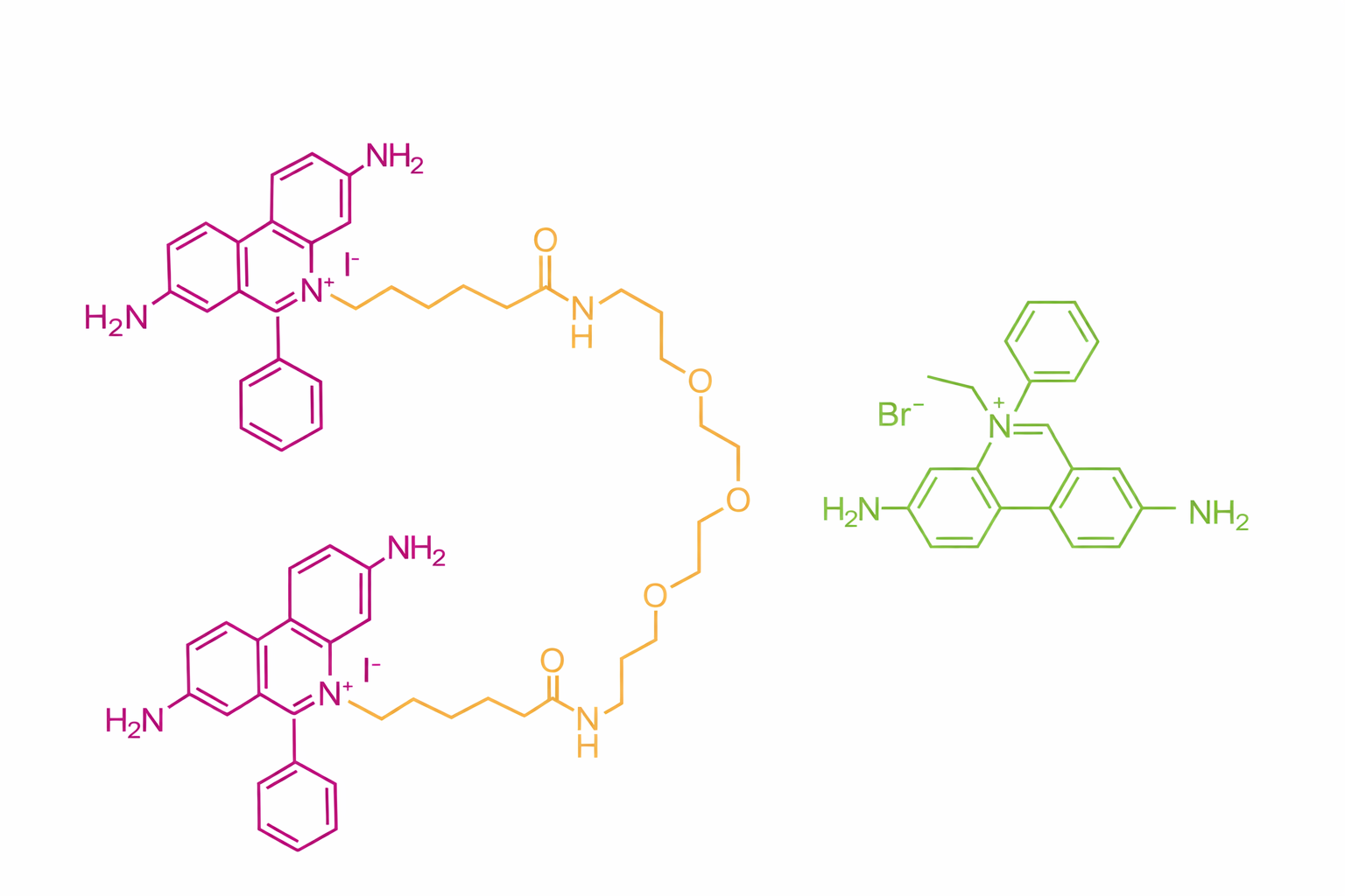
Figure 1. Comparison of the chemical structures of GelRed and ethidium bromide (EtBr) (left: GelRed; right: EtBr).
IV. Does GelRed require a destaining step?
Answer: Under most optimized conditions, GelRed yields low background and high contrast and typically does not require a dedicated destaining step. If background is elevated, it can be mitigated by reducing the working concentration, shortening staining time, increasing wash stringency, and/or optimizing imaging parameters.
V. Should GelRed be used as precast (pre-stain) or post-stain?
Answer: In most cases, post-staining is recommended:
(1) It reduces the likelihood that dye–DNA binding during electrophoresis will perturb migration.
(2) It more reliably produces sharp bands and higher resolution, particularly when accurate size determination is required.
(3) It is generally more robust across gel batches and buffer systems. Pre-staining can be used to streamline workflows or for high-throughput needs, but potential migration shifts and band-shape changes should be anticipated and minimized through condition optimization.
VI. Does GelRed affect DNA migration, and why?
Answer: Under certain conditions, GelRed can affect DNA migration, most commonly in pre-stain workflows. Dye–nucleic acid complex formation can alter effective charge density and hydrodynamic drag, leading to shifts in migration rate. Higher DNA loading and greater dye-binding fraction typically increase the likelihood and magnitude of migration shifts.
VII. How can GelRed-induced migration shifts be reduced?
Answer: The following strategies are commonly effective:
(1) Prefer post-staining to avoid continuous binding during electrophoresis.
(2) Dilute the stock solution strictly according to recommended working ratios to avoid excessive dye concentration.
(3) Reduce sample loading to prevent overloading-related binding saturation and abnormal migration.
(4) Apply a consistent staining strategy to both ladders and samples (e.g., all post-stained) to reduce systematic alignment bias.
VIII. Why do bands appear diffuse, smeared, or “overloaded,” and how should this be troubleshot?
Answer: Such artifacts typically reflect combined effects of sample loading, gel parameters, buffer conditions, and staining mode:
(1) Excessive loading: given GelRed’s high sensitivity, overloading more readily causes tailing or saturation-related diffusion, especially with DNA ladders.
(2) Migration perturbation from pre-staining: switching to post-staining is recommended to assess improvement.
(3) Mismatch between gel concentration and fragment size range: lower agarose concentration is generally preferred for larger fragments, whereas higher concentration improves resolution of smaller fragments.
(4) Buffer capacity and heat generation: prolonged high-voltage runs or insufficient buffering can increase diffusion and band distortion; using TBE buffer and/or reducing voltage or run distance may improve performance.
IX. Why is the fluorescence signal weak?
Answer: Common causes include:
(1) Working concentration too low and/or insufficient staining time.
(2) Imaging settings not matched (e.g., exposure, gain, or inappropriate filter/channel selection).
(3) Low nucleic acid amount and/or inadequate electrophoretic separation.
(4) Reduced excitation/collection efficiency due to light-source aging or filter degradation. A practical approach is to establish a baseline using a DNA standard of known concentration with fixed imaging parameters to distinguish staining-related issues from imaging-system limitations.
X. How should imaging filters or channels be selected?
Can the EtBr channel be used directly? Answer: In many systems, GelRed can be imaged using the EtBr channel or EtBr-compatible filters. Some imaging platforms also provide strong performance using SYBR-type or GelStar-type filter sets. Selection should be constrained by the available excitation source (UV vs. blue/visible) and filter configuration, followed by a one-time parameter scan to identify exposure settings that maximize signal-to-noise ratio.
XI. Is GelRed compatible with ssDNA and RNA?
Answer: GelRed can be used for ssDNA and RNA visualization, though signal intensity is often stronger with dsDNA under typical conditions. For RNA-focused applications, the following should be considered:
(1) RNA degradation can produce diffuse bands or trailing; troubleshooting should be coordinated with RNA extraction practices and RNase control.
(2) Use gel and buffer systems appropriate for RNA electrophoresis, and maintain consistent conditions across controls and samples.
XII. Is GelRed suitable for Southern blot and related blotting workflows?
Answer: GelRed can be used in such workflows, but post-staining is generally preferred to minimize potential effects of pre-staining on migration and downstream transfer efficiency. For applications requiring precise band localization for gel excision or membrane transfer, priority should be given to conditions that yield sharp bands, low background, and minimal migration shift.
XIII. Which downstream molecular biology operations are compatible with GelRed?
Answer: Under standard experimental conditions, GelRed is generally compatible with common downstream DNA workflows, including restriction digestion, ligation, cloning, and sequencing. If downstream reactions are highly sensitive to trace inhibitors, the following practices are recommended:
(1) Prefer post-staining to reduce dye binding during electrophoresis.
(2) Use standard gel extraction/column purification methods with adequate washing and optimized elution volumes.
(3) Include functional controls (e.g., ligation controls, PCR amplification controls) at critical steps to confirm the absence of inhibitory effects.
XIV. Can GelRed be used for polyacrylamide gel electrophoresis (PAGE)?
Answer: GelRed can be used for certain PAGE applications, but pre-staining is not universally recommended across PAGE systems. For PAGE, careful evaluation is advised with respect to:
(1) Gel format (denaturing vs. non-denaturing) and staining approach (post-staining preferred).
(2) Resolution and migration-accuracy requirements. A small-scale optimization experiment is recommended to verify imaging quality and migration stability before routine or high-throughput use.
XV. What are the main differences between GelRed and GelGreen?
Answer: Key differences include:
(1) Emission color: GelRed emits orange-red fluorescence, whereas GelGreen emits green fluorescence.
(2) Instrument and light-source compatibility: GelGreen is often better matched to blue-light imaging systems and is commonly selected to reduce UV exposure; GelRed typically shows strong compatibility with EtBr channels.
(3) Sensitivity and nucleic-acid preference: in some applications, GelRed may provide advantages for single-stranded nucleic acids; in practice, performance should be validated based on sample type and imaging configuration.
XVI. What is the difference between water-soluble GelRed and DMSO-based GelRed?
Answer: Differences primarily arise from the solvent system and associated safety-management considerations:
(1) Water-soluble formulations are often preferred to reduce solvent-related exposure risks and may be more operator-friendly.
(2) DMSO can enhance dermal permeability, necessitating more stringent personal protective measures and strict avoidance of skin contact. Both formulations share the same nucleic-acid visualization mechanism; selection should be guided by institutional safety requirements and compatibility with established workflows.
XVII. What are key safety and disposal considerations when using GelRed?
Answer: GelRed should be managed as a potentially hazardous chemical:
(1) Personal protective equipment: disposable gloves, lab coat, and eye protection; avoid direct contact with concentrated solutions and contaminated consumables.
(2) UV safety: when using UV transilluminators, wear UV-rated face shields or protective eyewear and minimize exposure time.
(3) Contamination control: designate dedicated staining/imaging areas and waste containers to prevent spread of dye and nucleic acid contamination.
(4) Waste disposal: dispose of liquids and contaminated materials in accordance with institutional EHS policies and applicable local regulations; requirements vary by organization and should follow the governing procedures at the user’s site.
References
[1] Ames, Bruce N., and Lois S. Gold. "The Causes and Prevention of Cancer: The Role of Environment." Biotherapy 11 (1998): 205-220.
[2] Oswald, N. (2008, March 03). Ethidium Bromide: The Alternatives [Web blog post]. Retrieved February 15, 2017.
[3] Berge, T., Jenkins, N. S., Hopkirk, R. B., Waring, M. J., Edwardson, J. M., & Henderson, R. M. (2002). Structural perturbations in DNA caused by bis-intercalation of ditercalinium visualised by atomic force microscopy. Nucleic Acids Research, 30(13), 2980-2986. Retrieved February 15, 2017.
[4] Crisafuli, F. A., Ramos, E. B., & Rocha, M. S. (2015). Characterizing the interaction between DNA and GelRed fluorescent stain. European Biophysics Journal, 44(1), 1-7.
5] Huang, Q., Baum, L., & Fu, W. L. (2010). Simple and practical staining of DNA with GelRed in agarose gel electrophoresis. Clinical Laboratory, 56, 149-152. Retrieved February 14, 2017.
[6] Kimball, J. (2010, December 21). Ames Test [Web blog post]. Retrieved February 14, 2017.
For more related articles, please see below:
[1] Five Key Strategies to Optimize Agarose Gel Electrophoresis with GelRed
[2] DNA Gel Stain Selection Guide
[3] Types, Mechanisms, and Selection Principles of Nucleic Acid Dyes for Gel Electrophoresis