
Lipid Droplet Biogenesis, Breakdown, Function, and Applications
Lipid Droplet Biogenesis, Breakdown, Function, and Applications
Lipid droplets (LDs) are dynamic organelles formed by a phospholipid monolayer surrounding a neutral lipid core. Their core components mainly include triacylglycerol (TAG) and cholesteryl esters. Lipid droplets are not only lipid storage structures, but also participate in energy metabolism, membrane lipid supply, lipotoxicity buffering, cellular stress responses, mitochondrial metabolic coupling, and ferroptosis regulation.
Keywords: lipid droplets; triacylglycerol; cholesteryl ester; Seipin; PLIN; lipolysis; lipophagy; mitochondria; ferroptosis
1 Basic Structure and Research Entry Points of Lipid Droplets
1.1 Structural Features of Lipid Droplets
Lipid droplets differ from double-membrane or single-membrane organelles such as mitochondria, the endoplasmic reticulum, and lysosomes. Their surface is a phospholipid monolayer, while the interior is a hydrophobic neutral lipid core that mainly stores TAG and cholesteryl esters. Multiple classes of functional proteins are distributed on the lipid droplet surface, including PLIN family proteins, lipid synthesis enzymes, lipolytic enzymes, membrane contact site proteins, and factors related to protein quality control.
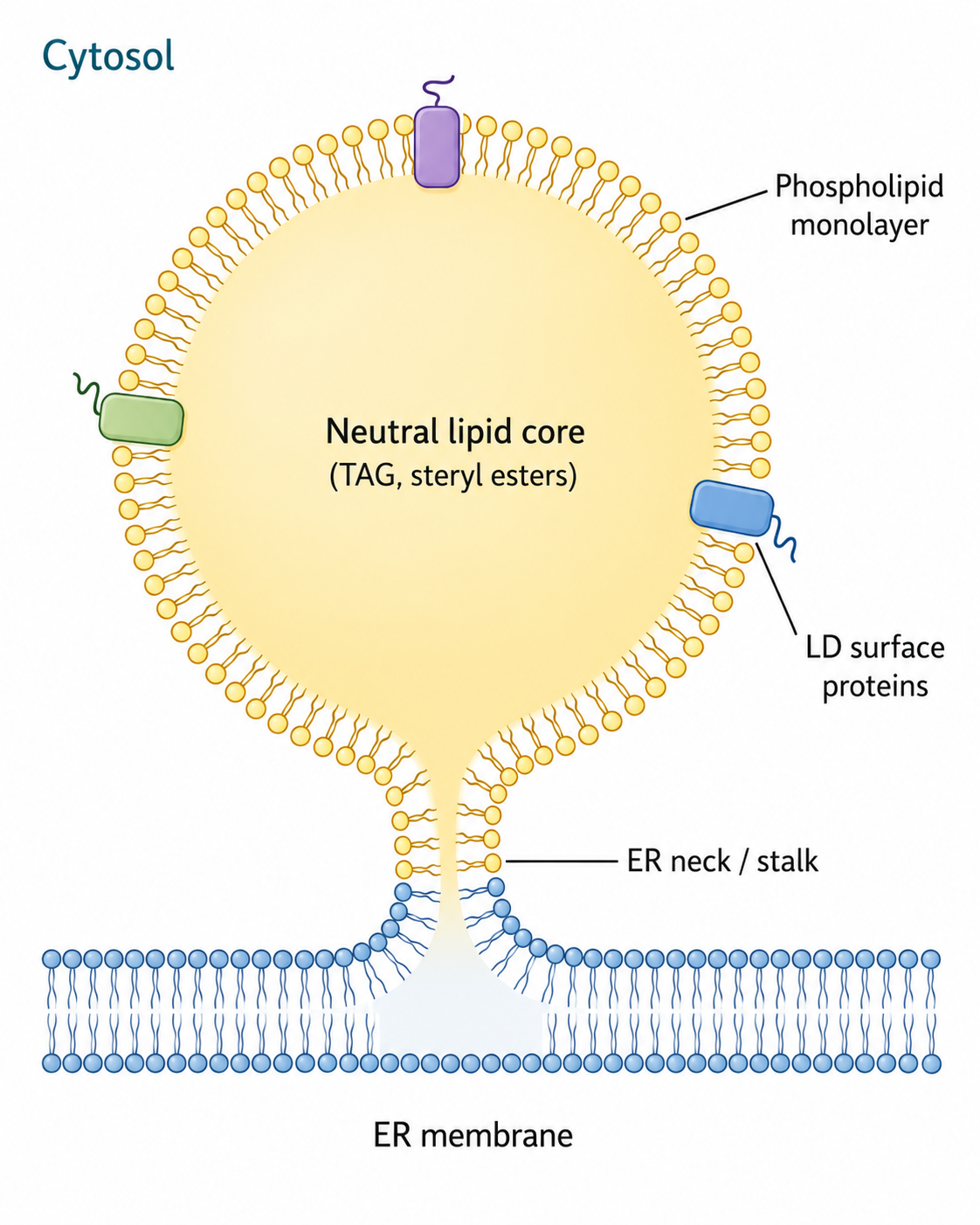
Figure 1. Basic Structure of a Lipid Droplet
Lipid droplet surface proteins can associate with the lipid droplet monolayer in different ways:
Binding Mode | Representative Feature | Functional Significance |
Hydrophobic hairpin structure | Inserts into the lipid droplet phospholipid monolayer | Common in some endoplasmic reticulum-derived proteins |
Lipidated domain | Enhances membrane binding through lipid modification | Stabilizes protein localization on the lipid droplet surface |
Amphipathic helix | Recognizes phospholipid arrangement on the lipid droplet surface | Participates in dynamic recruitment and release |
Protein-protein interaction | Depends on existing proteins on the lipid droplet surface for recruitment | Forms lipid droplet functional complexes |
1.2 Lipid Droplet Size and Morphology Differ by Cell Type
Lipid droplet size varies greatly. Lipid droplets in yeast and most non-adipocyte cells are usually small and mainly function in lipid buffering and local metabolic regulation. In adipocytes, lipid droplets can enlarge markedly during differentiation. Early-stage adipocytes often contain multiple small lipid droplets, whereas mature adipocytes can form large unilocular lipid droplets.
Cell Type/State | Lipid Droplet Morphology | Research Significance |
Yeast cells | Multiple small lipid droplets | Suitable for studying lipid droplet biogenesis and basic lipid metabolism mechanisms |
Fibroblasts | Dispersed small lipid droplets | Commonly used for lipid droplet staining, stress response, and metabolic research |
Early differentiating adipocytes | Multilocular lipid droplets | Reflects lipid droplet formation and fusion/expansion processes |
Mature adipocytes | Large unilocular lipid droplet | Reflects lipid storage and adipocyte maturation status |
Tumor cells/stressed cells | Increased lipid droplet number or redistribution | Commonly associated with hypoxia, acidic environment, ferroptosis resistance, and lipid reprogramming |
2 Lipid Basics: TAG, Phospholipids, and Fatty Acids
2.1 TAG and Phospholipids
Lipids include fats and lipoids. Fats mainly refer to triacylglycerol (TAG), which consists of one glycerol molecule esterified with three fatty acids and serves as an important cellular energy storage form. Lipoids include phospholipids, cholesterol, and cholesteryl esters, among which glycerophospholipids are important structural foundations of cellular membranes.
TAG is mainly stored in the lipid droplet core, whereas phospholipids are located in the monolayer membrane structure on the lipid droplet surface. These two components do not exist in isolation in cells. Lipid droplet formation, expansion, and breakdown all depend on coordination among TAG synthesis, phospholipid supply, and fatty acid metabolism.
2.2 Classification of Fatty Acids
Fatty acids are composed of carbon, hydrogen, and oxygen. According to whether the carbon chain contains double bonds, they can be divided into saturated fatty acids and unsaturated fatty acids. According to the number of double bonds, unsaturated fatty acids can be further divided into monounsaturated fatty acids and polyunsaturated fatty acids.
Type | Structural Feature | Metabolic Significance |
Saturated fatty acids (SFA) | No double bonds in the carbon chain | Excessive levels can easily cause lipotoxicity and altered membrane properties |
Monounsaturated fatty acids (MUFA) | Contain one double bond | Commonly used for TAG synthesis and lipid droplet storage |
Polyunsaturated fatty acids (PUFA) | Contain two or more double bonds | Prone to lipid peroxidation and closely associated with ferroptosis |
ω-3 fatty acids | The first double bond from the methyl end is at the third position | EPA, DHA, and others have important physiological functions |
ω-6 fatty acids | The first double bond from the methyl end is at the sixth position | Participate in membrane lipid composition and inflammatory mediator generation |
PUFA is not only an important component of membrane lipid structure, but also an important substrate for lipid peroxidation and ferroptosis. Lipid droplets can absorb and store PUFA by converting it into TAG form, thereby reducing the risk of PUFA exposure to membrane phospholipid peroxidation under certain conditions.
3 Lipid Droplet Biogenesis
3.1 The Endoplasmic Reticulum Is the Main Platform for Lipid Droplet Biogenesis
Lipid droplets mainly originate from the endoplasmic reticulum (ER). Neutral lipids gradually accumulate between the two leaflets of the ER membrane. When TAG and cholesteryl esters reach a certain local concentration, phase separation occurs, forming a neutral lipid "lens" structure. This structure then buds toward the cytosolic side, forming a lipid droplet with a phospholipid monolayer membrane.
Lipid droplet biogenesis can be summarized into four stages:
Stage | Key Event | Representative Molecules/Processes |
Nucleation | TAG accumulates within the ER membrane and undergoes phase separation | TAG, cholesteryl esters, Seipin/BSCL2 |
Budding | The neutral lipid lens forms a lipid droplet toward the cytosolic side | LDAF1, Ldo proteins, FIT2 |
Early expansion | TAG continues to enter the lipid droplet core, while phospholipids and proteins are supplied to the surface | Seipin complex, ER-LD connection |
Late expansion | Lipid droplets continue to grow and acquire metabolic enzymes and coating proteins | GPAT4, ATGL, PLIN family proteins |
3.2 The Seipin Complex Participates in Lipid Droplet Nucleation and Maturation
Seipin is encoded by the BSCL2 gene and is a key regulatory factor in lipid droplet biogenesis. Seipin localizes to ER-lipid droplet contact sites, stabilizes neutral lipid nucleation regions, and promotes normal control of lipid droplet size and number. Seipin dysfunction can lead to abnormal lipid droplet morphology, disrupted lipid distribution, and abnormal adipose tissue development.
The role of Seipin is not merely to "help generate lipid droplets." More importantly, it restricts abnormal dispersion of lipid droplets within the ER membrane, allowing neutral lipids to gather at appropriate sites and form lipid droplet structures that can mature. Therefore, Seipin deficiency is often associated with abnormal small lipid droplets, giant lipid droplets, or uneven lipid droplet distribution.
3.3 TAG Synthesis Is the Metabolic Basis for Lipid Droplet Expansion
TAG is mainly synthesized through the glycerol-3-phosphate pathway. Fatty acids first bind to coenzyme A to form acyl-CoA, and then undergo stepwise esterification with glycerol-3-phosphate, sequentially generating lysophosphatidic acid (LPA), phosphatidic acid (PA), and diacylglycerol (DAG). Finally, DAG is esterified with a third fatty acid to form TAG.
The basic process of TAG synthesis is as follows:
Reaction Step | Product | Key Significance |
Fatty acid activation | Acyl-CoA | Provides activated fatty acids for lipid synthesis |
Glycerol-3-phosphate esterification | LPA | Initial step of TAG synthesis |
Addition of the second fatty acid | PA | Connects phospholipid synthesis and TAG synthesis |
PA dephosphorylation | DAG | Important intermediate for TAG and membrane lipid synthesis |
Addition of the third fatty acid | TAG | Enters the lipid droplet core for storage |
DGAT1 and DGAT2 are important enzymes in the terminal step of TAG synthesis. When TAG synthesis is enhanced, lipid droplet number and size usually increase. When TAG synthesis is inhibited, free fatty acids and lipotoxic stress may increase.
4 Lipid Droplet Breakdown
Lipid droplet breakdown is not simply "fat disappearance," but the conversion of stored neutral lipids into usable fatty acids, glycerol, and cholesterol for energy metabolism, membrane lipid remodeling, or signaling molecule synthesis. Lipid droplet breakdown mainly includes two mechanisms: lipolysis and lipophagy.
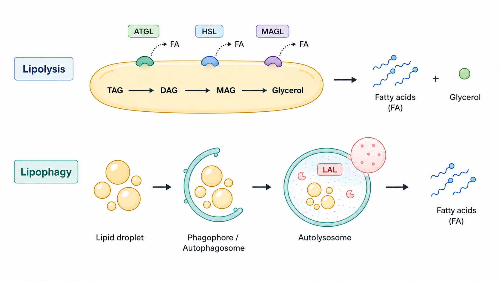
Figure 2. Lipolysis and Lipophagy Pathways of Lipid Droplets
4.1 Lipolysis
Lipolysis is the classical mode of lipid droplet breakdown and is mainly completed through sequential catalysis by ATGL, HSL, and MAGL. TAG is first hydrolyzed by ATGL to generate DAG. DAG is then hydrolyzed by HSL to generate MAG. Finally, MAG is hydrolyzed by MAGL to release glycerol and fatty acids.
Step | Key Enzyme | Reaction Result |
TAG→DAG | ATGL | Releases the first fatty acid |
DAG→MAG | HSL | Releases the second fatty acid |
MAG→glycerol + fatty acid | MAGL | Completes TAG degradation |
ATGL is the key enzyme that initiates lipolysis and is commonly regulated by CGI-58/ABHD5, PLIN family proteins, and hormonal signals. HSL is regulated by PKA phosphorylation in adipocytes and participates in mobilizing stored lipids. MAGL completes the terminal hydrolysis, allowing fatty acids to enter β-oxidation or other lipid metabolic pathways.
4.2 Lipophagy
Lipophagy is a form of selective autophagy. Cells can enclose part or all of a lipid droplet into an autophagosome, which then fuses with lysosomes. Neutral lipids in lipid droplets are degraded by lysosomal acid lipase and other hydrolases.
Lipophagy differs from lipolysis in that it is more dependent on the autophagy pathway and lysosomal function. It is often enhanced during starvation, metabolic stress, hepatic lipid accumulation, and certain pathological states. Abnormal lipophagy affects fatty acid supply, hepatic lipid homeostasis, and cellular energy metabolism.
4.3 Relationship Between Lipolysis and Lipophagy
Lipolysis and lipophagy are not mutually exclusive. In different cell types and metabolic states, both can participate in lipid droplet mobilization. Lipolysis is more suitable for rapid fatty acid release, while lipophagy is more suitable for processing lipid droplet structures and long-term metabolic remodeling through the lysosomal system.
Breakdown Mode | Main Location | Key Molecules | Feature |
Lipolysis | Lipid droplet surface | ATGL, HSL, MAGL, PLIN | Rapid mobilization of fatty acids |
Lipophagy | Autophagosome-lysosome system | LC3, p62, LAMP1, lysosomal acid lipase | Depends on autophagy and lysosomal degradation |
Cooperative action | Lipid droplet-autophagy-mitochondria axis | ATGL, autophagy proteins, β-oxidation-related enzymes | Maintains long-term energy homeostasis |
5 Major Functions of Lipid Droplets
5.1 Energy Storage and Fatty Acid Supply
The most fundamental function of lipid droplets is to store excess fatty acids and release fatty acids for mitochondrial β-oxidation when energy demand increases. Adipocytes, hepatocytes, skeletal muscle cells, and cardiomyocytes all rely on lipid droplets for lipid storage and mobilization.
5.2 Lipotoxicity Buffering
Excessive free fatty acids can cause abnormal membrane structure, endoplasmic reticulum stress, mitochondrial injury, and increased ROS. Lipid droplets can esterify excess fatty acids into TAG and store them, thereby reducing free fatty acid toxicity. This function is especially important in metabolic syndrome, fatty liver disease, diabetes, and tumor metabolic reprogramming.
5.3 Membrane Lipid Supply and Organelle Homeostasis
Lipid droplets can provide fatty acid sources for membrane lipid synthesis and form contact sites with the ER, mitochondria, peroxisomes, and lysosomes. Through these contact sites, lipid droplets participate in lipid transport, membrane lipid remodeling, and metabolic coordination among organelles.
5.4 Protein Quality Control and Stress Responses
Some lipid droplet surfaces can recruit molecules related to protein folding, degradation, and ubiquitination, participating in protein quality control. Under stress conditions, lipid droplets can also sequester hydrophobic molecules or abnormal lipids, reducing the risk of cellular injury.
5.5 Immune and Inflammatory Regulation
Lipid droplets are closely associated with inflammatory responses. After immune cell activation, lipid droplets often increase. Lipid droplets can participate in arachidonic acid metabolism and the generation of lipid mediators such as prostaglandins and leukotrienes. In macrophage foam cell formation, infection responses, and chronic inflammation, lipid droplets often serve as platforms where lipid metabolism and inflammatory signaling converge.
6 Roles of Lipid Droplets in Disease
6.1 Metabolic Diseases
Abnormal lipid droplet accumulation is closely associated with obesity, nonalcoholic fatty liver disease, insulin resistance, and type 2 diabetes. Lipid droplets themselves are not necessarily harmful. The key lies in whether lipid droplet storage capacity, lipolysis rate, fatty acid oxidation capacity, and lipotoxicity buffering capacity are properly matched.
6.2 Tumors
Tumor cells often adapt to hypoxia, acidic environments, and nutrient stress by enhancing lipid uptake, fatty acid synthesis, and lipid droplet storage. Lipid droplets can provide tumor cells with energy sources, membrane lipid materials, and antioxidant buffering capacity, and may also enhance tumor cell tolerance to ferroptosis, chemotherapy, or metabolic stress.
6.3 Neurodegenerative Diseases
In the nervous system, lipid droplets can appear during metabolic stress processes involving neurons, astrocytes, microglia, and oligodendrocytes. Abnormal lipid droplets may be related to mitochondrial injury, lipid peroxidation, inflammatory responses, and protein homeostasis imbalance. In neurodegenerative diseases, lipid droplets may act as protective buffering structures, but may also reflect lipid metabolic disorders and chronic cellular stress.
6.4 Infectious Diseases
Viruses and some bacteria can exploit host lipid droplets for replication, assembly, or energy supply. Lipid droplets can also participate in host innate immune responses and provide a platform for lipid mediator synthesis and antimicrobial responses. Therefore, in the context of infection, lipid droplets may be exploited by pathogens and may also participate in host defense.
Disease Type | Lipid Droplet Change | Possible Mechanism |
Obesity/fatty liver | Massive lipid droplet accumulation | Enhanced TAG storage and insufficient fatty acid oxidation |
Diabetes | Abnormal lipid droplet distribution and lipolysis | Lipotoxicity, insulin resistance, mitochondrial dysfunction |
Tumors | Increased lipid droplets or redistribution | Hypoxic adaptation, membrane lipid supply, ferroptosis resistance |
Neurodegenerative diseases | Abnormal lipid droplets in glial cells or neurons | Oxidative stress, mitochondrial injury, lipid peroxidation |
Infection | Lipid droplets are mobilized or remodeled | Pathogen replication and immune lipid mediator generation |
7 Lipid Droplet Visualization and Isolation
7.1 Lipid Droplet Visualization
Lipid droplet visualization mainly depends on immunofluorescence of lipid droplet coating proteins and lipophilic fluorescent dyes. PLIN family proteins are commonly used lipid droplet surface markers and are suitable for lipid droplet localization, number, and morphology analysis. Lipophilic dyes can directly label the neutral lipid core of lipid droplets.
Common visualization methods include:
Method | Representative Indicator/Dye | Feature |
Immunofluorescence of lipid droplet coating proteins | PLIN1, PLIN2, PLIN3, PLIN5 | Reflects lipid droplet surface proteins and functional status |
Neutral lipid fluorescent dyes | BODIPY, LipidTOX, Nile Red, LipidGreen | Simple operation, suitable for observing lipid droplet number and distribution |
Traditional lipid staining | Oil Red O, Sudan III | Commonly used for observing lipid deposition in tissues or cells |
Live-cell imaging | BODIPY, some LipidTOX dyes | Allows observation of dynamic lipid droplet changes |
Oil Red O and Sudan III are suitable for observing lipid deposition, but may affect lipid droplet structure during certain sample processing procedures. If the study focuses on precise lipid droplet localization, quantitative analysis, and colocalization, fluorescent probes and PLIN immunofluorescence are more suitable.
7.2 Lipid Droplet Isolation
Lipid droplets have low density and can be enriched by density gradient centrifugation after cell lysis. Isolated lipid droplets can be used for lipidomics, proteomics, and analysis of lipid droplet surface proteins. The key to lipid droplet isolation is to avoid strong mechanical disruption and detergent interference; otherwise, lipid droplet fusion, rupture, or loss of surface proteins may occur.
After lipid droplet isolation, the following analyses are often combined:
Detection Direction | Analysis Content |
Lipidomics | TAG, cholesteryl esters, phospholipids, PUFA content |
Proteomics | PLIN, lipolytic enzymes, membrane contact site proteins |
Western blot | Validation of lipid droplet marker enrichment |
Microscopic imaging | Examination of lipid droplet isolation integrity |
Enzyme activity detection | Analysis of lipolysis- or lipid metabolism-related enzyme activity |
8 Lipid Droplets and Mitochondria
8.1 Lipid Droplet-Mitochondria Contact
Functional contact sites exist between lipid droplets and mitochondria. Lipid droplets can supply fatty acids to mitochondria, while mitochondria generate ATP through β-oxidation. PLIN5 is an important lipid droplet coating protein in lipid droplet-mitochondria interaction and is often expressed in tissues with active oxidative metabolism, such as skeletal muscle, myocardium, and brown adipose tissue.
8.2 PLIN5-, Mfn2-, and Rab8a-Related Regulation
PLIN5 can promote fatty acid flow from lipid droplets to mitochondria and participate in physical connections between lipid droplets and mitochondria. PLIN1 is specifically expressed in adipocytes and can also be associated with the mitochondrial fusion protein Mfn2, affecting mitochondria-lipid droplet interaction. In skeletal muscle, the small GTPase Rab8a can interact with PLIN5, regulate lipid droplet-mitochondria contact, and respond to AMPK activation.
During starvation or increased energy demand, lipid droplet-mitochondria contact is enhanced, facilitating fatty acid transfer and β-oxidation. FATP4 and other proteins related to fatty acid transport/activation can also participate in lipid mobilization between lipid droplets and mitochondria.
Key Molecule | Main Function | Research Significance |
PLIN5 | Promotes lipid droplet-mitochondria contact | Connects lipid droplet storage with fatty acid oxidation |
Mfn2 | Participates in mitochondrial fusion and organelle contact | Affects mitochondria-lipid droplet interaction |
Rab8a | Regulates lipid droplet-mitochondria contact in skeletal muscle | Related to AMPK and energy demand |
FATP4 | Promotes fatty acid activation and transport | Enhances lipid droplet fatty acid utilization during starvation |
AMPK | Energy stress sensor | Regulates lipid droplet mobilization and fatty acid oxidation |
9 Lipid Droplets and Ferroptosis
9.1 Lipid Peroxidation and Ferroptosis
Ferroptosis is an iron-dependent form of cell death centered on lipid peroxidation accumulation. PUFA is easily oxidized and serves as an important substrate for lipid peroxidation. Fe²⁺ can promote free radical generation through the Fenton reaction, triggering chain reactions of phospholipid peroxidation and ultimately disrupting membrane stability.
Lipid peroxidation can occur through non-enzymatic and enzymatic pathways. In the non-enzymatic pathway, free radicals abstract hydrogen atoms from PUFA, generating lipid radicals and propagating oxidative chain reactions. In the enzymatic pathway, lipoxygenases (LOX) and related enzymes can catalyze PUFA oxidation to generate lipid peroxides. Lipid peroxidation products such as 4-HNE and MDA can further modify proteins and amplify cellular injury.
9.2 System Xc⁻, GSH, and GPX4 Axis
System Xc⁻ is composed of SLC7A11 and SLC3A2 and transports cystine into cells for glutathione (GSH) synthesis. GSH is the key reducing substrate required for GPX4 to exert its anti-lipid peroxidation function. GPX4 can reduce phospholipid hydroperoxides to relatively stable phospholipid alcohols, thereby blocking lipid peroxidation chain reactions.
Regulatory Axis | Key Molecules | Effect on Ferroptosis |
Cystine uptake | SLC7A11/SLC3A2 | Affects GSH synthesis |
Antioxidant system | GSH | Supports GPX4 in clearing lipid peroxides |
Lipid peroxide clearance | GPX4 | Inhibits the core execution process of ferroptosis |
Iron metabolism | Fe²⁺, ferritin, transferrin receptor | Enhances or limits free radical generation |
PUFA metabolism | ACSL4, LPCAT3, LOX | Affects formation of peroxidizable phospholipid substrates |
9.3 Dual Effects of Lipid Droplets on Ferroptosis
The relationship between lipid droplets and ferroptosis is context-dependent. On one hand, lipid droplets can esterify and store PUFA in TAG, reducing PUFA incorporation into membrane phospholipids and subsequent peroxidation, thereby providing a certain buffering effect against ferroptosis. On the other hand, lipid droplets may also serve as fatty acid reservoirs and provide substrate sources for lipid peroxidation under specific conditions.
In acidic tumor microenvironments, increased PUFA uptake can enhance ferroptosis sensitivity. If lipid droplets can effectively absorb PUFA, they can reduce PUFA peroxidation toxicity. If lipid droplet synthesis is inhibited, making it difficult for PUFA to be buffered in TAG, PUFA-induced ferroptosis may be enhanced. In some tumor models, a high ω-3 PUFA diet can enhance the killing effect of ferroptosis inducers, suggesting important translational value in the relationship among lipid droplets, PUFA, and ferroptosis.
10 Cell Cycle Arrest, Lipid Droplet Formation, and Ferroptosis Resistance
Cell cycle arrest can alter cellular lipid uptake and lipid storage status. Under cell cycle arrest, cells may enhance TAG accumulation and form more lipid droplets. Excessive PUFA retained in lipid droplets can reduce its entry into membrane phospholipid peroxidation pathways, thereby decreasing ferroptosis sensitivity.
This mechanism suggests that lipid droplets are not only metabolic storage structures, but may also influence cellular responses to therapeutic stress. For tumor cells, cell cycle arrest, lipid droplet accumulation, and ferroptosis resistance may form a protective metabolic state. Therefore, in ferroptosis-inducing therapy or metabolic therapy, combined intervention in lipid droplet synthesis, PUFA allocation, or TAG storage may increase cellular sensitivity to ferroptosis.
11 Application Directions of Lipid Droplet Research
11.1 Metabolic Disease Mechanism Research
Lipid droplets are core structures in obesity, fatty liver disease, diabetes, and atherosclerosis research. By detecting lipid droplet number, size, PLIN expression, TAG content, and lipolytic activity, lipid storage, lipotoxicity, and energy metabolic status can be evaluated.
11.2 Tumor Metabolism and Ferroptosis Research
Lipid droplets can affect tumor cell adaptation to hypoxia, acidic environments, drug pressure, and ferroptosis. Combined detection of lipid droplet staining, lipidomics, and ferroptosis indicators helps explain why tumor cells develop therapeutic tolerance under high lipid uptake or cell cycle arrest conditions.
11.3 Organelle Interaction Research
Lipid droplets have close contact with the ER, mitochondria, lysosomes, and peroxisomes. Lipid droplet-mitochondria colocalization, lipid droplet isolation, and detection of membrane contact site proteins can be used to study fatty acid transport, β-oxidation, and organelle metabolic coupling.
11.4 Drug Screening and Functional Evaluation
Changes in lipid droplet number and morphology can serve as phenotypic readouts for lipid metabolism drugs, antitumor drugs, ferroptosis modulators, and metabolic stress intervention agents. High-content imaging, flow cytometry analysis, and lipidomics can be used for lipid droplet-related drug screening.
Application Direction | Detection Content | Research Value |
Fatty liver/obesity | Lipid droplet number, size, TAG content | Evaluates lipid deposition and metabolic abnormalities |
Tumor metabolism | Lipid droplet accumulation, PUFA allocation, ferroptosis sensitivity | Analyzes metabolic adaptation and therapeutic tolerance |
Neurodegenerative diseases | Glial cell lipid droplets, oxidative stress, lipid peroxidation | Explains chronic neural stress and cell injury |
Organelle interaction | Lipid droplet-mitochondria colocalization, PLIN5, Mfn2 | Studies fatty acid mobilization and β-oxidation |
Drug screening | Lipid droplet staining, high-content imaging, lipolysis indicators | Establishes evaluation systems for lipid metabolism-modulating drugs |
12 Product Selection Related to Lipid Droplet Biogenesis, Breakdown, Function, and Application Research
Product Module | Cat. No. | Product Name | Grade & Purity | Application Positioning |
Lipid droplet fluorescence staining | BODIPY 493/503 methyl bromide | ≥98% | Labels neutral lipids and lipid droplet structures; suitable for observing lipid droplet number, size, and intracellular distribution | |
Lipid droplet fluorescence staining | 2,6-Diiodo-BODIPY 493/503 |
| BODIPY-type lipid fluorescent probe; can be used for lipid droplet/neutral lipid-related fluorescence detection | |
Lipid fluorescence staining | LipidGreen 2 | ≥98% | Used for fluorescent detection of intracellular lipid structures; suitable for lipid droplet visualization and lipid accumulation analysis | |
Lipid peroxidation detection | BODIPY 581/591 C11 (Lipid Peroxidation Sensor) | BioReagent, biological stain, suitable for fluorescence analysis, for microscopy, ≥98% | Detects lipid ROS and lipid peroxidation; suitable for lipid droplet-PUFA-ferroptosis mechanism research | |
Traditional lipid staining | Oil red O(C.1.26125) | Biological stain | Detects neutral lipid deposition in cells or tissues; suitable for adipocyte differentiation, fatty liver, and foam cell research | |
Traditional lipid staining | Oil red O | 10 mM in DMSO | Solution-type Oil Red O, suitable for optimizing lipid deposition staining conditions | |
Lipid droplet coating protein | Perilipin 2/PLIN2/ADFP Antibody | See COA | Detects PLIN2/ADFP; suitable for lipid droplet labeling in non-adipocyte cells, IF colocalization, and WB analysis | |
Lipid droplet coating protein detection | Human Perilipin-2 (PLIN2) ELISA Kit | BioReagent | Quantitatively detects PLIN2 levels and assists in evaluating lipid droplet accumulation and changes in lipid droplet coating proteins | |
Lipid droplet induction substrate | Oleic acid | BioReagent, Moligand™, for cell culture | Commonly used in cell lipid droplet induction models to promote TAG synthesis and lipid droplet formation | |
Lipid droplet induction substrate | Oleic acid | Moligand™, 10 mM in DMSO | Solution-type oleic acid, suitable for cell treatment and lipid droplet accumulation model establishment | |
Lipid droplet induction substrate | Oleic acid | Moligand™, ≥96% | Used for fatty acid treatment, lipid droplet induction, and lipid metabolism intervention experiments | |
Lipid droplet induction substrate | Oleic acid | Moligand™, ≥99%(HPLC) | High-purity oleic acid, suitable for lipid droplet induction experiments requiring higher fatty acid purity | |
Lipid droplet induction substrate | Oleic acid | ≥98% | Used for lipid droplet formation, TAG accumulation, and fatty acid metabolism research | |
Fatty acid standard/analysis | Oleic acid | Analytical standard, Moligand™, ≥99%(GC) | Can be used for fatty acid quantification, lipid analysis method establishment, or standard control | |
Lipid droplet induction substrate | Oleic acid | USP, ≥98% | Can be used for fatty acid treatment and lipid droplet accumulation models; suitable for experiments requiring pharmacopeial grade | |
Lipid droplet induction substrate | Oleic acid | Moligand™, ≥85% | Can be used for routine fatty acid treatment and preliminary lipid droplet induction experiments | |
TAG synthesis intervention | AZD7687 | Moligand™, ≥98% | Inhibits DGAT1 to study TAG synthesis, lipid droplet expansion, and lipid storage regulation | |
TAG synthesis intervention | JNJ-DGAT1-A |
| Used for DGAT1-related TAG synthesis and lipid droplet biogenesis mechanism research | |
TAG synthesis intervention | LCQ-908 | Moligand™ | Inhibits DGAT1 to analyze lipid droplet formation, lipid deposition, and metabolic intervention effects | |
TAG synthesis intervention | JNJ DGAT2-A | ≥98% | Inhibits DGAT2 to analyze the terminal step of TAG synthesis, lipid droplet formation, and hepatic lipid deposition | |
TAG synthesis intervention | JNJ-DGAT2-A | Moligand™, 10 mM in DMSO | Solution-type DGAT2 inhibitor, suitable for cell lipid droplet formation intervention experiments | |
TAG synthesis intervention | PF 06424439 | ≥98%(HPLC) | Used for DGAT2-mediated lipid droplet biogenesis, lipid metabolism reprogramming, and lipotoxicity research | |
Lipolysis detection | Human Adipose Triglyceride Lipase (ATGL) ELISA Kit | BioReagent | Detects ATGL levels and assists in evaluating lipolysis-related changes; initial TAG hydrolysis and lipid droplet mobilization should be interpreted together with ATGL activity, CGI-58/PLIN regulatory status, and glycerol/free fatty acid release | |
Lipolysis detection | Human Hormone Sensitive Lipase (HSL) ELISA Kit | BioReagent | Detects HSL levels and assists in analyzing lipolysis-related changes; hormone-regulated lipolysis should be interpreted together with HSL phosphorylation, lipid droplet localization changes, and glycerol/free fatty acid release | |
Lipophagy | Recombinant LC3B Antibody | Recombinant, ExactAb™, validated, see COA | Detects LC3B and assists in analyzing autophagosome marker changes; lipid droplet entry into autophagosomes or lipophagy should be interpreted together with lipid droplet staining/PLIN labeling, LC3 colocalization, LAMP1/lysosome colocalization, and autophagic flux blockade experiments | |
Lipophagy detection | Human Microtubule-associated Protein Light Chain 3B(LC3B) ELISA Kit | BioReagent | Quantitatively detects LC3B levels and assists in evaluating autophagy/lipophagy pathway changes | |
Lysosomal degradation | LAMP1 /CD107a Antibody | KD Validation | Labels lysosomes and is suitable for lipid droplet-lysosome colocalization and lipophagy research | |
Lysosomal degradation | LAMP1 Mouse mAb | KD Validation | Detects LAMP1 and analyzes lysosome-mediated lipid droplet degradation | |
Lysosomal degradation | LAMP1 Mouse mAb | Carrier-free, ExactAb™, azide-free, validated, ≥95%(SDS-PAGE), 1.0 mg/mL | Used for IF/WB detection of LAMP1; suitable for lipid droplet-lysosome fusion analysis | |
Lysosomal degradation | LAMP1/CD107a Mouse mAb | See COA | Used for lysosome labeling and assisting in determining lipid droplet degradation through the autophagy-lysosome pathway | |
Lysosomal degradation | Recombinant LAMP1 /CD107a Antibody | KD Validation | Detects LAMP1/CD107a; suitable for lipophagy and lysosomal function research | |
Lysosomal degradation | Recombinant LAMP1 Antibody | Recombinant, ExactAb™, validated, 0.2 mg/mL | Labels lysosomes and analyzes lipid droplet-lysosome fusion and lipophagic degradation | |
Lysosome-related detection | SAR428926 (anti-LAMP1) | Animal-free, carrier-free, recombinant, ExactAb™, low endotoxin, azide-free, validated, ≥95%(SDS-PAGE&SEC-HPLC), see COA | Can be used for LAMP1-related detection or lysosomal marker research; should be selected according to the specific experimental system | |
Lipid droplet-mitochondria interaction | Recombinant Human TOMM20 Protein | Carrier-free, His tag, ≥95%(SDS-PAGE), see COA | Can be used for TOMM20-related antibody validation or mitochondrial marker research | |
Lipid droplet-mitochondria interaction | Recombinant TOMM20 Antibody | Recombinant, ExactAb™, validated, 0.3 mg/mL | Labels the mitochondrial outer membrane; suitable for lipid droplet-mitochondria colocalization and fatty acid oxidation-related research | |
Lipid droplet-mitochondria interaction | Recombinant TOMM20 Antibody | Recombinant, ExactAb™, validated, see COA | Detects TOMM20 and assists in analyzing lipid droplet-mitochondria contact and changes in mitochondrial distribution | |
Lipid droplet surface phospholipids/membrane lipid remodeling | Phospholipid PL1 |
| Can be used for research related to lipid droplet surface phospholipids, membrane lipid composition, or lipid model systems | |
Membrane phospholipid remodeling | Recombinant Phospholipid Scramblase 1 Antibody | KD Validation | Detects PLSCR1 and is suitable for membrane phospholipid rearrangement, lipid stress, and cell membrane lipid homeostasis research | |
Membrane phospholipid remodeling/mitochondria-related lipid regulation | Recombinant Phospholipid Scramblase 3 Antibody | KD Validation | Detects PLSCR3 and can be used for mitochondria-related membrane lipid remodeling and lipid stress mechanism research | |
Membrane phospholipid remodeling | Phospholipid Scramblase 11 Antibody | KD Validation | Detects PLSCR11 and is suitable for membrane phospholipid distribution and lipid homeostasis-related research |
Lipid droplets are dynamic organelles connecting lipid storage, energy metabolism, organelle interaction, and cellular stress responses. Their biogenesis depends on neutral lipid nucleation and budding in the endoplasmic reticulum, while their breakdown depends on lipolysis and lipophagy. Their functions involve lipotoxicity buffering, fatty acid oxidation, membrane lipid supply, inflammatory regulation, and control of ferroptosis sensitivity.
References
[1] Zadoorian A, Du X, Yang H. Lipid droplet biogenesis and functions in health and disease. Nat Rev Endocrinol. 2023;19(8):443-459.
[2] Klemm RW, Carvalho P. Lipid Droplets Big and Small: Basic Mechanisms That Make Them All. Annu Rev Cell Dev Biol. 2024;40(1):143-168.