
Professional Guide to Streptavidin-Modified Proteins
Professional Guide to Streptavidin-Modified Proteins
I. Molecular Engineering Fundamentals
1. Structure and Valency
vStreptavidin is a homotetramer (each subunit contains one biotin-binding site), allowing up to four ligands per tetramer in theory.
vMultivalency yields higher apparent affinity and stronger surface anchoring, benefiting signal amplification and resistance to wash-off.
2. Thermodynamic/Kinetic Considerations
vThe extremely low Kd corresponds to a very slow dissociation rate (koff ≈ 0); once bound, the interaction behaves quasi-irreversibly.
vDesign implications: favor single-endpoint readouts or rigorous regeneration protocols (extreme pH/denaturants). It is not suitable for online affinity chromatography requiring reversible exchange.
3. Sources of Nonspecific Background and Mitigation
vHigh-pI proteins and anionic/glycan receptor interactions → prefer streptavidin over avidin.
vSurface/matrix adsorption → raise ionic strength (150–300 mM NaCl), apply surface blocking (BSA/casein/fish gelatin), shorten incubations, and optimize wash conditions.
vHydrophobic patch exposure after crosslinking → control degree of labeling (DoL) and use mild buffers.
II. Avidin vs. Streptavidin: Selection Criteria
Avidin is a highly glycosylated tetrameric glycoprotein composed of four identical subunits, each ~16.4 kDa (total protein mass ~66 kDa). Each subunit contains one binding site for biotin (vitamin H/B7) and carries an N-linked oligosaccharide at an Asn residue. The avidin tetramer is strongly basic, with an isoelectric point of approximately pI ≈ 10. The biotin–avidin interaction ranks among the strongest known noncovalent interactions, with a dissociation constant of about Kd ≈ 1.3 × 10⁻¹⁵ M.Streptavidin is a non-glycosylated tetrameric protein derived from Streptomyces avidinii (~13–14 kDa per subunit; ~60 kDa total). It has an isoelectric point of pI ~5–6 and exhibits an equally strong affinity for biotin (Kd ~10⁻¹⁴–10⁻¹⁵ M). Owing to its near-neutral surface charge and lack of glycans, it typically yields lower nonspecific background and is thus well suited to background-sensitive applications such as immunoassays, nucleic acid capture, and surface immobilization.
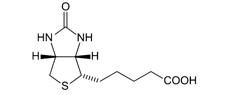
Figure 1. Chemical structure of biotin.
Dimension | ||
Glycosylation | Glycoprotein (highly glycosylated) | Non-glycosylated |
Isoelectric point (pI) | ~10 (strongly cationic) | ~5–6 (weakly acidic) |
Nonspecific binding | Prone to interact with anions/glycan receptors; higher background | Significantly reduced |
Typical use cases | Situations demanding extreme stability | Preferred for routine immunoassays and imaging |
III. Conjugation Chemistries (Three Common Routes)
1. NHS–Maleimide Bridging (SMCC/Sulfo-SMCC)
1)Use cases
Broadly applicable to protein–protein conjugation; one partner must provide –NH₂ groups (e.g., streptavidin Lys/N-termini), the other –SH (native or introduced).
2)Mechanism
NHS esters form amide bonds with primary amines; maleimides undergo selective Michael addition with thiols (–SH).
3)Reagents
SMCC (or Sulfo-SMCC), Traut’s reagent (2-iminothiolane, for thiolation if needed), TCEP, L-cysteine, PBS/HEPES (pH 7.2–7.5; avoid Tris during NHS steps).
4)Standard procedure
vNHS activation (SMCC end): Streptavidin 1–2 mg/mL + SMCC 5–10 eq; PBS/HEPES, pH 7.2–7.5, RT 0.5–2 h, light-protected.
vDesalting: PD-10 or 30–100 kDa ultrafiltration to obtain maleimide-streptavidin intermediate.
vPrepare –SH protein: If the counterpart lacks thiols, introduce –SH with Traut’s reagent (pH 7.8, 30 min). TCEP is for pre-reduction only; remove completely before coupling—no residual TCEP during conjugation.
vCoupling: Intermediate : thiol-containing protein = 1 : (1–3); pH 6.5–7.0, RT 45–90 min.
vCapping: Add L-cysteine 2–5 mM for 10 min to quench remaining maleimide.
vPurification/buffer exchange: Ultrafiltration or SEC to remove small molecules and aggregates; formulate in PBS + 150 mM NaCl (optionally 5–10% glycerol).
2. Periodate Oxidation/Reductive Amination (Preferred for Glycoproteins: HRP, Ferritin)
1)Use cases
Conjugation of glycoproteins such as HRP or ferritin with streptavidin (labeling predominantly on glycans, typically distal to the catalytic center).
2)Advantages
No need to introduce thiols; gentle for enzymes; conjugation sites on glycans are usually away from active sites.
3)Reagents
NaIO₄, NaBH₃CN (or NaBH₄), acetate buffer pH 6.0–6.5, EDTA.
4)Standard procedure
vOxidation: HRP 1 mg/mL + NaIO₄ 5 mM, ice bath, light-protected, 15–30 min.
vImmediate desalting: PD-10 into acetate buffer pH 6.0–6.5.
vCondensation: Add streptavidin (molar ratio 1:1–1:2), RT 30–60 min.
vReduction: Add NaBH₃CN 20–30 mM, RT 1–2 h or 4 °C overnight.
vPurification/formulation: Ultrafiltration/SEC; storage buffer may contain trace EDTA and 5–10% glycerol.
3. Glutaraldehyde Crosslinking (Prefer Two-Step Method; Surface Immobilization/Macromolecular Assembly)
1)Use cases
General amine–amine crosslinking, surface immobilization, or assembly with large carriers; economical with broad material compatibility.
2)Strategy
vOne-step: React two proteins directly with low glutaraldehyde; simple but often heterogeneous/aggregative.
vTwo-step: First activate one partner (create an “aldehyde-active layer”), remove free glutaraldehyde, then couple—typically lowers aggregation.
3)Reagents
Glutaraldehyde (25% stock, dilute before use), ethanolamine/glycine, phosphate buffers/carbonate buffers (avoid Tris).
4)Standard two-step procedure
vActivate carrier: Carrier protein 1–2 mg/mL + 0.02–0.05% glutaraldehyde, RT 10–20 min.
vDesalting: Remove free glutaraldehyde to obtain the “aldehyde-active layer.”
vCoupling: Add streptavidin (1:1–1:3), RT 30–60 min.
vQuench: Ethanolamine or glycine 50 mM for 10–15 min to fully quench residual aldehydes.
vPurification/formulation: Ultrafiltration/SEC; PBS + 150 mM NaCl (optionally 5–10% glycerol).
IV. Workflow Selection and Overall Design
1) Application-to-Parameter Mapping
Application | Preferred chemistry | Recommended DoL | Readout | Notes |
ELISA/CLIA amplification | SMCC or reductive amination (HRP) | Low–medium (reduce background) | Colorimetric/chemiluminescent | Track specific activity and linear range |
Fluorescence imaging/flow cytometry | SMCC (for fluorescent proteins/dyes) | Medium–high (boost brightness) | IF/FC | Avoid self-quenching and aggregation |
Solid-phase capture/sorting | Two-step glutaraldehyde or SMCC | Low–medium | Recovery/specificity | Surface chemistry and blocking are critical |
EM labeling (ferritin) | Reductive amination | Low–medium | EM contrast | Control particle size and uniformity |
2) Quick Buffer Compatibility Matrix
System | NHS step | Maleimide step | Reductive amination | Glutaraldehyde |
✅ | ✅ | ⚠ (pH 6–7) | ✅ | |
❌ (competes with NHS) | ⚠ (keep pH ≤7.0; avoid alkaline) | ❌ (primary amines compete; forms Schiff bases) | ❌ (quenches aldehydes) | |
Acetate (pH 6–6.5) | ❌ | ⚠ | ✅ (preferred for condensation) | ✅ |
Ionic strength (≤300 mM) | ✅ | ✅ | ✅ | ✅ |
V. FAQs and Optimization Tips
Q1: High background/severe nonspecific binding?
A: Prefer streptavidin; increase salt (e.g., 150–300 mM NaCl); include carrier/blocking proteins; avoid high-pI carriers or glycan-mediated off-target interactions; shorten incubation and strengthen washes.
Q2: Product aggregation or precipitation?
A: Lower crosslinker equivalents/reaction time; conduct reactions at neutral, moderately low-salt conditions; switch to two-step glutaraldehyde or the NHS–maleimide route; add mild nonionic surfactants (0.01–0.05%).
Q3: Activity loss?
A: Potential site disruption or over-modification; reduce activation level and keep pH in a mild range; for enzymes, prioritize periodate/reductive amination and control oxidation time with light protection.
Q4: How to set the coupling density (DoL)?
A: Be application-driven: use lower DoL for quantitative assays to minimize background; moderately raise DoL for imaging amplification. Optimize empirically by functional titration (signal-to-noise/linear range).
VI. Safety and Compatibility Notes
vNaIO₄, NaBH₃CN, glutaraldehyde: Handle in a fume hood with gloves/goggles; dispose according to SDS. Do not use NaBH₃CN under strongly acidic conditions (avoid excessively low pH/strong acids); it is safe and standard to use in mildly acidic buffers (e.g., acetate pH 5–7).
vAvoid Tris in NHS steps; avoid sodium azide in HRP systems (inhibits enzyme activity).
vTCEP is for pre-reduction only; it must be absent during maleimide reactions.
Aladdin: https://www.aladdinsci.com/