
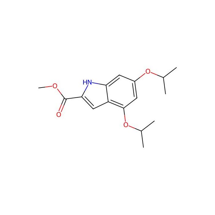
Determine the necessary mass, volume, or concentration for preparing a solution.
XST-14 - ≥99% , CAS No.2607143-50-8
AVAILABLE TO ORDER
GRADE & PURITY ≥99%
Storage
Store at -20°C
Shipped In
Ice chest + Ice pads
≥99% for sensitive chromatographic and analytical workflows requiring minimal baseline interference.
Store at -20°C Ships Ice chest + Ice pads Check lot-specific COA for exact specifications.
SDS, COA, datasheet, and spec sheet available for download. Lot-specific COA accessible via lot number lookup.
Cited in 0 peer-reviewed publications across chromatography, organic synthesis, and cross-coupling reactions.
XST-14 is a potent, competitive and highly selective ULK1 inhibitor with an IC 50 of 26.6 nM. XST-14 induces autophagy inhibition by reducing the phosphorylation of the ULK1 downstream substrate. XST-14 induces apoptosis in hepatocellular carcinoma (HCC) cells and has antitumor effects
In Vitro
XST-14 inhibits ULK1 (IC 50 =13.6 nM), MAP2K1/MEK1 (IC 50 =721.8 nM), MAPK14/p38 alpha (IC 50 =283.9 nM), TGFBR2 (IC 50 =809.3 nM), ACVR1/ALK2 (IC 50 =183.8 nM), ULK2 (IC 50 =70.9 nM) and CAMK2A (IC 50 =66.3 nM) by the 10-point titration results from SelectScreen Kinase Profiling Services. XST-14 (20-80 μM; for 24 h) leads a decrease in cell proliferation activity. XST-14 (5 μM; for 24 h) induces apoptosis in HepG2 and human primary HCC cells. XST-14 (5 μM; for 12 h) strongly inhibits the conversion of LC3-I to LC3-II in CHO cells stably expressing GFP-LC3. XST-14 (5 μM; for 12 h) inhibits the Ser249 phosphorylation of PIK3C3 and Ser15 phosphorylation of BECN1. MCE has not independently confirmed the accuracy of these methods. They are for reference only. Cell Proliferation AssayCell Line: HepG2 cells Concentration: 20, 40, 60, 80 μM Incubation Time: 24 hours Result: Led a decrease in cell proliferation activity. Apoptosis AnalysisCell Line: HepG2 and human primary cells Concentration: 5 μM Incubation Time: 24 hours Result: Induced apoptosis in HepG2 and human primary HCC cells. Cell Autophagy AssayCell Line: CHO, HepG2 cells stably expressing GFP-LC3 Concentration: 5 μM Incubation Time: 12 hours Result: Strongly inhibited the conversion of LC3-I to LC3-II in CHO cells. Dramatically decreased the number of GFP-LC3 puncta in HepG2 cells. Decreased autophagosome formation and blocked autophagosome/lysosome fusion in HepG2 cells. Western Blot AnalysisCell Line: HepG2 cells Concentration: 5 μM Incubation Time: 12 hours Result: Inhibited the Ser249 phosphorylation of PIK3C3 and Ser15 phosphorylation of BECN1.
In Vivo
XST-14 (15, 30 mg/kg/day; IP; for 4 consecutive weeks) displays anti-HCC efficacies, resulting in decreased tumor weights and suppressed tumor growth of HCC cells in nude mice . XST-14 (2 mg/kg for IV; 10 mg/kg for IP) has a T 1/2 of 2.31 hours for IV and a T 1/2 of 2.69 hours for IP . MCE has not independently confirmed the accuracy of these methods. They are for reference only. Animal Model: Nude mice bearing HepG2 tumor xenografts Dosage: 15, 30 mg/kg Administration: IP; daily; for 4 consecutive weeks Result: Displayed anti-HCC efficacies, resulting in decreased tumor weights and suppressed tumor growth of HCC cells in nude mice. Animal Model: Sprague-Dawley rat Dosage: 2 mg/kg for IV; 10 mg/kg for IP (Pharmacokinetic Analysis) Administration: IV or IP Result: Had a T 1/2 of 2.31 hours, a CL of 26.28 mL/min•kg, and and an AUC of 1269 hr•ng/mL for IV. Had a T 1/2 of 2.69 hours, a C max of 2033 ng/mL, and an AUC of 5979 hr•ng/mL for IP.
Form:Solid
IC50& Target:CaMK II ULK1 26.6 nM (IC 50 )
| Canonical Smiles | O=C(OC)C1=CC2=C(OC(C)C)C=C(OC(C)C)C=C2N1 |
|---|---|
| Molecular Weight | 291.34 |
Comprehensive hazard, handling, storage, and regulatory compliance document.
Download SDS →Lot-specific quality data. Enter your lot number to retrieve the exact COA.
Look up COA →Full quality attributes and acceptance criteria for this grade.
View spec sheet →| Solubility | DMSO : 250 mg/mL (858.10 mM; Need ultrasonic) |
|---|