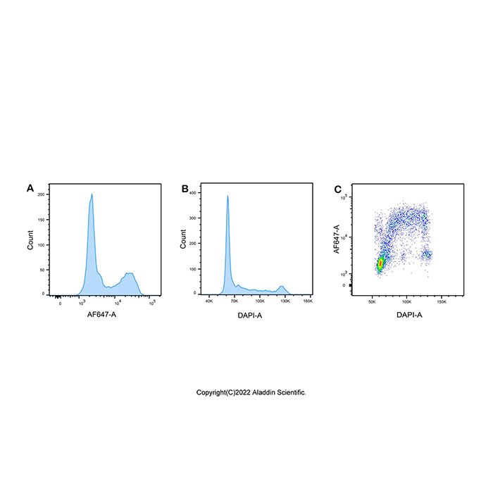
Cell proliferation assays are widely used in the evaluation of cell viability, genotoxicity, and the efficacy of antitumor drugs. Direct detection of DNA synthesis in cells is considered the most accurate method for assessing cell proliferation. EdU (5-ethynyl-2′-deoxyuridine) is a novel thymidine (thymine deoxyribonucleoside) analogue. During DNA synthesis, EdU can be incorporated into newly synthesized DNA in place of thymidine. The ethynyl group on EdU can undergo a covalent reaction with fluorescently labeled small-molecule azide probes (such as Azide AF 488, Azide AF 555, Azide AF 594, Azide AF 647, etc.) via Cu(I)-catalyzed click chemistry, forming a stable triazole ring. This reaction is highly efficient and is referred to as the Click reaction. Through this process, newly synthesized DNA is labeled with the corresponding fluorescent probes, enabling the detection of proliferating cells using appropriate fluorescence detection equipment.
E1373493 | Component | 50 T | 100 T | 200 T | Storage conditions | Quantity Per Test |
E1373493A | EdU(10 mM) | 100 μL | 200 μL | 400 μL | -20℃.Store in the dark. | 2 μL per 1.0-2.0x 10⁶ cells |
E1373493B | AF647 azide | 250 μL | 500 μL | 1000 μL | -20℃.Store in the dark. | 5 μL per 1.0-2.0x 10⁶ cells |
E1373493C | Click Reaction Buffer | 12 mL | 24 mL | 48 mL | -20℃.Store in the dark. | 240 μL per 1.0-2.0x 10⁶ cells |
E1373493D | CuSO4 | 250 μL | 500 μL | 1000 μL | -20℃. | 5 μL per 1.0-2.0x 10⁶ cells |
E1373493E | Click Additive | 991 mg | 1982 mg | 3964 mg | -20℃.Store in the dark. | 250 μL per 1.0-2.0x 10⁶ cells |
E1373493F | DAPI Staining Solution(1000×) | 25 μL | 50 μL | 100 μL | -20℃.Store in the dark. | 0.5 μL per 1.0-2.0x 10⁶ cells |
Note: The recommended number of cells to stain per test is 1.0-2.0x 10⁶ cells. The amount of Click reaction solution can be adjusted according to the experimental samples.
Usage Protocol
1. Preparation
1) Preparation of Click Additive Solution:
For a 50-test kit: Add 12.5 mL of pre-chilled deionized water to the tube. Mix thoroughly until completely dissolved to obtain the Click Additive Solution. For a 100-test kit: Add 25 mL of pre-chilled deionized water to the tube. Mix thoroughly until completely dissolved to obtain the Click Additive Solution. For a 200-test kit: Add 50 mL of pre-chilled deionized water to the tube. Mix thoroughly until completely dissolved to obtain the Click Additive Solution. After preparation, aliquot the solution as needed and store at -20°C. If a white precipitate forms after dissolution, invert the tube repeatedly until it is fully dissolved before use. If the solution turns brown, it indicates degradation of the active component; discard it.
2) Upon initial dissolution of the Click Reaction Buffer, aliquot it according to the number of samples per experiment and store at -20°C.
2. EdU Labeling of Cells
It is recommended to use a final EdU concentration of 10 μM (1×). A 1:500 dilution of EdU (10 mM) in cell culture medium yields a 2× EdU working solution (20 μM). Mix an equal volume of pre-warmed (37°C) 2× EdU working solution (20 μM) with the cell suspension to achieve a final 1× EdU concentration. Incubate in a 37°C, 5% CO₂ incubator. Factors such as cell culture medium, cell density, cell type, and other experimental conditions may affect labeling efficiency. Therefore, the optimal EdU concentration and labeling duration must be empirically determined based on the cell type under investigation.
3. Fixation and Permeabilization
1) Harvest cells and centrifuge at 300 ×g for 5 min. Wash cells twice with PBS containing 2% FBS.
2) Fix cells with 4% paraformaldehyde solution. Mix thoroughly and incubate for 15 min at room temperature protected from light.
3) Collect cells and centrifuge at 300 × g for 5 min. Wash cells twice.
4) Resuspend cells in PBS containing 0.3% Triton X-100. Mix well and incubate for 15 min at room temperature.
5) Centrifuge at 300 × g for 5 min and wash cells twice.
4. Fluorescent Labeling
1) This protocol is based on a 500 μL reaction system per 2 × 10⁶ cells. The volume of the Click reaction mixture can be adjusted according to the experimental sample size.
2) Centrifuge the cells at 300 ×g for 5 minutes. Add 500 μL of Click reaction mixture per sample, mix gently, and incubate for 30 minutes at room temperature protected from light.
3) After the reaction, wash the cells twice with PBS containing 2% FBS.
4) Dilute the DAPI Staining Solution (1000×) to 1× using PBS containing 2% FBS. Add 250 μL of the diluted DAPI solution to each sample and incubate for 5 minutes at room temperature.
5) Add an additional 250 μL of PBS containing 2% FBS, mix gently, and proceed to detection using an appropriate flow cytometry instrument.
Component | Number of samples |
1 | 2 | 4 | 5 | 10 | 25 | 50 |
Click Reaction Buffer | 240 μL | 480 μL | 960 μL | 1200 μL | 2400 μL | 6000 μL | 12000 μL |
CuSO4 | 5 μL | 10 μL | 20 μL | 25 μL | 50 μL | 125 μL | 250 μL |
Azide 647 | 5 μL | 10 μL | 20 μL | 25 μL | 50 μL | 125 μL | 250 μL |
Click Additive Solution | 250 μL | 500 μL | 1000 μL | 1250 μL | 2500 μL | 6250 μL | 12500 μL |
Total volume | 500 μL | 1000 μL | 2000 μL | 2500 μL | 5000 μL | 12500 μL | 25000 μL |
Precautions
1. Strictly adhere to the component order and volumes specified in the table above when preparing the Click reaction mixture, as deviations may affect subsequent experimental results.
2. The Click reaction mixture must be used within 15 minutes of preparation.
3. To avoid fluorescence quenching, perform detection as soon as possible after sample preparation.