
Subcellular localization analysis
Subcellular localization analysis
The target proteins are labeled with specific marker proteins such as green fluorescent protein (GFP) or β-glucosidase (GUS), and their location is subsequently detected by immunofluorescence or GUS staining.
Principle
Currently, the most commonly used is fusion reporter gene localization, in which the target protein is fused to the N-terminus or C-terminus of a fluorescent protein, and the fusion protein is expressed in the recipient cell by transient transformation or stable genetic transformation techniques, and the target protein is localized to the target organelle along with the fluorescent protein. Subsequently, the fusion protein will emit fluorescence of a specific color under the laser irradiation of a confocal microscope, thus pinpointing the location of the target protein.
Operation method
Subcellular localization of target proteins in rice plants
Principle
Currently, the most commonly used is fusion reporter gene localization, in which the target protein is fused to the N-terminus or C-terminus of a fluorescent protein, and the fusion protein is expressed in the recipient cells by transient transformation or stable genetic transformation techniques, and the target protein is localized to the target organelle together with the fluorescent protein. Subsequently, the fusion protein will emit fluorescence of a specific color under the laser irradiation of a confocal microscope, thus pinpointing the location of the target protein.
Materials and Instruments
This experimental method is introduced with the help of GFP localization in rice as an example: Move 0. Reagent preparation 1) Enzymatic solution (10 ml) 0.6 M mannitol 1.093 g 10 mM MES (pH=5.7) 1 ml (100 mM master mix) 1.5% Cellulose R-10 0.15 g 0.75% Macerozyme R-10 0.075 g 0.1% BSA 0.01 g 1 mM CaCl2 0.1 ml (100 mM master mix) 0.25 g/ml Carboxybenzylpenicillin 2 μl β- ME 4 μl ddH2O, 10 ml, 55 °C, 10 min, natural cooling (must be used now).
1, carrier and plant materials: PAN580-GFP vector, seeds of indica rice 9311, etc.;
2, the main reagents: mannitol, MES, cellulose R-10, Macerozyme R-10, BSA, carboxybenzylpenicillin, β-ME, PEG 4,000, CaCl
2
NaCl, KCl, MgCl
2
, glucose, etc;
3. Required supplies and instruments: 50 ml centrifuge tube, 50 ml syringe, razor blade, nylon filter cloth (200 mesh), 0.45 μm microporous filter membrane, blood cell counting plate, slides, coverslips, low-temperature centrifuge, vacuum pump, spectrophotometer, and laser confocal microscope.
2) PEG 4000 (10 ml)
0.6 M mannitol | 1.093 g |
100 mM CaCl2 | 0.111 g |
40% PEG 4000 | 4 g |
ddH2O to 10 ml, adjust the pH of the solution to 7.5-8.0 with 1M KOH, filter and sterilize the solution, then dispense into 1.5 ml centrifuge tubes and store in the refrigerator at -20 °C.
3) W5 (100 ml)
154 mM NaCl | 0.9 g |
125 mM CaCl2 | 1.39 g |
5 mM KCl | 5 ml (100 mM mother liquor) |
5 mM glucose | 0.09 g |
2 mM MES | 2 ml (100 mM mother liquor) 1 M KOH |
1M KOH adjust the pH value of the solution to 5.8, autoclave for 20 min and store at room temperature.
4) MMG (10 ml)
15 mM MgCl2 | 1.5 ml (100 mM mother liquor) |
4 mM MES | 0.4 ml (100 mM mother liquor) |
0.6 M Mannitol | 1.093 g |
1M KOH adjust the pH value of the solution to 5.8, autoclave for 20 min, and store at room temperature.
1. Target protein prediction
The localization of target proteins can be predicted by some online websites, and here we recommend a few common prediction websites for plants:
WoLF PSORT: Protein Subcellular Localization Prediction Tool (genscript.com)
iPSORT Home Page (hgc.jp)
Services list (dtu.dk)
TargetP 2.0 - DTU Health Tech - Bioinformatic Services
2. Constructing a vector containing the target gene
The vector used in this experiment is PAN580 vector with GFP tag, and its vector diagram is shown below:
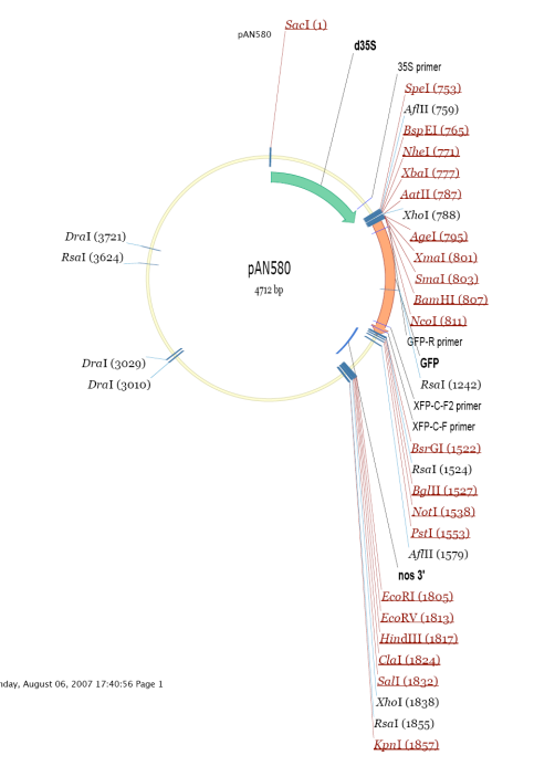
The vector construction method is double digestion + homologous recombination (the primers for the gene upstream of GFP need to remove the stop codon when designing), and the plasmid is macroextracted after the sequencing is correct, and the concentration of the final plasmid should be more than 1,000 ng/ul, and it is recommended that it should not be less than 800 ng/ul.
3. Rice planting
Select a number of indica rice 9311 seeds with the same size and full appearance, soak the seeds at 30 ℃ for 2 days, germinate for 12 h, and then sow the seeds, and cultivate them in a light incubator with 10 h of light/14 h of darkness, 28 ℃, and 10,000 lx of light intensity for a few days until the seedlings grow to 10 cm in height.
4. Protoplast extraction
1) Seedlings are uprooted, cut off the roots and cleaned. Starting from the root base, the white firm stem part is selected and cut into shortest possible segments, weighed and poured into a clean 100 ml conical flask. 1 g of seedling segments should be added to 10 ml of the enzyme solution;
2) Vacuum for 45~60 min, incubate at 28 ℃ and 40 rpm in an incubator with shaking for 4~5 h (avoid light);
3) Filter the enzyme solution through a nylon membrane (200 mesh) into a sterilized 50 ml centrifuge tube and place it in an incubator at 28 ℃;
4) Add 10 ml of W5 solution into the filter residue and incubate at 28 ℃ and 80 rpm for 1 h to fully release the protoplasts;
5) Filter again through a nylon membrane (200 mesh), mix thoroughly with the filtrate from step 3, and add an equal volume of W5 solution, centrifuge at 150 g for 5 min at room temperature (gentle mode is selected for both elevation and deceleration), and discard the supernatant;
6) Add 1 ml of resuspension solution to resuspend the protoplasts, then slowly add 20 ml of 20% sucrose solution to suspend the protoplasts in sucrose solution, and centrifuge the protoplasts at 120 g for 5 min (select gentle mode for both elevation and deceleration speeds);
7) Collect the protoplasts at the stratified interface, wash with W5 solution, incubate on ice for 30 min, centrifuge at 100 g for 5 min to precipitate the protoplasts, discard the supernatant, and remove the residual liquid as much as possible, then gently resuspend the protoplasts by adding 1 ml of MMG solution, and adjust the density of the protoplasts to 2×10⁵ cells/ml under the microscope.
5. Plasmid transformation and observation results
(1) Add 10 μl (10-20 μg) of recombinant plasmid containing the target fragment into a sterilized 2.0 ml EP tube, carefully add 150 μl of protoplasts, shake gently and mix well;
(2) Add 150 μl of 40% PEG, shake gently and incubate for 20 min at 28 ℃;
(3) Slowly add 500 μl W5 solution, mix carefully, centrifuge at 150 g for 4 min (pay attention to the direction of the centrifuge tube), and remove the PEG with the tip of the gun;
(4) Slowly add 100 μl of W5 solution and resuspend, incubate in the dark at 28 ℃ for more than 12 hours;
(5) Observe the expression of fluorescent protein under laser confocal microscope.
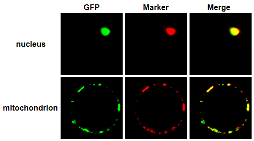
The following figure shows the localization of the two proteins in rice protoplasts, which were studied in our laboratory, in terms of cytosolic and mitochondrial localization. This figure is for study and communication purposes only. Figure shown:
Caveat
1、When constructing the vector, we need to pay attention to the fusion position of the GFP tag, if there is a signal peptide in the target sequence, we need to avoid fusing fluorescent proteins with this end of the vector, because different fusion methods may observe different localization, which may affect the experimental results;2, the expression of different species will be slightly different, so there will be different localization in different receptor species, when selecting the receptor, should try to select and the sequence of the target protein or gene source of similar species materials (such as rice protein is often selected in rice, onion epidermis and tobacco);3, it is recommended to provide GFP empty load as a blank control, to prove that the fluorescent protein can exercise its function normally in the experiment, and at the same time to exclude the target protein from being affected by fluorescent signals in the experimental operation;4、It is recommended to add a bright field channel to show that the protoplasts are living cells with good activity status, and at the same time to show that the fluorescent signals are generated by the fusion protein rather than cell debris.5、The PAN580 vector used in this experiment carries a 35 S strong promoter, and it is recommended to construct a vector with GFP fused to the gene's own promoter at the same time, so as to avoid the localization results being interfered by the gene expression level. Common Problems 1. Low rice protoplast yield and viability: the low protoplast yield may be due to the failure of the enzyme solution to contact the plant incision, such as rice stems are not easy to be immersed in the enzyme solution, so it is necessary to cut them into as small as possible, and increase the contact area between the enzyme solution and the incision by means of vacuuming, so as to promote the enzyme solution to enter into the plant tissues; the low viability of protoplasts may be due to the lack of tight protection from light in the course of the operation, and the light will cause damage to the protoplast membrane, resulting in the low viability. The lower survival rate of protoplasts may be due to the lack of strict light protection during the operation, light will cause damage to the protoplast membrane, resulting in a lower survival rate. 2. No fluorescence can be observed: cells of different species may have different effects on gene expression, it is recommended to change the receptor material to observe fluorescence, such as tobacco. For more product details, please visit Aladdin Scientific website.