Transplacental transmission is the main mode of intrauterine transmission. Intrauterine transmission refers to the process by which pathogenic microorganisms, etc., pass from the mother into the blood and/or cells and tissues of the fetus during pregnancy, and is the basis of intrauterine infection. The placenta is an important organ for the exchange of materials between the fetus and the mother, and the placental barrier, composed of trophoblast cells, capillary endothelial cells and the basement membrane between the two, is the necessary route for nutrients, as well as certain drugs, viruses and hormones, to enter the fetus from the mother.
Operation method
Animal Models of Placenta-Transmitted Diseases (Placental Barrier Models)
Principle
The cell polarity model of placental barrier was constructed to isolate and characterize placental trophoblast cells by trypsin digestion + density gradient centrifugation + magnetic bead separation, and to establish a primary trophoblast cell culture system.
Materials and Instruments
Experimental material: Move The basic process of the animal model of transplacental transmission (placental barrier model) can be divided into the following steps: Caveat A. In vitro Transwell upper and lower chambers co-polar culture, simulating the structural level of in vivo placenta, inoculate the trophoblast cells and placental capillary endothelial cells in the upper and lower part of the microporous membrane respectively, instead of just a simple mixing culture of the two kinds of cells, to establish the co-polar culture model. The growth of cells was observed continuously by phase contrast microscopy; the ultrastructure of cells was observed by scanning and transmission electron microscopy. Based on the specificity of in vivo placenta for immunoglobulin transport and the polarity of secreting certain hormones, the function of the constructed cell polarity model was characterized.B. Continuous observation of cell growth by phase contrast microscopy and ultrastructure of cells by scanning and transmission electron microscopy: Cells on the 7th day of co-culture were taken with cell transwells, fixed and dehydrated; the specimens for scanning electron microscopy were dried and sprayed with gold, and a large number of microvilli on the surface of the trophectoderm cells and at the cell junctions were observed; specimens for transmission electron microscopy were cut into 2 mm x 2 mm x 2 mm pieces, and 1% agar jelly was added to the specimens for scanning electron microscope. For transmission electron microscopy, the specimen was cut into 2 mm x 2 mm x 2 mm pieces, embedded in 1% agarose, embedded in Epon812 resin, routinely trimmed, vertically sectioned, stained, and observed by electron microscopy that various connections were formed between the cells, and polar epithelial structures, such as connective complexes, were seen near the top.C. Add a certain amount of rabbit immunoglobulin (IgG, lgM) from the upper chamber of the Transwell, and detect the content of IgG and IgM in the lower chamber of the Transwell by ELISA, and set up a blank control. Only IgG was detected in the lower chamber (equivalent to the fetal circulation), and IgM was not detected.D. Placental growth hormone was measured in the upper and lower chambers of the transwell by RIA to determine the function of trophoblast cells in the polarized release of certain hormones. Placental growth hormone is expected to be detected only in the upper chamber of the Transwell, but not in the lower chamber. For more product details, please visit Aladdin Scientific website.
① Transwell;
② Polycarbonate membrane with 3.0 μm pore size;
③ 12-well culture plate;
③ 12-well culture plate; ④ DMEM medium;
⑤ Cold acetone;
⑥ OCT.
A. Separate and characterize placental trophoblast cells by trypsin digestion + density gradient centrifugation + magnetic bead separation, and establish a primary trophoblast cell culture system.
B. Separate and characterize placental capillary endothelial cells by collagenase digestion + magnetic bead separation, and establish a primary placental capillary endothelial cell culture system.
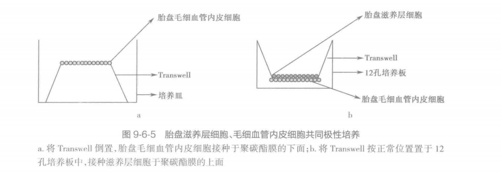
C. Establishment of co-polarized culture model of trophoblast and capillary endothelial cells The co-culture model requires the use of a Transwell with a polycarbonate membrane with a 3.0 μm pore size throughout the bottom of the Transwell, which is used in conjunction with a 12-well plate. The Transwell is used in conjunction with a 12-well culture plate. The polycarbonate membrane is first coated with Matrigel and placed upside down in a Petri dish, inoculated with placental capillary endothelial cells underneath the polycarbonate membrane (Fig. 9-6-5a) at a cell density of 1 × 10/cm2, and added to the DMEM medium. 24 hours later the Transwell is placed in its normal position in the 12-well culture plate, and trophoblast cells are inoculated on top of the polycarbonate membrane (Fig. 9-6-5b). (Figure 9-6-5b), the cell density was 1 × 10/cm2, the medium was unchanged, and the medium was changed once every 2 days. 7 days later, the polycarbonate membrane was fixed in cold acetone, embedded in OCT, and sliced on the polycarbonate membrane, and the growth of the two kinds of cells was observed by HE staining; meanwhile, the slices were used for rabbit immunohistochemistry staining, electron microscopy to observe the ultrastructure, and so on.