This product is a dye reagent for visualizing protein bands in SDS-PAGE gel electrophoresis. It offers convenience and high sensitivity, comparable to silver staining (detection limit: 0.1 ng/band), without requiring additional special steps. Simply treat your protein samples as you would with standard loading buffer. After electrophoresis, results can be directly visualized using a gel imager's UV or LED light source.
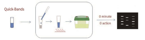
Protocol
1. Reconstitution: Before use, resuspend the contents of the brown tube with the specified volume of Resuspension Buffer (provided on the tube label) to prepare the Quick-Bands Protein Sample Treatment Solution.
Note: Resuspension Buffer may form precipitates at 4°C. Allow it to warm to room temperature until precipitates dissolve completely.
2. Sample Mixing: Mix the prepared Quick-Bands Protein Sample Treatment Solution with your protein sample at a 1:2 ratio (e.g., 3 µL Quick-Bands + 6 µL protein sample).
3. Heat Denaturation: Heat the mixed sample at 90-100°C for 5 minutes. For cell or tissue samples, extend the heating time to 10 minutes. Ensure the temperature exceeds 90°C for proper denaturation.
Note for Precast Gels: Most precast gels (except Bis-Tris system gels) can proceed directly to step 4. For Bis-Tris system precast gels (e.g., from Life Technologies), add Enhancing Buffer to a final concentration of 20% of the treated sample volume (e.g., add 2 µL Enhancing Buffer to 10 µL treated sample). Enhancing Buffer must be purchased separately.
4. Electrophoresis: Load the processed samples directly onto the gel for electrophoresis (no additional Loading Buffer is required). If needed, load 3 µL of a ready-to-use fluorescent protein marker per well.
5. Visualization & Imaging: After electrophoresis, place the gel on a transilluminator for observation and photography. Compatible light sources include UV lamps, blue LED lights, or other gel imaging systems. If using a visible light source, the gel does not need to be removed from its plastic/glass plates, as visible light can penetrate them.
6. Optional Coomassie Staining: Gels treated with Quick-Bands can subsequently be stained with Coomassie Blue if desired, following standard Coomassie staining protocols.
Usage Tips
1. Gels stained with Quick-Bands can still undergo conventional Coomassie Blue staining.
2. For imaging gels between glass plates, do not use UV light. Use reflected green light only with an exposure time of 6 seconds, as glass blocks UV transmission.
3. Quick-Bands is compatible with Western Blotting and does not interfere with subsequent antigen-antibody reactions. After transfer, both the gel and the membrane can be directly visualized under UV or LED light to assess transfer efficiency.
4. Quick-Bands is suitable for: checking protein expression via SDS-PAGE, assessing protein purity, tracking target proteins during purification, visualizing control samples in Western Blot experiments, and detecting immunoprecipitation results.
5. Quick-Bands is NOT suitable for excising gel bands intended for protein sequencing, mass spectrometry analysis, or antibody production.
6. You can use either fluorescently labeled protein molecular weight standards or protein molecular weight standards treated with Quick-Bands (and not pre-treated with other loading buffers).
Storage & Stability
1. Upon receipt, store the product immediately at -20°C protected from light.
2. Quick-Bands can be stored at -20°C for 12 months, at 4°C for 8 weeks, or at room temperature for 1 week.
3. If Quick-Bands is stored at room temperature for over one week, add DTT to a final concentration of 20 mM.
Factors Affecting Performance
1. pH: Low pH may reduce the staining efficiency of Quick-Bands. If the sample pH is below 5, adjust it to pH 7 or higher.
2. Staining with Quick-Bands is generally unaffected under standard experimental conditions.
3. Reagents that can interfere with Quick-Bands staining include: Glycerol (>20%), Urea (>8 M), DTT (>50 mM), L-Arg (>0.5 M).
Frequently Asked Questions (FAQ)
Q: Why can't I stain my purchased protein molecular weight standard with Quick-Bands?
A: Most ready-to-use protein markers are not compatible with Quick-Bands. Do not treat these markers with Quick-Bands. You can purchase fluorescent protein markers from our company or use Quick-Bands to treat protein marker powders (e.g., lyophilized markers) not pre-treated with other loading buffers.
Q: How stable is Quick-Bands?
A: Quick-Bands itself is stable. However, the deterioration of reducing agents (like DTT) in the system over time can cause some protein bands to become less sharp and bright. Typically, Quick-Bands can be stored at room temperature for 1 week. If stored longer, adding fresh DTT (final concentration not exceeding 20 mM) can restore band sharpness and intensity.
Q: Do post-translational protein modifications affect Quick-Bands?
A: No.
Q: Why do protein bands sometimes appear less sharp and bright?
A: This is likely due to protein oxidation. Add a reducing agent (e.g., DTT) to the system. The final concentration of DTT should not exceed 20 mM.
Q: Can samples treated with Quick-Bands be used for Western Blotting?
A: Based on extensive testing, Quick-Bands does not interfere with Western Blot experiments.
Q: Why does Quick-Bands stored at 4°C sometimes form a precipitate?
A: The precipitate is SDS. Warm the solution to room temperature to dissolve it.
Experimental Data Presentation
.png)
.png)

