GRADE & PURITYAzide Free?Azide-free — without sodium azide preservative. Use in conjugations, cell work, or assays where azide is toxic or inhibitory.Ex:498nm?Fluorescence excitation maximum at 498 nm — the wavelength of light that best excites this fluorophore. Match it to your instrument's laser or filter set for the strongest signal.Em:517nm?Fluorescence emission maximum at 517 nm — the wavelength this fluorophore emits after excitation. Use it to pick the right detection filter and avoid channel cross-talk.Streptavidin Concentration: 1 mg/mL
Azide Free, Ex:498nm, Em:517nm, Streptavidin Concentration: 1 mg/mL Azide Free,Em:517nm,Ex:498nm for sensitive chromatographic and analytical workflows requiring minimal baseline interference.
🌡
Storage & shipping
Store at 2-8°C,Protected from light,Do not freeze Ships Wet ice,Do not freeze Check lot-specific COA for exact specifications.
📋
Quality documents
SDS, COA, datasheet, and spec sheet available for download. Lot-specific COA accessible via lot number lookup.
📚
Literature proof
Cited in 0 peer-reviewed publications across chromatography, organic synthesis, and cross-coupling reactions.
Overview
Background
Streptavidin is a tetrameric bacterial protein isolated from Streptomyces avidinii providing 4 high-affinity biotin binding sites. Streptavidin homo-tetramers have an extraordinarily high affinity for biotin. With a dissociation constant on the order of ≈10⁻¹⁴ mol/L, the binding of biotin to streptavidin is one of the strongest non-covalent interactions known in nature. Unlike egg-white avidin, which has a net positive charge at neutral pH and contains about 7% carbohydrate, streptavidin has almost no net charge at neutral pH, does not contain carbohydrate, and exhibits lower non-specific background. Streptavidin conjugates are widely used together with a conjugate of biotin for specific detection of a variety of proteins, protein motifs, nucleic acids and other molecules. This FITC-streptavidin conjugate was prepared by highly purified Streptavidin and free FITC was removed. Streptavidin (FITC) is a useful second-step reagent for the indirect immunofluorescent staining of cells in combination with biotinylated primary antibodies for flow cytometric analysis. Excitation at 488nm light leads to a fluorescence emission maximum of 520 nm.
Recommended Usage:
Every lot of Streptavidin-FITC is tested by flow cytometry using biotinylated primary antibodies. From this testing it is recommended that between 0.02 and 0.25 µg of streptavidin be used per 106 cells in a 100 µl staining volume.
Specifications
Product Name
Streptavidin protein (FITC) used for Flow Cytometry; IF; various applications
Shipped at 4℃. Store at 4°C and protected from prolonged exposure to light. Do not freeze.
Images
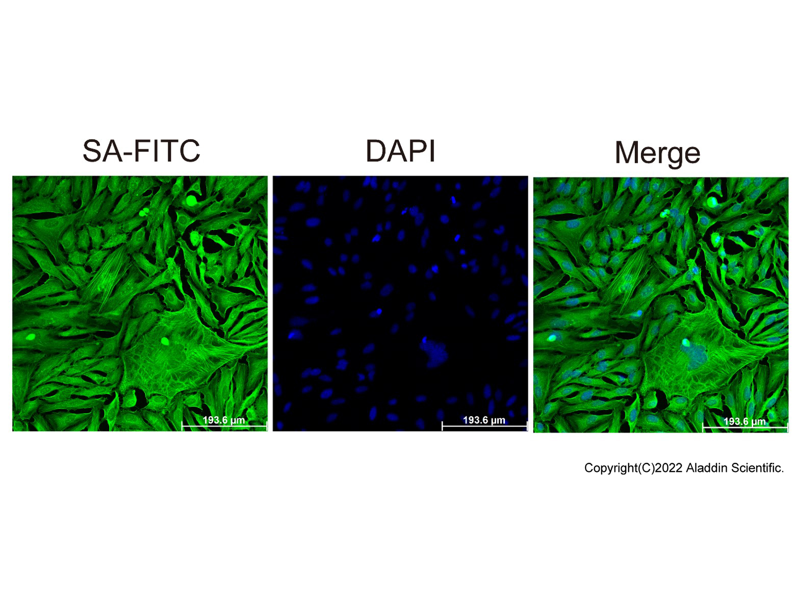
ICC/IF-Streptavidin protein (FITC) (rp155876) ICC/IF with Streptavidin protein (FITC) (rp155876) stained HeLa cells. The cells were methanol fixed (5 min) and blocked with 1% BSA . The cells were then incubated with Biotin-γ-actin antibody overnight at 4°C. Then Streptavidin protein (FITC) rp155876 (green) was used at 1:1000 for 1h. DAPI was used to stain the cell nuclei (blue) .
ICC/IF-Streptavidin protein (FITC) (rp155876) ICC/IF with Streptavidin protein (FITC) (rp155876) stained HeLa cells. The cells were methanol fixed (5 min) and blocked with 1% BSA . The cells were then incubated with Biotin-γ-actin antibody overnight at 4°C. Then Streptavidin protein (FITC) rp155876 (green) was used at 1:1000 for 1h. DAPI was used to stain the cell nuclei (blue) .
Flow-Streptavidin protein (FITC) (rp155876) BALB/c splenocytes stained with Biotin-CD45R antibody, followed by Streptavidin protein (FITC) (rp155876) at 1/2500 dilution.
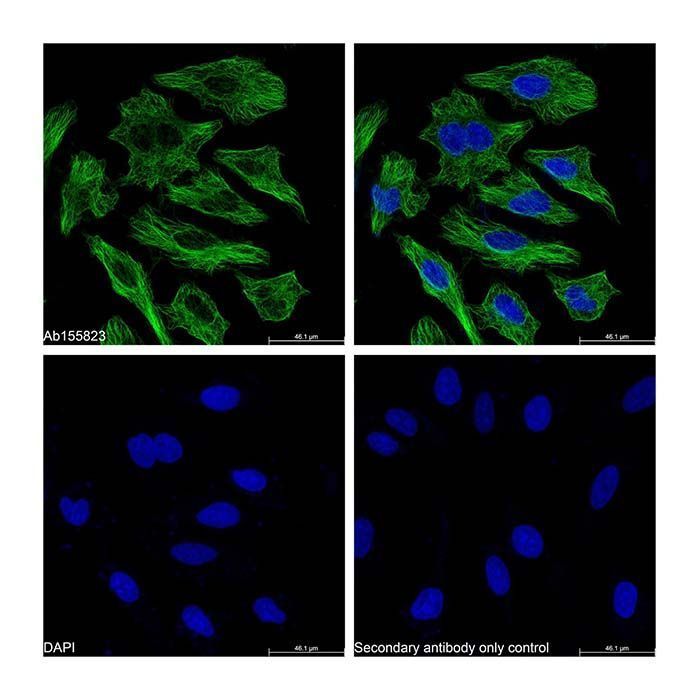
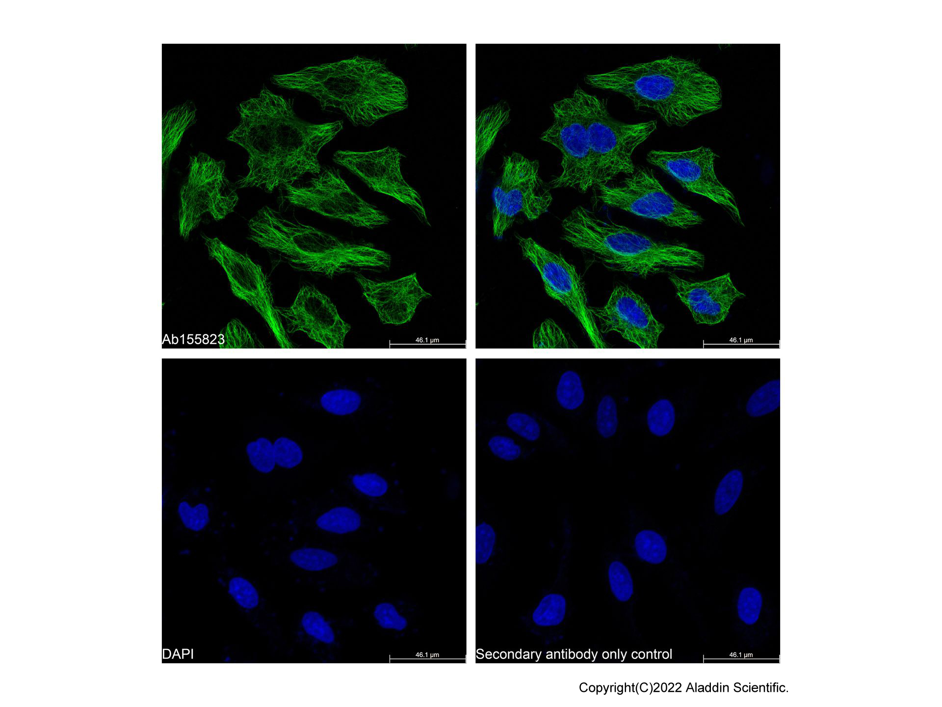
Streptavidin Protein (FITC) (rp155876) - IF IF analysis of beta Tubulin (green) in HeLa cells. The cells were fixed and permeabilized with 100% methanol for 5 minutes, and blocked with 2% BSA for 1 hour at room temperature. Cells were stained with beta Tubulin Mouse mAb (Ab155823) at 1/2000 dilution in blocking buffer overnight at 4°C, and then incubated with Goat Anti-Mouse IgG H&L (Biotin) (Ab179002) at a dilution of 1/1000 for 1 hour at room temperature in the dark. Then incubated with Streptavidin Protein (FITC) (rp155876) at a dilution of 1/1000 for 1 hour at room temperature in the dark (green). Cells were counterstained with DAPI (blue). PBS instead of the primary antibody was used as the secondary antibody only control. Images were taken on the confocal laser scanning microscope.
Application
Application
Dilution info
Flow Cytometry
1:500-1:5000
IF/ICC
1:50-1:500
Documentation
📋 Safety Data Sheet (SDS)
Comprehensive hazard, handling, storage, and regulatory compliance document.
We use cookies to ensure the website functions properly and, where permitted, to improve your experience. You can manage your preferences at any time in Settings. Learn more in our Cookie Policy.
Shall we send you a message when we have discounts available?
Remind me later
Thank you! Please check your email inbox to confirm.
Products are supplied to verified businesses, institutions, and qualified professionals for research and development use only. Not for use in humans, animals, diagnosis, or therapy.



 ZJ24F1216185