
fluorescence immunochromatography
fluorescence immunochromatography
Fluorescent Immunoassay (FIA) is an analytical technique based on antibody-antigen specific recognition to detect and quantify mobile target molecules by fluorescent signals. The technique combines several disciplines such as immunology, fluorescence and biochemistry, and has the advantages of high sensitivity, high specificity, rapidity and simplicity.
Principle
Fluorescence immunochromatography uses a strip of fibrous chromatographic material with a detection line (coated antigen, T-line) and a quality control line (anti-antibody, C-line) as the stationary phase, a test solution as the mobile phase, and a fluorescently labeled antibody or antigen immobilized on a connecting pad, which moves the analyte to be tested over the chromatographic strip by capillary action.
For macromolecular antigens (proteins, viruses, etc.) with multiple antigenic determinants, a "sandwich" type double-antibody sandwich immunochromatographic method is usually used, i.e., the analyte to be tested binds to the fluorescent-labeled antibody first under the action of the mobile phase, and then binds to the encapsulated antibody to form a double-antibody sandwich of the "sandwich" type when it reaches the detection line.
When the test is performed, the sample solution will be chromatographed upward under capillary effect, and two kinds of results will appear: if it is negative, the sample does not bind to the fluorescent antibody, and the fluorescent antibody binds to the sample-specific coupler, and the result of the accompanying instrument will be negative; if it is positive, the sample and the fluorescent antibody react first, and the fluorescent antibody no longer binds to the sample-specific coupler in the test area when the antigen content of the sample reaches a certain amount, and the result of the accompanying instrument will be positive. The test result of the companion instrument is positive.
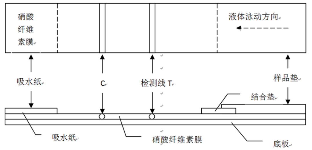
Diagram of the structure of fluorescent immunochromatographic test strips
Operation method
Fluorescence immunochromatography
Principle
Fluorescence immunochromatography uses a strip of fibrous chromatographic material with a detection line (coated antigen, T-line) and a quality control line (anti-antibody, C-line) as the stationary phase, a test solution as the mobile phase, and a fluorescently labeled antibody or antigen immobilized on a connecting pad, which moves the analyte to be tested over the chromatographic strip by capillary action. For macromolecular antigens (proteins, viruses, etc.) with multiple antigenic determinants, a "sandwich" type double-antibody sandwich immunochromatographic method is usually used, i.e., the analyte to be tested binds to the fluorescent-labeled antibody first under the action of the mobile phase, and then binds to the encapsulated antibody to form a double-antibody sandwich of the "sandwich" type when it reaches the detection line. When the test is performed, the sample solution will be chromatographed upward under capillary effect, and two kinds of results will appear: if it is negative, the sample does not bind to the fluorescent antibody, and the fluorescent antibody binds to the sample-specific coupler, and the result of the accompanying instrument will be negative; if it is positive, the sample and the fluorescent antibody react first, and the fluorescent antibody no longer binds to the sample-specific coupler in the test area when the antigen content of the sample reaches a certain amount, and the result of the accompanying instrument will be positive. instrument test result is positive. Diagram of the structure of fluorescent immunochromatographic test strips
Materials and Instruments
Reagents: nitrocellulose membrane (NC membrane), sample pad, polyester membrane, PVC backing, absorbent paper, positive anti-mouse IgG, samples to be tested, fluorescent microspheres, Move (1) Sample preparation: The samples to be tested (e.g. serum, urine, cell extracts, etc.) will be treated and diluted as necessary to meet the needs of subsequent experiments. (2) Preparation of the kit: apply the treated fluorescent microspheres to the binding pad with a certain amount of spraying; process the NC membrane, spray the T-line and C-line at the appropriate position, and dry it overnight in an incubator at 30 ℃; then paste it onto the PVC board in the order of sample pad, binding pad, NC membrane, and absorbent pad in sequence, and then cut it into 4 mm wide strips of test strips with a high-speed chopping machine, assembled on the shell, and dry and stored in a vacuum packaging machine. (3) Sample addition: add the prepared sample into the kit, and bind with fluorescent labeled antibody to form antigen-antibody complex. (4) Washing: Through the washing step, the non-specifically bound substances are removed and the specifically bound complexes are retained. (5) Signal Detection: Through a fluorescence detector, the fluorescence signal of the fluorescein substrate is detected to quantify the content of the target molecule. (6) Data analysis: Based on the fluorescence signal intensity of the sample, combined with the standard curve or other calculation methods, the content of the target molecule is calculated. Caveat (1) Disinfection of instruments: instruments required for the experiment should be wiped clean with alcohol cotton pads and preferably autoclaved to avoid cross-contamination and affecting the experimental results.(2) Pay attention to your own protection: appropriate protective measures should be taken during collection, disposal, storage, mixing of samples and testing.(3) The reagent kit should be complete: please do not open the reagent kit (test card) before use, and do not use the reagent kit with obvious damage or the test card with broken package; in addition, the reagent kit (test card) is a single-use in vitro diagnostic reagent, so do not reuse it.(4) Reagents and ID cards should be matched: reagents of different lot numbers should not be mixed, and ID cards and kits (test cards) should not be used with mixed lot numbers.(5) Use the matching instrument: the reagent kit (test card) and its components are only applicable to the matching rapid analyzer.(6) Pay attention to avoid damage or contamination of the instrument: Do not insert the reagent kit (test card) whose surface is wetted by other liquids into the detector, otherwise it will contaminate or damage the instrument; please dispose of the used reagent kit (test card) properly and do not dispose of it casually.(7) Temperature requirements: Avoid the high temperature of the experimental environment, the reagent kit (test card) stored at low temperature needs to be restored to room temperature before opening, so as to avoid moisture absorption.(8) Standardize the operation of the instrument: the reagent kit (test card) and the supporting analyzer should be used to avoid vibration and electromagnetic environment (vibration of the instrument itself in normal use is a normal phenomenon); do not pull out the ID card when the test is in progress.(9) Requirements on testing time: To ensure accurate test results, the off-line reaction time and result reading time of the kit (test card) should be carried out in strict accordance with the instruction manual of the kit, and the results will be invalid after exceeding the specified time. Common Problems (1) Weak or no signal: This may be due to improper sample handling, unstabilized reagents, poor antibody quality, incomplete washing step, insufficient concentration of fluorescein substrate, etc. (2) Non-specific binding: may be due to the presence of cross-reactants in the sample, poor quality of antibodies in the kit, expired reagents, etc. (3) Standard curve does not meet the requirements: it may be due to improper preparation of standards, insufficient concentration of fluorescein substrate, inaccurate reading of photometer, etc. (4) Poor experimental reproducibility: it may be due to inconsistent experimental conditions, differences in antibody batches, inconsistent quality of fluorescein substrate and other reasons. (5) Unstable experimental results: may be due to unstable laboratory conditions such as temperature, humidity, light, etc., improper storage of reagent kits, poor stability of fluorescein substrate and other reasons. For more product details, please visit Aladdin Scientific website.
Instruments: Immunofluorescence detector, Spot film gold sprayer, ultrasonic cleaner, constant temperature incubator.