In vitro modeling of the blood-brain barrier
In vitro modeling of the blood-brain barrier
Summary
The Blood-Brain Barrier (BBB) is a highly specialized biological barrier composed of microvascular endothelial cells, basement membranes, and surrounding astrocytes, which protects the Central Nervous System (CNS) from external harmful substances.
Human brain microvascular endothelial cells are an important cell type in the construction of the blood-brain barrier. Their main role is to restrict the passage of most drugs, pathogens and other harmful substances through intercellular tight junctions and specialized transport channels, while retaining selective permeability to neurotrophins and metabolites.
Principle
Human brain microvascular endothelial cells are co-grown with surrounding astrocytes and other neural tissue cells through in vitro culture to form a biological barrier similar to the real blood-brain barrier.
Appliance
Research in the areas of drug permeability, neurotrophin transport mechanisms, neuroinflammatory and neurodegenerative diseases.
Operation method
Construction of the blood-brain barrier by human brain microvascular endothelial cells
Principle
Human brain microvascular endothelial cells are co-grown with surrounding astrocytes and other neural tissue cells through in vitro culture to form a biological barrier similar to the real blood-brain barrier.
Materials and Instruments
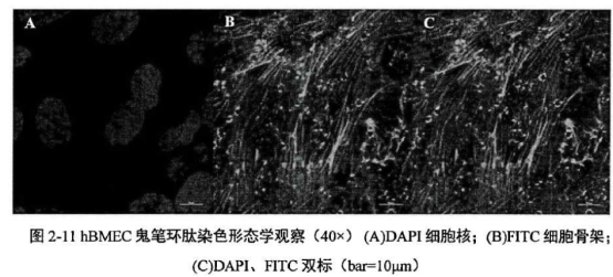
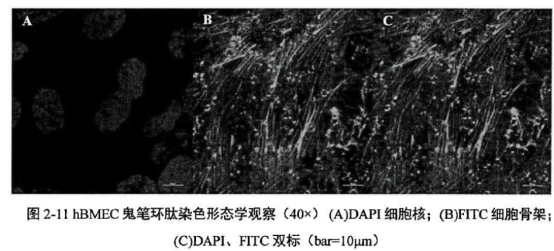
hBMEC, DMEM culture medium, cell culture incubator Move 1. hBMEC were cultured in DMEM culture medium containing 10% FBS and 1% double antibody at 37 ℃ in a 5% CO2 cell culture incubator, and the culture medium was changed every 2 days. hBMEC were passaged after 2 days of 100% growth, digested with 0.25% EDTA-trypsin and blown into single floating cells, which started to attach to the wall about 1 h after inoculation with new flasks. 2. Collagen type I cells were cultured in Transwell chambers, and the culture medium was changed every 2 days. 2. Collagen type I coating Transwells (1) Prepare 100 mL of 0.006 mol/L (0.36 g/L) HAC solution: take 34.5 ul of glacial acetic acid and add it to 100 ml of ddH20, mix it well, then use a 0.22 μm filter to remove the bacteria and wait for use. (2) Prepare 0.1 mg/mL Collagen Storage Solution: Take 1 mL of 5 mg/mL Collagen and add 49 mL of prepared, filtered and sterilized 0.006 mol/L HAC solution to a 50 mL centrifuge tube, gently invert it, and dispense it into 15 mL centrifuge tubes to store at 4 °C. (3) Preparation of working solution and Transwell encapsulation: The Transwell is encapsulated at 10 μg/cm2 . Dilute the stock solution into 0.06 mg/mL working solution, i.e., 1000 μL of 0.1 mg/mL stock solution was added to 800 μL of 0.006mol/L HAC. (4) Add 187 μL of collagen working solution to each well of the Transwell to ensure that the collagen solution spreads over the surface of the vessel, and dry overnight at room temperature on the ultra-clean bench. Rinse with DMEM culture solution before inoculating cells. The collagen-coated Transwells should be stored in a 12-well plate at 4 °C for at least 3 months. 3. Establishment of the monolayer hBMEC barrier model The hBMEC grown to 100% confluence in T25 cell vials were digested with 0.25% EDTA-trypsin, resuspended in DMEM culture medium containing 10% FBS and 1% double antibody, and inoculated into the upper chamber of Collagen type I-coated Transwells at a density of 500 μL at a density of 5x10⁵/mL, and the lower chamber was inoculated with 1.5 mL of DMEM. Add 1.5 mL of DMEM in the lower chamber to ensure that the liquid level inside and outside of the Transwell is even, and change the complete culture medium every 2 days. 4. 4. Observation on the growth of single-layer hBMEC barrier model (1) HE staining of monolayer hBMEC: Observe whether the cells are successfully inoculated and grown on the polymeric membrane, as well as the number and density of cells by HE staining. hBMEC was inoculated in Transwell for 4~6 days, one well was fixed with 4% paraformaldehyde for 15 min at room temperature, rinsed with ddH20 for 3 times and then stained by HE staining, and then cut off the polymeric membrane and fixed it on the slide, and then observe the growth of cells on the membrane by ordinary light microscope. Cell growth on the membrane was observed by normal light microscope. (2) Monolayer hBMEC ghost peptide staining: Since HE staining could only observe whether the cells grew on the polymeric membrane and the approximate density, and could not reflect the microstructure of hBMEC well, the cytoskeleton was stained with ghost peptide to observe the closeness of the cell-to-cell growth and the microstructure of the cells on the polymeric membrane. That is, 4% paraformaldehyde was fixed for 15 min at room temperature, PBS was rinsed for 3 times, and then 100~200 μL of the cytoskeleton staining solution was added to each well. Incubate at 37 ℃ for 30 min, and then cut off the polymeric membrane and fix it on slides for observation by confocal microscopy. 5. Determination of barrier function Starting from the fourth day after inoculation, 500 ul of culture medium was added to each of the upper and lower chambers at the time of fluid change every two days, which could form an obvious level difference (about 0.5 m). If no level difference could be formed in the Transwell without inoculation, it meant that the cells had been grown to the confluence and were sufficiently compact. The transmembrane resistance (TEER) of both sides of the Transwell was measured using a cytoresistivity meter before each change of fluid, and the TEER over time curve was plotted. When the curve rose significantly and entered the plateau period, it indicated that the barrier had formed. Caveat 1) Select human brain microvascular endothelial cells from reliable sources to ensure the reliability and stability of the model;2) Establish appropriate cell culture conditions, including the formulation of culture medium, temperature, humidity and gas content;3) Strictly control the cell density and culture time during the culture process to ensure the consistency and stability of the model;4) After the model is built, it should be verified and evaluated to ensure its ability to simulate the real blood-brain barrier.References:Pang Bo. Establishment of in vitro blood-brain barrier cell model by co-culture of human brain microvascular endothelial cells and astrocytes[D]. Nanjing Agricultural University, 2018.DOI:10.27244/d.cnki.gnjnu.2018.000460.Cha Yufeng, Fu Xiaozhong, Zhang Shun, Luo Min, Ou Yu, Dong Yongxi, Wang Aimin, Wang Yonglin. Co-culture of rat brain microvascular endothelial cells with pericytes and astrocytes to establish an in vitro blood-brain barrier model[J]. Chinese Pharmacology Bulletin, 2015,31(05):730-735.HU Limin, FAN Xiang, ZHANG Yanjun, ZHANG Boli, MEI Jianxun, GAO Xiumei. Establishment of an in vitro model of blood-brain barrier by co-culture of rat brain microvascular endothelial cells and astrocytes[J]. Tianjin Traditional Chinese Medicine, 2005(02):149-151. For more product details, please visit Aladdin Scientific website.
EDTA-trypsin, HAC solution, glacial acetic acid, ddH
2
0
Collagen storage solution, working solution and 0.4 μm pore size Transwell chambers
Plates and dishes, T25 cell culture flasks
4% paraformaldehyde, HE Stain, Ghost Pen Cyclic Peptide Stain
Microscope, slides, coverslips