
Western blotting assay for protein detection
Western blotting assay for protein detection
Western blotting, i.e. protein immunoblotting.
Principle
The basic principle of Western blotting for protein detection is that the protein separated by polyacrylamide gel is transferred to nitrocellulose membrane, and then the membrane is interacted with specific antibody, which can be further detected after binding to the bands of protein on the membrane, such as enzyme-labeled secondary antibody or biotin-labeled secondary antibody, which can be further developed with the color-emitting substrate.
Operation method
Protein immunoblotting
Principle
The basic principle of Western blotting for protein detection is that the protein separated by polyacrylamide gel is transferred to nitrocellulose membrane, and then the membrane is interacted with specific antibody, which can be further detected after binding to the bands of protein on the membrane, such as enzyme-labeled secondary antibody or biotin-labeled secondary antibody, which can be further developed with the color-emitting substrate.
Materials and Instruments
Equipment: Move The basic procedure of the Western blotting experiment for protein detection can be divided into the following steps: (i) Protein extraction A Wash the cultured cells with PBS once.B 0.025% trypsin was used to digest the cells until the cells were loose, and the trypsin was poured off.C PBS blown cells and collected in centrifuge tube.D Transfer to Eppendorf tube and wash once with PBS.E Add 100 μl of cell lysate and beat well.F Ice bath for 20 min.G Centrifuge at 13000 r/min for 5 min.H Aspirate the supernatant and add an equal amount of sample buffer.I Boil and denature for 5 min and store at -20 ℃. (ii) Electrophoresis A Filling the gel Place the electrophoresis glass plate on the electrophoresis tank, fill 15 ml of 8% separating gel, cover the separating gel with 5 ml of n-butanol or 0.1% SDS; after the gel solidifies, pour off the covering material, and rinse the upper part of the gel with deionized water for the total protein extract of the cells; fill 8 ml of the layered gel with the separating gel, and insert the comb.B Sampling 1 ml of sampling buffer add 9 μl of mercaptoethanol; the sample to be tested and sampling buffer mixed at 1:2, boiling at 100 ℃ for 5 min; after solidification of the layer gel, pull out the comb, electrophoresis buffer rinsing the comb empty; will be the protein molecular mass standard reference and the sample sample on the sample.C Electrophoresis The sample side was connected to the negative electrode with a current of 10 mA. After the sample entered the separation gel, the current was increased to 20 mA and electrophoresis was carried out for about 4-6 h until the dye reached the bottom of the separation gel. (iii) Membrane transfer A Remove the gel, cut off the upper left corner for marking, and equilibrate the gel, PVDF membrane and filter paper in membrane transfer buffer for 30 min.B Install the membrane transfer device (Figure 8-4) as follows. Caveat Western blotting combines the high resolution of gel electrophoresis with the specific sensitivity of solid-phase immunoassay, allowing the detection of proteins down to 1-5 ng. When transferring the membrane, it should be noted that the nitrocellulose membrane and the 3 MM filter paper should be moistened with transfer buffer beforehand, and both the membrane and the filter paper should not be held directly by hand because the oils and secretions on the skin will prevent the transfer of proteins from the gel to the membrane. The size of the filter paper and membrane should be cut down to the same size as the gel. If the area of the filter paper or membrane is larger than that of the gel, the edges of the filter paper and membrane may come into contact with each other and cause a short circuit. After the membrane is interacted with primary and secondary antibodies, it should be rinsed clean with buffer. According to the different molecular weights of proteins, different membrane transfer devices can be used, and the time of membrane transfer varies with the different membrane transfer devices, such as using a semi-dry carbon plate transfer electrophoresis tank, 150 mA only needs to be transferred for about 2 h. The membrane transfer time can be adjusted according to the different molecular weights of proteins. For more product details, please visit Aladdin Scientific website.
Nitrocellulose membrane, Whatman 3 MM filter paper, Electrotransfer meter.
Reagents:
① primary antibody
② Secondary antibody
③ Sheep serum
① Primary antibody ② Secondary antibody ③ Sheep serum ④ ECL development kit
⑤ Transfer buffer
(12 g of Tris base, 57.65 g of glycine dissolved in methanol, then add water to 1 L and adjust the pH to 8.3).
⑥ TTBS 10 mmol/L Tris (pH 8.3), 150 mmol/L NaCl, 0.05% Tween-20.
(vii) 0.5% Reichun Red S dye solution (Reichun Red S0.5).
(0.5 g of Li Chun red S, 1 ml of glacial acetic acid, dissolve and add water to 100 ml, prepare before use.)
⑧ Closed buffer
(Skimmed milk powder 10 g; TTBS 70 ml; fully dissolved and fixed to 100 ml, add 10% sodium azide 200 μl (final concentration of 0.02%), 4 ℃ storage).
⑨ DAB color development solution
(0.01 mol/L TBS (pH 7.6) 9 ml; DAB, 6 mg; 3 g/L NiCl2 or CoCl2, 1 ml; filtered through filter paper and added with 10 μl of 30% H2O2, mixed well and used immediately).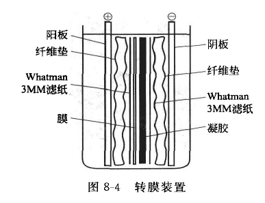
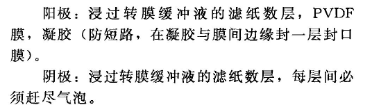 C Turn on the power, constant current, select the current according to the membrane size, ≤ 0.8 mA/c㎡, membrane transfer for 6 hours.D Remove the PVDF membrane, mark the cutout at the upper left corner, and wash the membrane with TTBS for 5 min, 3 times to remove Tris and glycine.E Lichun red S staining for 5 min, water decolorization for 2 min, you can see the electrophoretic bands on the membrane, according to the protein molecular mass standard reference, in the middle of the corresponding molecular mass with the measured protein actin cut the membrane into two halves, and then decolorization for 10 min, so that it fades completely.F Put the membrane into a heat-sealable plastic bag, add the sealing solution according to the area of the membrane, 0.1 ml/c ㎡, seal it, and leave it at 4 ℃ overnight. (iv) Antibody reaction A After overnight, wash the membrane with TTBS for 10 min, 3 times.B Add the primary antibody diluted with blocking solution, the antibody dilution is 1:200~1:2000 (depending on the potency of the antibody), and incubate at 25 ℃ with gentle shaking for 1 h. The primary antibody is diluted with the blocking solution, the antibody dilution is 1:200~1:2000 (depending on the potency of the antibody).C Wash the membrane with TTBS for 10 min, 3 times.D Transfer the membrane into another heat-sealed plastic bag, add the secondary antibody working solution (1:1000), 0.1 ml/c㎡, incubate at 25 ℃ for 45 min with gentle shaking.E Wash the membrane with TTBS for 10 min, 3 times.F ECL kit developed.