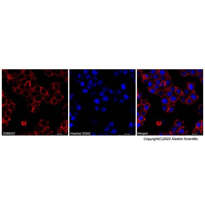
DiD, DiO, DiI, DiR and DiS dyes are a family of lipophilic fluorescent dyes used for staining cell membranes and other lipid-soluble biological structures. Their fluorescence intensity is greatly enhanced upon membrane binding, and these dyes exhibit high quenching constants and long excited-state lifetimes. Once cells are stained, the dyes diffuse across the entire cell membrane, and at optimal concentrations, uniformly stain the whole membrane. They display distinct fluorescence colors: DiI (orange fluorescence), DiO (green fluorescence), DiD (red fluorescence) and DiR (deep-red fluorescence), enabling multicolor imaging and flow cytometry analysis of live cells. DiI and DiO can be used with standard FITC and TRITC filter sets, respectively. DiD is excitable by a 633 nm He-Ne laser and has longer excitation and emission wavelengths than DiI, making it more valuable for cell and tissue staining. The infrared fluorescence of DiR penetrates cells and tissues, allowing for in vivo imaging and tracing applications.
Product Parameters
Ex/Em (nm): 549/565 nm
Note: Di-series dyes are lipophilic and tend to precipitate in aqueous buffers. It is recommended to add a surfactant such as F127 or Tween-20 to a final concentration of 0.1%.
Protocol
1. Preparation of DiD, DiO, DiI, DiR and DiS Cell Membrane Staining Solutions
(1) Preparation of stock solutions in DMSO or EtOH:Prepare stock solutions at a concentration of 1-5 mM using DMSO or ethanol.
Note: Unused stock solutions should be stored at -20°C and avoid repeated freeze-thaw cycles.
(2) Preparation of working solutions:Dilute the stock solution with an appropriate buffer (e.g., serum-free medium, HBSS or PBS) to prepare a working solution at 1-5 µM.
Note: The final working concentration should be optimized based on specific cell types and experimental conditions. Optimal conditions may be determined starting from 10 times the recommended concentration.
2. Staining of Suspension Cells
(1) Add suspension cells at a density of 1 × 10⁶/mL to the working solution.
(2) Incubate the cells at 37°C for 2-20 minutes; optimal incubation time varies by cell type.
(3) Centrifuge the stained cell suspension at 1000-1500 rpm for 5 minutes.
(4) Discard the supernatant and gently add pre-warmed (37°C) culture medium.
(5) Repeat steps (3) and (4) at least twice.
3. Staining of Adherent Cells
(1) Culture adherent cells under sterile conditions.
(2) Remove coverslips from the culture medium, aspirate excess medium, and place coverslips in a humid environment.
(3) Add 100 μL of dye working solution to one corner of the coverslip and gently tilt to ensure uniform coverage of all cells.
(4) Incubate the cells at 37°C for 2-20 minutes; optimal incubation time varies by cell type.
(5) Aspirate the dye working solution and wash the coverslip 2-3 times with pre-warmed medium. For each wash, cover cells with medium, incubate for 5-10 minutes, then aspirate the medium completely.
4. Microscopy Detection
(1) Filter sets for DiI: XF32 (Omega), 31002 (Chroma)
(2) For simultaneous detection of multiple dyes, use the following filter sets:a. DiI & DiO: Omega XF52, Chroma 51004b. DiI & DiD: Omega XF92, Chroma 51007c. DiI, DiO & DiD: Omega XF93, Chroma 61005
5. Flow Cytometry Detection
Cells stained with DiD, DiO, DiI, DiR and DiS can be detected using conventional flow cytometers with FL1, FL2, FL3 and FL4 channels, respectively.

 ZJ26F0535136